© Mary Ann Liebert, Inc. Pp. 281–287
DOI: 10.1089/pho.2007.2149
Antimicrobial Photodynamic Action on Dentin
Using a Light-Emitting Diode Light Source
Juçaíra S.M. Giusti, Ph.D.,
1Lourdes Santos-Pinto, Ph.D.,
1Antonio C. Pizzolito, Ph.D.,
2Kristian Helmerson, Ph.D.,
3Eurico Carvalho-Filho, B.Sc.,
4Cristina Kurachi, Ph.D.,
4and Vanderlei S. Bagnato, Ph.D.
4Abstract
Objective:
The aim of this study was the evaluation of two different photosensitizers activated by red light
emitted by light-emitting diodes (LEDs) in the decontamination of carious bovine dentin.
Materials and Methods:
Fifteen bovine incisors were used to obtain dentin samples which were immersed in
brain-heart infusion culture medium supplemented with 1% glucose, 2% sucrose, and 1% young primary
cul-ture of
Lactobacillus acidophilus
10
8CFU/mL and
Streptococcus mutans
10
8CFU/mL for caries induction. Three
different concentrations of the Photogem solution, a hematoporphyrin derivative (1, 2, and 3 mg/mL) and two
different concentrations of toluidine blue O (TBO), a basic dye (0.025 and 0.1 mg/mL) were used. To activate
the photosensitizers two different light exposure times were used: 60 sec and 120 sec, corresponding
respec-tively to the doses of 24 J/cm
2and 48 J/cm
2.
Results:
After counting the numbers of CFU per milligram of carious dentin, we observed that the use of LED
energy in association with Photogem or TBO was effective for bacterial reduction in carious dentin, and that
the greatest effect on
S. mutans
and
L. acidophilus
was obtained with TBO at 0.1 mg/mL and a dose of 48 J/cm
2.
It was also observed that the overall toxicity of TBO was higher than that of Photogem, and that the
photo-toxicity of TBO was higher than that of Photogem.
Conclusion:
Based on our data we propose a mathematical model for the photodynamic effect when different
photosensitizer concentrations and light doses are used.
281 Introduction
P
HOTODYNAMIC THERAPY(PDT) is a technique that involvesthe activation of certain dyes (photosensitizers) by light in the presence of tissue oxygen, resulting in the production of reactive radicals capable of inducing cell death. Until re-cently, the main application of PDT was to treat malignant and some benign lesions;1,2however, the efficacy of this
pro-cedure for microbial reduction has been demonstrated in several studies.3–6This emerging class of applications, which
we refer to as antimicrobial photodynamic action (APDA), shows great potential.7–10
Currently, lasers are the most common light sources used to activate the photosensitizers. However, the development of bright light-emitting diodes (LEDs) provides an alterna-tive light source for PDT.11–13The light produced by an LED
has characteristics different from those of laser light: it is a
narrow-band non-coherent type of energy that is not dif-fraction-limited. In this respect it is similar to the light of a suitably filtered mercury lamp. LED devices have, in gen-eral, a lower cost and simpler technology compared to other laser devices, and moreover, they can be readily assembled in several configurations that are suitable for different types of anatomical illumination.
Studies from the 1990s have shown that a large number of types of oral bacteria, including periodontal pathogenic and cariogenic bacteria, are susceptible to photodynamic therapy.14–18However, the use of LED-based light sources
to activate photosensitizers for use against oral microorgan-isms is a recent development. Study of their use is impor-tant to promote the clinical use of LEDs instead of lasers for therapeutic applications in dentistry.19
Our study evaluated the efficiency of APDA using two different photosensitizers activated by LED energy for
de-1Faculdade de Odontologia de Araraquara , 2Faculdade de Ciências Farmacêuticas de Araraquara, Universidade do Estado de São Paulo (UNESP), Araraquara, SP, Brazil.
3National Institute of Standard and Technology (NIST), Gaithersburg, Maryland.
contaminating carious bovine dentin. The investigation of the decontamination induced in a tissue environment is rel-evant since the photosensitizer’s distribution and its inter-action with light will vary depending on the culture medium.
Materials and Methods
Fifteen bovine incisors without any visible structural anomalies were used in this study. The periodontal tissue was scaled and the buccal enamel was removed using a con-ical diamond bur in a high-speed handpiece. From each tooth, four dentin fragments were obtained using a titanium trephine drill. Each dentin fragment was air-dried for 60 sec and weighed; the average weight was 23 mg. The fragments were autoclaved for 20 min at 121°C and transferred to dividual test tubes, each containing 2 mL of a braheart in-fusion (BHI) culture medium 1 g/100 mL supplemented with glucose and 2 g/100 mL sucrose PA (Pro-analysis). For each 50 mL of medium solution, 5 mL of 108CFU/mL Lactobacil-lus acidophiLactobacil-lus(ATCC #ITAL-523) and 5 mL of 108CFU/mL Streptococcus mutans (ATCC #25175) were added to induce bacterial colonization. The test tubes were maintained in a micro-aerobic environment at 37°C for 14 d, with the solu-tion changed every 48 h. Following the 14-day period, the specimens were maintained under refrigeration at 4°C until treatment.
The specimens of the experimental groups were washed in sterile saline solution and then immersed for 60 sec in a solution containing either Photogem®(Photogem, Moscow,
Russia), a hematoporphyrin derivative, or toluidine blue O (TBO) (Sigma-Aldrich Chemie GmbH, Steinheim, Germany), a basic dye. Three different concentrations of Photogem so-lution, 1 mg/mL, 2 mg/mL, and 3 mg/mL, and two differ-ent concdiffer-entrations of TBO, 0.025 mg/mL and 0.1 mg/mL, were used. These solutions were prepared with distilled wa-ter at pH values of 7.4 for Photogem and 5.1 for TBO, as de-termined in previous studies using these
photosensitiz-ers.13,20
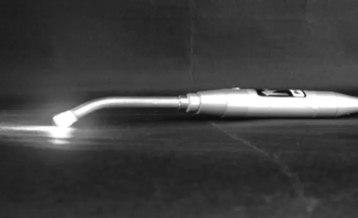
A homemade light source based on an LED emitter (LXHL-PD01; Luxeon, San Jose, CA, USA) for photosensi-tizer activation was developed. The generated light was op-tically collimated to a spot size 0.8 cm in diameter. The to-tal irradiance was 400 mW/cm2from an output power of
200 mW. The wavelength was centered at 630 nm with a bandwidth of⫾10 nm. To keep the semiconductor junction temperature below 60°C, the emitter was mounted on a heat sink constructed of a metallic base as shown in Fig. 1. Two fluence levels were investigated, 24 J/cm2and 48 J/cm2, with
exposure times of 60 sec and 120 sec, respectively.
In the control group, after the caries induction and with-out any treatment, the specimens were placed in 5 mL of saline solution. The individual action of the photosensitizer (Photogem or TBO) and of illumination was investigated at all experimental parameters. There were experimental groups using Photogem or TBO under different concentra-tions and no light exposure, and some non-sensitized spec-imens were illuminated with both fluences. For each group three samples were evaluated.
After treatment the specimens were placed into individ-ual test tubes with 5 mL of saline solution and stirred in a vortexer (60 Hz; Heidolph, Kelheim, Germany) for 60 sec. From this solution, an aliquot of 0.5 mL was transferred to
another test tube with 4.5 mL of saline solution, followed by two similar serial dilutions; then 0.1 mL of each solution was put into Petri dishes containing modified BHI culture medium with agar, after which the plates were incubated micro-aerobically at 37°C for 48 h.
The number of bacterial colony-forming units (CFU) was counted using a digital counter (Gallenkamp, Leicestershire, UK). The number of CFU was divided by the weight of each sample to obtain the number of CFU per milligram (CFU/mg) of the carious dentin. In order to compare the re-sults between the different photosensitizer concentrations, the measurements were normalized to the CFU/mg of the control group (non-sensitized and non-illuminated) and was defined as the survival factor (SF). An SF of 1 means that 100% of the colonies survived the treatment, and an SF of 0 represents complete elimination of the colonies. We calcu-lated the percentage killed using a simple equation: % killed⫽(1– SF)⫻100%.
We developed an empirical expression for the variations in SF for different Photogem and TBO concentrations (C) and fluences (D). The complexity of a biological system such as this makes establishment of a mathematical relation to allow quantification, comparison, and extrapolation of the observa-tions was difficult. This relation, besides being empirical, has features that may assure its validity within certain limits.
The SF variation in the absence of light corresponds to the dark toxicity of the two photosensitizers at each concentra-tion. Our expression may present a solution within a range from 0 to 1. In samples in which no photosensitizer was used (C⫽0) complete survival (SF⫽1) is expected. On the other hand, even if the photosensitizer has a very low dark tox-icity, a concentration approaching infinity must result in complete elimination of the colonies (SF⫽0). These two lim-its are incorporated into equation [1], for the dependence of SF upon the drug concentration when no light is present.
SF(D⫽0)⫽ [1]
For very small C/C0, this expression can be mathematically
expanded, resulting in SF (D⫽0)⫽1 – C/C0, which implies
a linear toxicity effect proportional to the concentration used. Here D is the fluence (in this case, zero), C is the concen-tration expressed in milligrams per milliliter, and C0is a
con-1
ᎏ
1⫹ ᎏ C C 0 ᎏ
stant related to the dark toxicity of the photosensitizer. The value of C0can be determined through a mathematical
ad-justment of equation [1] using experimental data for each photosensitizer used. C0is expected to depend intrinsically
upon the photosensitizer.
Since the presence of light amplifies the toxic effect of the drug, we expect the toxicity to increase with increasing light dose. We added a term to equation [1] that represents the enhancing effect of the light, resulting in equation [2]:
SF(D,C)⫽
[2]
Here the function G(D/D0) must have the property of
in-creasing value with D, and having the value of unity at D⫽
O. The simplest function that can be represented is G(D/D0)⫽1⫹D/D0, where D0is a constant related to the
phototoxicity, and it was again determined by a linear re-gression fitting the experimental data. A higher value means that a higher light dose is necessary to kill the microorgan-isms. Alternatively, lower values of D0imply a stronger
ef-fect of light in the APDA application. So, finally another ex-pression can be defined, equation [3]:
SF(D,C)⫽ [3]
Results
The results obtained for the SF are shown in Fig. 2, where each data point is the average of three specimens. The error bars correspond to the standard deviation of the data from the average. The solid lines correspond to the best adjust-ment for the data using equation [2]. Fig. 2A and D (dark toxicity) show that the concentrations of photosensitizers used produced toxicity at different levels. Equation [2] can be adjusted to the experimental data resulting from D⫽0, to C0⬇0.845 mg/mlfor Photogem and C0⬇0.127 mg/ml for
TBO. This indicates that TBO has a higher dark toxicity. Ex-posure to LED light at the dose of 24 J/cm2 resulted in a
smaller SF, as shown in Fig. 2B and E. Taking into consid-eration the value of C0, equation [2] can be compared with
the data, and the best agreement is obtained for D0⬇9.923 J/cm2 for Photogem and D
0⬇2.081 J/cm2 for TBO. Since
higher values of D0mean lower phototoxicity, TBO shows
greater phototoxicity than Photogem.
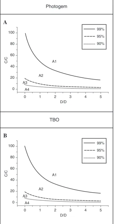
It is possible to generate from equation [2] plots relating the concentration of Photogem or TBO to the light dose needed to achieve different levels of SF (Fig. 3A and B). The plots are divided into regions separated by various curves. Region A1 corresponds to the light dose and drug concen-tration required to produce a SF ⬍0.01; A2 yields 0.01⬍SF⬍
0.05; A3 yields 0.05⬍SF⬍0.1, and A4 yields 0.1⬍SF⬍1. Using these plots, one can determine the proper combina-tion of photosensitizer concentracombina-tion and light dose to kill 90%, 95%, or 99% of the microorganisms, corresponding to SF values of 0.1, 0.05, or 0.01, respectively.
The solid lines in Fig. 2 correspond to the best mathemat-ical adjustment using equation [3], to obtain optimal values of C0and D0for each photosensitizer tested.
1
ᎏᎏ
1⫹ ᎏ C C
0
ᎏ
冢
1⫹ ᎏD D
0
ᎏ
冣
1
ᎏᎏ
1⫹ ᎏ C C
0
ᎏ G
冢
ᎏD D
0
ᎏ
冣
Discussion
Photodynamic action has been used to kill oral microor-ganisms since the beginning of the 1990s, when studies dem-onstrated that some photosensitizers show an affinity for bacterial walls and can be photoactivated to cause the de-sired damage.21–23 Excited photosensitizer molecules can
transfer energy to nearby molecules, resulting in the forma-tion of reactive molecules as singlet oxygen, superoxide, and other free radicals, capable of causing damage and even death of cells and bacteria.5,7,24,25
Ito, in 1977,26showed that TBO was photodynamically
ac-tive, promoting yeast cell death without inducing any ge-netic alterations, and that the cellular membrane was the re-action site for singlet oxygen. The effect on the yeast cells was to increase permeability and loss of control over per-meability, resulting in an imbalance of intracellular sub-stances, resulting in cellular death without any apparent chromosomal damage. In 1995, Paardekooper et al.27
ob-served that toluidine blue entered into cells during illumi-nation because of a sudden change in the cellular membrane, resulting in damage to the cellular membrane and intracel-lular structures.
Studies of biofilms15 have shown the efficacy of TBO at
concentrations of 0.01% and 0.1% when illuminated with HeNe laser energy, for reduction of the numbers of Strepto-coccus sanguis, Porphyromonas gingivalis, Actinobacillus actino-mycetemcomitans, and Fusobacterium nucleatum. An apparent bactericidal effect was observed after exposure to laser en-ergy for 30 sec, with a light dose of 219 mJ and fluence of 16.5 J/cm2. Burns et al.,28in 1993, observed that when a
sus-pension of cariogenic bacteria (Streptococcus mutans, S. sobri-nus, Lactobacillus caseiand Actinomyces viscosus) was mixed with TBO at 50 g/mL and exposed to 7.3 mW of HeNe laser
energy, a considerable amount of cellular death was accom-plished with a fluence of 33.6 J/cm2. In the case of TBO, the
results of our study are similar to those of Burns et al.28If
equation [2] is used with C0and D0values determined for
TBO from our data (0.13 mg/mL and 9 J/cm2, respectively),
then C ⬇0.05 mg/mL and D⫽34 J/cm2, and an SF of 5.6⫻
10–2is obtained for S. mutans. This is a much lower value
than the reduction of viability observed by Burns et al.28This
difference arises mainly from intrinsic differences between the two light sources used. While Burns et al. used 500 mW/cm2of light centered at 632 nm,28our experiment
uti-lized approximately 400 mW/cm2 distributed in a range
from 615–638 nm, covering a much broader spectral band, and possibly influencing the resulting bactericidal effect. Okamoto et al.16also reported the death of various species
of Streptococcususing a HeNe laser dose of 720 mJ and a flu-ence of 5.7 J/cm2with TBO at 7.5 g/mL.
In 2005 Zanin et al.19evaluated the antimicrobial effect of
TBO in combination with either a HeNe laser or a LED, on the viability and architecture of Streptococcus mutansbiofilms and observed that the bactericidal effect was light dose-de-pendent, an effect we also observed in this study. The re-ductions in viability up to 99.99% with both light sources were observed using energy densities between 49 and 294 J/cm2, a pre-irradiation time of 5 min, and TBO
concentra-tion of 100 mg/L.
A
0.0
Photogem concentration (mg/mL)
Photogem
TBO
Su
rviv
a
l f
a
ctor
0.5 1.0 1.5 2.0 2.5 3.0
1.0
0.8
0.6
0.4
0.0 0.2
B
0.0
Photogem concentration (mg/mL)
Su
rviv
a
l f
a
ctor
0.5 1.0 1.5 2.0 2.5 3.0
1.0
0.8
0.6
0.4
0.0 0.2
C
0.0
Photogem concentration (mg/mL)
Su
rviv
a
l f
a
ctor
0.5 1.0 1.5 2.0 2.5 3.0
1.0
0.8
0.6
0.4
0.0 0.2
D
0.00
TBO concentration (mg/mL)
Su
rviv
a
l f
a
ctor
0.02 0.04 0.06 0.08 0.10 1.3
1.2
1.1
1.0
0.8
0.7
0.6
0.5
0.4
0.3
0.2 0.9
E
0.00
TBO concentration (mg/mL)
Su
rviv
a
l f
a
ctor
0.02 0.04 0.06 0.08 0.10 1.0
0.8
0.6
0.4
0.0 0.2
F
0.00
TBO concentration (mg/mL)
Su
rviv
a
l f
a
ctor
0.02 0.04 0.06 0.08 0.10 1.0
0.8
0.6
0.4
0.0 0.2
derivatives show a bactericidal effect on S. mutansand other microorganisms.20,24,30 Additionally, hematoporphyrin
de-rivatives were the first drugs authorized by the U.S. Food and Drug Administration for clinical use in PDT.7Their
cy-totoxic effect is mainly carried out by the production of sin-glet oxygen. Gram-negative bacteria seem to be more resis-tant to this treatment, probably due to their more complex cell wall.26,31
Figures 2A and D show the dark toxicity of Photogem and TBO. The toxicity of both photosensitizers in the absence of light increased with increasing concentration, but the effect
is more pronounced for TBO. Figures 2B, E, C, and F show the phototoxicity of the photosensitizers activated with LED light with doses of 24 and 48 J/cm2. These graphs also
indi-cate a higher phototoxicity of TBO. Malik et al.24and
Dob-son and WilDob-son,15using hematoporphyrin at 0.005% and a
HeNe laser emitting 7.3 mW at 5.5 J/cm2, did not report any
favorable results for gram-negative microorganisms, al-though Wilson et al.22noticed lethal photosensitization of P. gingivalis with hematoporphyrin when these microorgan-isms were in suspension, but not in biofilms.
Many studies have shown TBO’s effectiveness as a pho-tosensitizer, but were performed with bacteria in suspen-sion.5,8,13,16,17,22However, similar studies demonstrated that
the reduction of S. mutans in carious dentin was less than that seen in suspension, and was also less than that attained in a collagen matrix. In that study, it was concluded that the time of contact between the bacteria and TBO was a critical factor.32
In 2003, Williams et al.13noted 100% death of S. mutansin
a plankton suspension, using a diode laser emitting at 633⫾
2 nm with fluences ranging from 0.4–4.8 J with TBO as the photosensitizer. In 2004 the same authors32using the same
light source, with fluences varying from 1.8–14.4 J and TBO at 10 g/mL obtained a reduction of S. mutansin a collagen
matrix and carious human dentin, although the dentin re-sults were more variable than those observed in the collagen matrix. Those results are similar to the results found in our study, although the presence of the biofilm on the dentin it-self negatively affect the efficacy of the technique, because the success of the process depends on photosensitizer diffu-sion throughout the dentin, and on light penetration and scattering in the tissue.
We believe that an important contribution of our study is the use of the empirical mathematical equation [2] relating the light dose, photosensitizer concentration, and bacterici-dal effect. Our experimental results indicate that there was bacterial reduction at all investigated parameters using the photosensitizers Photogem and TBO for treatment of S. mu-tansand L. acidophilus. Equation [2] allows the extrapolation of our results to parameters not yet investigated. In Fig. 3A and B we plot the SF determined by equation [2] as a func-tion of light dose for different concentrafunc-tions of Photogem and TBO. We note the general effect that toxicity, with and without exposure to light, increases with increasing concen-tration of photosensitizer. In addition, it is apparent that light alone, without any photosensitizer, has no bactericidal ef-fect. The effect for a large range of parameters is more evi-dent in Fig. 2A and B, where we can readily determine the combination of light dosage and photosensitizer concentra-tion to achieve the desired level of bacterial reducconcentra-tion.
Conclusion
In conclusion, we have shown that the use of LED light in association with Photogem or TBO was effective for bacte-rial reduction of S. mutansand L. acidophilusin carious dentin. The greatest toxic photodynamic effect was obtained with TBO at 0.1 mg/mL and 48 J/cm2of illumination. It was also
observed that the dark toxicity of TBO is higher than of Pho-togem. Similarly, the phototoxicity of TBO is higher than that of Photogem.
A
0
D/D A1
99%
95%
90%
A2
A3
A4
Photogem
TBO
C/C
1 2 3 4 5
0
D/D
1 2 3 4 5
100
80
60
40
0 20
C/C
100
80
60
40
0 20
B
99%
95%
90%
A1
A2
A3
A4
We used an empirical mathematical expression to corre-late the measured bactericidal effect with drug concentration and fluence. The use of such a formula provides a great deal of data about the expected results for a large variety of pa-rameters.
This expression may represent a simple tool to evaluate the efficacy of APDA for different microorganism species, and this concept may be used in the dosimetry planning of future antimicrobial clinical applications. The introduction of constants that quantify dark toxicity and phototoxicity is a good way to compare different photosensitizers operating under equivalent conditions.
Acknowledgments
The authors greatly acknowledge financial support from CAPES, CePOF (Program CEPID–FAPESP) and PIBIC-CNPq.
References
1. Al-Watban, F.A., and Zhang, X.Y. (2005). Photodynamic therapy of human undifferentiated thyroid carcinoma-bear-ing nude mice uscarcinoma-bear-ing topical 5-aminolevulinic acid. Pho-tomed. Laser Surg. 23, 206–211.
2. Tardivo, J.P., Sel Goglio, A., Pachoal, L.H., and Baptista, M.S. (2006). New photodynamic therapy protocol to treat AIDS-related Kaposi’s sarcoma. Photomed. Laser Surg. 24, 528–531.
3. Bedwell, J., Holton, J., Vaira, D., Macrobert, A.J., and Bown, S.G. (1990). In vitrokilling of Helicobacter pyloriwith photo-dynamic therapy. Lancet. 335, 1287.
4. Wilson, M. (1994). Bactericidal effect of laser light and its po-tential use in the treatment of plaque-related diseases. Int. Dent. J. 44, 181–189.
5. Sibata, C.H., Colussi, V.C., Oleinick, N.L., and Kinsella, T.J. (2000). Photodynamic therapy: a new concept in medical treatment. Braz. J. Med. Biol. Res. 33, 869–880.
6. Smucler, R., and Jatsová, E. (2005). Comparative study of a aminolevulic acid photodynamic therapy plus pulsed dye laser versus pulsed dye laser alone in treatment of viral warts. Photomed. Laser Surg. 23, 202–205.
7. Wainwright, M. (1998). Photodynamic antimicrobial che-motherapy (PACT). J. Antimicrob. Chemother. 42, 13–18. 8. Qin, Y., Luan, X., Bi, L., He, G., Bsai, X., Zhou, C., and Zhang,
Z. (2008). Toluidine blue-mediated photoinactivations of pe-riodontal pathogens from supragengival plaques. Lasers Med. Sci. 23, 49–54.
9. Oliveira, R.R., Schwartz-Filho, H.O., Novaes, A.B. Jr., and Taba, M. Jr. (2007). Antimicrobial photodynamic therapy in the non-surgical treatment of aggressive periodontitis: a pre-liminary randomized controlled clinical study. J. Periodon-tol. 78, 965–973.
10. Munin, E., Giroldo, L.M., Alves, L.P., and Costa, M.S. (2007). Study of germ tube formation by Candida albicansafter pho-todynamic antimicrobial chemotherapy (PACT). J Pho-tochem. Photobiol. B. 88, 16–20.
11. Lui, H., Hoobs, L., Tope, W.D., et al. (2004). Photodynamic therapy of multiple nonmelanoma skin cancers with verteporfin and light-emitting diodes. Arch. Dermatol. 140, 26– 32.
12. Yang, C.H., Lee, J.C., Chen, C.H., Hui, C.Y., Hong, H.S., and Kuo, H.W. (2003). Photodynamic therapy for bowenoid papulosis using a novel incoherent light-emitting diode de-vice. Br. J. Dermatol 149, 1292–1308.
13. Williams, J.A., Pearson, G.J., Colles, M.J., and Wilson, M. (2003). The effect of variable energy input from a novel light source on the photoactivated bactericidal action of toluidine blue O on Streptococcus mutans. Caries Res. 37, 190–193.
14. Burns, T., Wilson, M., and Pearson, G.J. (1995). Effect of den-tine and collagen on the lethal photosensitization of Strep-tococcus mutans. Caries Res. 29, 192–197.
15. Dobson, J., and Wilson, M. (1992). Sensitization of oral bac-teria in biofilms to killing by light from low-power laser. Arch. Oral. Biol. 37, 883–887.
16. Okamoto, H., Iwase, T., and Morioka, T. (1992). Dye-medi-ated bactericidal effect of He-Ne laser irradiation on oral mi-croorganisms. Lasers Surg. Med. 12, 450–458.
17. Sarkar, S., and Wilson, M. (1993). Lethal photosensitization of bacteria in subgingival plaque from patients with chronic periodontitis. J. Periodontal Res. 28, 204–210.
18. Soukos, S., Wilson, M., Burns, T., and Speigh, P.M. (1996). Photodynamic effects of toluidine blue on human oral ker-atinocytes and fibroblasts and Streptococcus sanguis evalu-ated in vitro. Lasers Surg. Med. 18, 253–259.
19. Zanin, I.C., Gonçalves, R.B., Vjunior, A.B., Hope, C.K., and Pratten, J. (2005). Susceptibility of Streptococcus mutans
biofilms to photodynamic therapy: an in vitrostudy. J. An-timicrob. Chemother. 56, 324–330.
20. Bertolini, G., Benedetto, S., Dall’acqua, M., Vazzoler, M., and Jori, G. (1984). Hematoporphyrin-sensitized photoinactiva-tion of Streptococcus faecalis. Photochem. Photobiol. 39, 811–816.
21. Wilson, M. (1993). Photolysis of oral bacteria and its poten-tial use in the treatment of caries and periodontal disease. J. Appl. Bacteriol. 7, 5299–5306.
22. Wilson, M., Dobson, J., and Sarkar, S. (1993). Sensitization of periodontopathogenic bacteria to killing by light from a low-power laser. Oral Microbiol. Immunol. 8, 182–187. 23. Wilson, M., Burns, T., Pratten, J., and Pearson, G.J. (1995).
Bacteria in supragingival plaque samples can be killed by low-power laser light in the presence of a photosensitizer. J. Appl. Bacteriol. 78, 569–574.
24. Malik, Z., Judith, H., and Nitzan, Y. (1990). New trends in photobiology (invited review) bactericidal effects of photoactivated porphyrins—an alternative approach to antimicrobial drugs. J. Photochem. Photobiol. B. Biol. 5, 281–293.
25. Tamietti, B.F., Machado, A.H., Maftoum-Costa, M., Da Silva, N.S., Tedesco, A.C., and Pacheco-Soares, C. (2007). Analysis of mitochondrial activity related to cell death after PDT with AlPCS(4) . Photomed. Laser Surg. 25, 175–179.
26. Ito, T. (1977). Toluidine blue: the mode of photodynamic ac-tion in yeast cells. Photochem. Photobiol. 25, 47–53. 27. Paardekooper, M., Bruijne, A.W., Van Steveninck, J., and
Van Den Broek, P.J.A. (1995). Intracellular damage in yeast cells caused by photodynamic treatment with toluidine blue. Photochem. Photobiol. 61, 84–89.
28. Burns, T., Wilson, M., and Pearson, G.J. (1993). Sensitization of cariogenic bacteria to kill by light from helium-neon laser. J. Med. Microbiol. 38, 401–405.
29. Venezio, F.R., Divincenzo, C., Sherman, R., et al. (1985). Bactericidal effects of photoradiation therapy with hematopophyrin derivative. J. Infect. Dis. 151, 166–169. 30. Nitzan, Y., Gutterman, M., Malik, Z.B. and Ehrenberg, B.
(1992). Inactivation of gram-negative bacteria by photosen-sitized porphyrins. Photochem. Photobiol. 55, 89–96. 31. Merchat, M., Bertolini, G., Giacomini, P., Villanueva, A., and
effi-cient photosensitizers of gram-positive and gram-negative bacteria. J. Photochem. Photobiol. B. Biol. 32, 153–157. 32. Williams, J.A., Pearson, G.J., Colles, M.J., and Wilson, M.
(2004). The photo-activated antibacterial action of toluidine blue O in a collagen matrix and carious dentine. Caries Res. 38, 530–536.
Address reprint requests to: Prof. Juçaíra Stella Martins Giusti Rua Paulino Botelho de Abreu Sampaio 728, São Carlos, SP, CEP 13561-060, Brazil