Anna Carolina Borges Pereira da Costa(a)
José Chibebe Junior(a) Cristiane Aparecida Pereira(a) Ana Karina da Silva Machado(a) Milton Beltrame Junior(b) Juliana Campos Junqueira(a) Antonio Olavo Cardoso Jorge(a)
(a) Department of Biosciences and Oral Diagnosis, School of Dentistry of São José dos Campos, UNESP - Univ. Estadual Paulista, São José dos Campos, SP, Brazil.
(b) Instituto de Pesquisa e Desenvolvimento- IP&D, Universidade do Vale do Paraíba - UNIVAP, São José dos Campos, SP, Brazil
Corresponding author:
Anna Carolina Borges Pereira da Costa Universidade Estadual Paulista (UNESP) Rua Francisco José Longo, 777, São Dimas São José dos Campos - SP - Brazil CEP: 12245-000
E-mail: [email protected]
Received for publication on Jun 12, 2010 Accepted for publication on Aug 06, 2010
Susceptibility of planktonic cultures of
Streptococcus mutans
to photodynamic
therapy with a light-emitting diode
Abstract: The objective of this study was to evaluate the effect of photo-dynamic therapy with erythrosine and rose bengal using a light-emitting
diode (LED) on planktonic cultures of S. mutans. Ten S. mutans strains,
including nine clinical strains and one reference strain (ATCC 35688),
were used. Suspensions containing 106 cells/mL were prepared for each
strain and were tested under different experimental conditions: a) LED irradiation in the presence of rose bengal as a photosensitizer (RB+L+); b) LED irradiation in the presence of erythrosine as a photosensitizer (E+L+); c) LED irradiation only (P-L+); d) treatment with rose bengal
only (RB+L-); e) treatment with erythrosine only (E+L-); and f) no LED
irradiation or photosensitizer treatment, which served as a control group (P-L-). After treatment, the strains were seeded onto BHI agar for deter-mination of the number of colony-forming units (CFU/mL). The results
were submitted to analysis of variance and the Tukey test (p≤ 0.05). The
number of CFU/mL was significantly lower in the groups submitted to
photodynamic therapy (RB+L+ and E+L+) compared to control (P-L-),
with a reduction of 6.86 log10 in the RB+L+ group and of 5.16 log10 in the E+L+ group. Photodynamic therapy with rose bengal and erythrosine exerted an antimicrobial effect on all S. mutans strains studied.
Descriptors:Streptococcus mutans; Photochemotherapy; Rose Bengal; Erythrosine.
Introduction
Mutans streptococci have raised interest because of their role in the
etiology of caries. Streptococcus mutans and S. sobrinus are important
agents involved in dental biofilm formation and are responsible for caries
lesions.1 The dental biofilm is formed by the aggregation of
microorgan-isms held together by a polysaccharide matrix.2 The ability of S. mutans
to form a biofilm is mainly due to the secretion of glucosyltransferases that form polysaccharides, which confer adhesion properties to hard sur-faces.3
non-toxic dye (photosensitizer) that is activated by exposure to visible light of an adequate wavelength in the presence of oxygen.6
The photosensitizers of the xanthene group, rose bengal and erythrosine, are cyclic compounds that contain three aromatic rings in a linear arrange-ment and an oxygen atom in the center of the ring, which absorbs light in the visible spectrum.6 Rose
bengal is used in ophthalmology for the diagnosis of eye diseases. Its photodynamic mechanism consists of the formation of 80% singlet oxygen and 20% superoxide anion, with absorption of light in the 450-600 nm range.6,7 Erythrosine absorbs light in
the visible region (500-550 nm) and is therefore able to initiate photochemical reactions. Its main appli-cation in dentistry is the demonstration of the pres-ence of dental biofilms.4
A light source emitting light in the visible region and at a wavelength adequate for photosensitization is recommended for the irradiation of the dyes. The light-emitting diodes (LEDs) used in dentistry for the light-curing of restorative materials have been suggested as an alternative to the use of lasers be-cause of their low cost and simplicity.5
Current options to reduce the population of car-iogenic microorganisms are restricted to mechanical removal and the use of antiseptics. PDT might be an excellent alternative, or accessory therapy, for the control of caries. The objective of the present study was to evaluate the effect of PDT with erythrosine and rose bengal using an LED on planktonic cul-tures of S. mutans.
Material and Methods
The project was approved by the Ethics Commit-tee of the School of Dentistry of São José dos Cam-pos, Universidade Estadual Paulista (UNESP).
Photosensitizer and light source
Rose bengal and erythrosine (Sigma-Aldrich, Steinheim, Germany), both at a concentration of
2 µM, were used for the sensitization of S. mutans
isolates. Each photosensitizer solution was prepared by dissolving the dye in phosphate-buffered saline (PBS), pH 7.4, and followed by filtration through a
sterile 0.22-µm Millipore membrane (Millipore, São
Paulo, Brazil). After filtration, the photosensitizer solutions were stored in the dark.
The light source used was an LED (MMOptics, São Carlos, SP, Brazil) with a wavelength of 440-460 nm, an output power of 200 mW and an illu-minated area of 0.38 cm2. A fluence of 95 J. cm-2
(energy of 36 J and time of 180 s) and a fluence rate of 526 mW.cm-2 were used.
The temperature monitoring at the bottom of the well was carried out with an infrared thermometer (MX4, Raytek, Sorocaba, Brazil), and no increase in the temperature was observed after the LED ir-radiation.
Microorganisms
Nine S. mutans strains previously isolated and identified from the oral cavity of healthy individuals and one reference strain (ATCC 35688) of S. mu-tans maintained in our laboratory stock collection were included in the study.
Standard suspensions of each isolate containing 106 cells/mL were prepared. For this purpose,
iso-lates were seeded onto brain heart infusion (BHI) agar (Difco, Detroit, USA) and incubated for 48 h
at 37°C under microaerophilic conditions. After
in-cubation, the microorganisms were cultured in BHI broth (Difco) for 18 h at 37°C under microaero-philic conditions. All incubations were carried out at 37°C and at a partial pressure of 5% CO2. The
bacterial cultures were then centrifuged at 1300 x g
for 10 min, and the supernatant was discarded. This procedure was repeated, and the sediment was re-suspended in 10 mL sterile PBS.
The number of cells in each suspension was mea-sured in a spectrophotometer (B582, Micronal, São Paulo, Brazil) at a wavelength of 398 nm and an op-tical density of 0.620.
Using the standard suspension, 600 assays were carried out (60 assays per strain). The assays were divided into the following experimental conditions (n = 10):
a. LED irradiation using rose bengal as
photosensi-tizer (RB+L+);
b. LED irradiation using erythrosine as
photosensi-tizer (E+L+);
d. treatment with rose bengal only (RB+L-);
e. treatment with erythrosine only (E+L-); and
f. no LED irradiation or photosensitizer treatment
(P-L-).
In vitro photosensitization
According to the experimental conditions de-scribed above, 0.1 mL of the bacterial suspension was added to each well of sterile 96-well flat-bot-tom microtiter plates (Costar Corning, New York, USA). Next, 0.1 mL of the photosensitizer was
add-ed for groups RB+L+ or E+L+ and RB+L- or E+L-,
whereas 0.1 mL PBS was added for groups P-L+
and P-L-. The plates were shaken for 5 min (pre-irradiation time) in an orbital shaker (Solab, Piraci-caba, Brazil). The well contents of groups RB+L+ or E+L+ and P-L+ were then irradiated according to the protocol described above. The distance between the light source and the bacterial cells was approxi-mately 6 mm. Irradiation was performed under aseptic conditions under a laminar flow hood in the dark. The plates were covered with a matte black screen with an orifice whose diameter corresponded to the size of the well entrance in order to prevent the spreading of light to neighboring wells.
After irradiation, serial dilutions were prepared, and 0.1 mL aliquots of each dilution were seeded in duplicate onto BHI agar plates and incubated for 48 h at 37°C under microaerophilic conditions. Af-ter incubation, the number of colony-forming units per milliliter (CFU/mL) was determined. The results were log transformed and analyzed by analysis of variance (ANOVA) and the Tukey test. A p val-ue ≤ 0.05 was considered to indicate a statistically significant difference.
Results
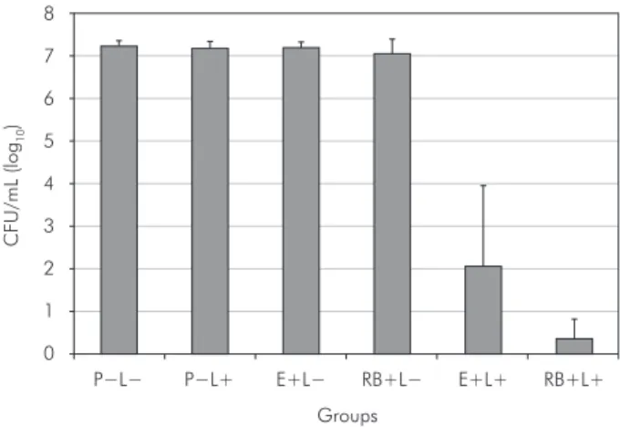
Figure 1 shows the mean and standard
devia-tion of the number of log10 CFU/mL obtained for the
groups studied. PDT (E+L+ and RB+L+) reduced S.
mutans viability, whereas no reduction in microor-ganisms was observed for the groups treated with the photosensitizer only (E+L- and RB+L-) or
ir-radiated in the absence of the dye (P-L+) when
com-pared to the control group (P-L-) (Table 1).
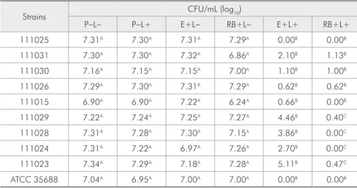
A significant difference was observed between
the groups submitted to PDT (E+L+ and RB+L+) and the other groups (Table 1). In addition, a sig-nificant difference between the E+L+ and RB+L+ groups was found for four strains, with a greater re-duction of log10 CFU in the groups treated with rose bengal and irradiated with the LED (Table 1).
When compared to the control group (P-L-), there was a reduction of 5.16 log10 CFU/mL in the E+L+ group and of 6.86 log10 CFU/mL in the RB+L+ group.
Discussion
The present results demonstrated the antimi-crobial efficacy of PDT using erythrosine and rose bengal against the ten S. mutans strains studied, in-cluding one reference strain (ATCC 35688) and nine
S. mutans strains previously isolated from the oral cavities of different individuals. We chose to use these clinical strains of S. mutans so that the effects of PDT would be more biologically relevant. No sig-nificant difference in the number of CFU/mL was
observed between the control group (P-L-) and the
groups treated in the dark with the photosensitizers
only (RB+L- and E+L-) or the group irradiated in
the absence of the dye (P-L+). These findings agree
with the basic principles of PDT, in which applica-tion of the dye or the light source alone has no
anti-CFU/mL (log
10
)
Groups
P−L− P−L+ E+L− RB+L− E+L+ RB+L+
0 1 2 3 4 5 6 7 8
bacterial effect.8
In the present study, rose bengal was used as a photosensitizer at a concentration of 2 µM for the
treatment of S. mutans suspensions containing
106 cells/mL. Paulino et al.7 investigated the toxicity
of rose bengal at concentrations of 0-10 µM to S. mutans at a cell density of 103 CFU/mL, and they
showed that rose bengal is not toxic at
concentra-tions lower than 5.0 µM. In another study, the same
authors observed that the application of rose bengal
concentrations lower than 5.0 µM in the dark were
also not toxic to fibroblasts.9
A study on the safety of topical rose bengal showed that the dye was unable to penetrate be-yond the epidermis. Thus, the authors proposed the topical dermatological use of rose bengal for various diagnostic applications of the dye, such as the use of 9.8 mM rose bengal for the diagnosis of dry eye, a concentration approximately 5,000 times higher than that used in the present study.10
Erythrosine is used in dental practice for the detection of dental biofilms at concentrations of 9-25 mM, i.e., at concentrations approximately 10,000 times higher than that used in the present study.11 Erythrosine presents advantages over other
photosensitizers since it is not toxic to the host and has already been approved for use in dentistry.4,12
Moreover, we did not find in the literature any pa-pers describing the effect of PDT with erythrosine
and blue a LED on planktonic cultures of S. mutans.
Studies have demonstrated that the use of LEDs alone exerts little or no microbicidal activity.5,13 This
light source is employed for the light-curing of re-storative materials and is therefore not damaging to oral tissues. In addition, LEDs present the following advantages over lasers: they are smaller and lighter equipment; are lower cost; have a broad spectrum output, resulting in greater flexibility during irradia-tion; and are easy to operate.14,15
A significant reduction in S. mutans was ob-served in the groups submitted to PDT (RB+L+ and E+L+) when compared to the other groups. Accord-ing to literature, rose bengal and erythrosine absorb light in the 450-600 nm and 500-550 nm ranges, respectively, and the light source used in the pres-ent study emits light in the 440-460 range. These data could explain the fact that better results were obtained for rose bengal than for erythrosine.4,7 The
association between the dyes and light resulted in cellular death, probably due to the generation of re-active oxygen species. The use of LEDs in PDT is advantageous primarily because of their safety for use in dentistry and their availability in the dentist’s office.
Significant differences between PDT with rose bengal and erythrosine were observed for four S. mutans strains, with the results suggesting that rose bengal was more effective in photodynamic
inacti-Strains CFU/mL (log10)
P-L- P-L+ E+L- RB+L- E+L+ RB+L+
111025 7.31A 7.30A 7.31A 7.29A 0.00B 0.00B
111031 7.30A 7.30A 7.32A 6.86A 2.10B 1.13B
111030 7.16A 7.15A 7.15A 7.00A 1.10B 1.00B
111026 7.29A 7.30A 7.31A 7.29A 0.62B 0.62B
111015 6.90A 6.90A 7.22A 6.24A 0.66B 0.00B
111029 7.22A 7.24A 7.25A 7.27A 4.46B 0.40C
111028 7.31A 7.28A 7.30A 7.15A 3.86B 0.00C
111024 7.31A 7.22A 6.97A 7.26A 2.70B 0.00C
111023 7.34A 7.29A 7.18A 7.28A 5.11B 0.47C
ATCC 35688 7.04A 6.95A 7.00A 7.00A 0.00B 0.00B
A, B and C: the differences between values marked with different letters are statistically significant (Tukey test,
p ≤ 0.05).
vation than erythrosine was. Rose bengal is a type II photosensitizer that exhibits easy photocatalytic conversion of triplet oxygen into singlet oxygen. The latter reactive oxygen species causes damage to unsaturated fatty acids present in lipid membranes that undergo peroxidation reactions, to enzymes by specific reactions with the amino acids methionine, tryptophan and tyrosine, and to nucleic acids by ini-tial interaction with guanine bases.16 These effects
result in bacterial death since antioxidant enzymes such as superoxide dismutase and catalase protect against some oxygenated radicals but not against singlet oxygen.14
In the present study, a mean reduction of 6.86 log10 CFU/mL was observed for bacterial sus-pensions treated with rose bengal and irradiated with the LED. Paulino et al.9 also demonstrated
the photodynamic activity of rose bengal on plank-tonic cultures of S. mutans, with a concentration of 0.5 µM of the photosensitizer resulting in a 3-log10 reduction of cells irradiated with a hand-held photo-polymerizer. Comparing the results of these works, the most suitable source light for rose bengal-medi-ated PDT was decided to be the LED emitting blue light, which demonstrated a higher reduction in the number of cells than the handheld photopolymer-izer did.
The microbial reduction observed in the group treated with erythrosine and irradiated with the LED was 5.16 log10. This result agrees with the studies of Wood et al.4 and Metcalf et al.17 Wood et al.4 evaluated the photodynamic activity of
erythro-sine, methylene blue and photophrin irradiated with a tungsten-filament lamp on S. mutans biofilms. Erythrosine (22 µM) was found to be the most ef-fective photosensitizer, resulting in a 2.2-log10 re-duction for 24-h biofilms and a 3.0-log10 reduction for 288-h biofilms. Metcalf et al.17 demonstrated
that fractionation of white light during irradiation of S. mutans biofilms treated with 22 µM eryth-rosine increased bacterial killing by 1.7 log10 when compared to continuous light irradiation.
LEDs have been used in PDT as an alternative light source to lasers. Bevilacqua et al.13 reported
ef-ficacy (7.91-log10 reduction) in the killing of
plank-tonic S. mutans cultures irradiated with an LED at
a wavelength of 640 nm in the presence of toluidine blue as photosensitizer. Zanin et al.5 evaluated the in vitro susceptibility of S. mutans biofilms to PDT. Af-ter pre-irradiation treatment with toluidine blue for
5, 15 and 30min, the biofilms were irradiated with
an LED (620-660 nm) and HeNe laser (632.8 nm). The reductions in cell viability ranged from 3.12 to 4.29 log10 and from 2.10 to 3.1 log10, respectively. However, the LED combined with toluidine blue was more effective than treatment with the HeNe laser, thus indicating the potential of LEDs in PDT for the treatment of oral diseases since the safety of LEDs have been demonstrated for oral tissues.
The application of antimicrobial photodynamic therapy is promising because the development of re-sistance after multiple treatments is unlikely due to the interaction of singlet oxygen and free radicals with various cellular structures and different meta-bolic pathways. In addition, PDT is equally effective against antibiotic-resistant and antibiotic-suscepti-ble bacteria and is not mutagenic.14,18
The oral cavity is an adequate site for the applica-tion of PDT because it is accessible to illuminaapplica-tion.14
Additional advantages of PDT include reductions in treatment time and in the occurrence of side effects, which are frequently observed after the systemic administration of antibiotics, and the lower cost of
treatment.19 PDT is an alternative or complementary
tool for the treatment of oral diseases such as caries in situations in which antimicrobial therapy is inef-fective or not recommended and in which mechani-cal removal is not possible.
Conclusion
In vitro PDT with rose bengal and erythrosine using an LED exerted antimicrobial activity on planktonic cultures of S. mutans.
Acknowledgements
References
1. Ge Y, Caufield PW, Fish GS, Li Y. Streptococcus mutans and Streptococcus sanguinis colonization correlated with caries experience in children. Caries Res. 2008;42(6):444-8. 2. Ahn SJ, Ahn SJ, Wen ZT, Brady J, Burne RA. Characteristics
of biofilm formation by Streptococcus mutans in the presence of saliva. Infect Immun. 2008 Sep;76(9):4259-68.
3. Almeida PF, Franca MP, Santos SP, Moreira RS, Tunes UR. Microbiota estreptocócica associada com a formação inicial da placa dental. Rev Cienc Med Biol. 2002 Nov;1(1):33-41. 4. Wood S, Metcalf D, Devine D, Robinson C. Erythrosine is
a potential photosensitizer for the photodynamic therapy of oral plaque biofilms. J Antimicrob Chemother. 2006 Apr;57(4):680-4.
5. Zanin ICJ, Gonçalves RB, Brugnera Jr A, Hope CK, Pratten J. Susceptibility of Streptococcus mutans biofilms to photo-dynamic therapy: an in vitro study. J Antimicrob Chemother. 2005 Aug;56(2):324-30.
6. Perussi JR. Inativação fotodinâmica de microrganismos. Quim Nova. 2007 Aug;304(4):988-94.
7. Paulino TP, Magalhães PP, Thedei Jr G, Tedesco AC, Cian-caglini P. Use of visible light-based photodynamic therapy to bacterial photoinactivation. Biochem Mol Biol Educ. 2005 Jan;33(1):46-9.
8. Pleatzer K, Krammer B, Berlanda J, Berr F, Kiesslich T. Pho-tophysics and photochemistry of photodynamic therapy: fun-damental aspects. Lasers Med Sci. 2009 Mar;24(2):259-68. 9. Paulino TP, Ribeiro KF, Thedei Jr G, Tedesco AC, Ciancaglini
P. Use of hand held photopolymerizer to photoinactivate Strep-tococcus mutans. Arch Oral Biol. 2005 Mar;50(3):353-9.
10. Wachter E, Dees C, Harkins J, Scott T, Petersen M, Rush RE, et al. Topical rose bengal: pre-clinical evaluation of pharma-cokinetics and safety. Lasers Surg Med. 2003;32(2):101-10. 11. Marsh PD, Bevis RA, Newman HN, Hallsworth AS, Robinson
C, Weatherell JA, et al. Antibacterial activity of some plaque-disclosing agents and dyes. Caries Res. 1989;23(5):348-50. 12. Allaker RP, Douglas CWI. Novel anti-microbial therapies
for dental plaque-related diseases. Int J Antimicrob Agents. 2009 Jan;33(1):8-13.
13. Bevilacqua IM, Nicolau RA, Khouri S, Brugnera Jr A, Teodoro GR, Zângaro RA, et al. The impact of photodynamic therapy on the viability of Streptococcus mutans in a planktonic cul-ture. Photomed Laser Surg. 2007 Dec;256(6):513-8. 14. Konopka K, Goslinski T. Photodynamic therapy in dentistry.
J Dent Res. 2007 Aug;86(8):694-707.
15. Meisel P, Kocher T. Photodynamic therapy for periodontal diseases: state of the art. J Photochem Photobiol B. 2005 May;79(2):159-70.
16. Banks JG, Board RG, Carter J, Dodge AD. The cytotoxic and photodynamic inactivation of micro-organisms by rose Bengal. J Appl Bacteriol. 1985 Apr;58(4):391-400.
17. Metcalf D, Robinson C, Devine D, Wood S. Enhancement of erythrosine-mediated photodynamic therapy of Strepto-coccus mutans biofilms by light fractionation. J Antimicrob Chemother. 2006 Jul;58(1):190-2.
18. Maisch T. Anti-microbial photodynamic therapy: useful in the future? Lasers Med Sci. 2007 Jun;22(2):83-91.