Volume 2013, Article ID 307136,7pages http://dx.doi.org/10.1155/2013/307136
Review Article
Bone Substitutes for Peri-Implant Defects
of Postextraction Implants
Pâmela Letícia Santos,
1Jéssica Lemos Gulinelli,
1Cristino da Silva Telles,
2Walter Betoni
Júnior,
2Roberta Okamoto,
3Vivian Chiacchio Buchignani,
1and Thallita Pereira Queiroz
41Department of Oral Biology Postgraduation, Universidade do Sagrado Corac¸˜ao (USC), Bauru, SP, Brazil
2School of Implantology of Cuiaba, Brazil
3Department of Basic Sciences, School of Dentistry of Arac¸atuba, SP, Brazil
4Department of Health Sciences, Implantology Post Graduation Course, Dental School,
University Center of Araraquara, UNIARA, SP, Brazil
Correspondence should be addressed to Pˆamela Let´ıcia Santos; [email protected]
Received 20 September 2013; Revised 8 November 2013; Accepted 11 November 2013
Academic Editor: Traian V. Chirila
Copyright © 2013 Pˆamela Let´ıcia Santos et al. his is an open access article distributed under the Creative Commons Attribution License, which permits unrestricted use, distribution, and reproduction in any medium, provided the original work is properly cited.
Placement of implants in fresh sockets is an alternative to try to reduce physiological resorption of alveolar ridge ater tooth extraction. his surgery can be used to preserve the bone architecture and also accelerate the restorative procedure. However, the diastasis observed between bone and implant may inluence osseointegration. So, autogenous bone grat and/or biomaterials have been used to ill this gap. Considering the importance of bone repair for treatment with implants placed immediately ater tooth extraction, this study aimed to present a literature review about biomaterials surrounding immediate dental implants. he search included 56 articles published from 1969 to 2012. he results were based on data analysis and discussion. It was observed that implant ixation immediately ater extraction is a reliable alternative to reduce the treatment length of prosthetic restoration. In general, the biomaterial should be used to increase bone/implant contact and enhance osseointegration.
1. Introduction
Although alveolar repair ater tooth extraction can be con-ducted by blood clot, this repair is not complete due to physi-ological resorption [1]. Studies demonstrated that vertical and horizontal dimensions are reduced around 11–22% and 29– 63%, respectively, due to alveolar resorption ater 6 months following tooth extraction [2]. his atrophy is more intense in the buccal surface (about 0.8 mm) during the irst 3 months [3].
he insertion of immediate implants in atrophic sockets is a challenge to achieve satisfactory esthetics and function [4]. In this sense, in 1976, Schulte and Heimke [5] presented the immediate implants that are placed in fresh sockets.
However, the diastasis observed between bone and implant ater dental extraction may inluence osseointegra-tion [6]. So, autogenous bone grats and/or biomaterials have
been used in those gaps to correct bone defects and provide appropriate stability.
Considering the importance of stability of immediate implants, this study presented a literature review about the most common biomaterials used for immediate dental im-plants.
2. Material and Method
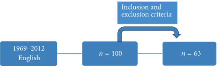
English
Inclusion and exclusion criteria
n = 100 n = 63 1969–2012
Figure 1: Method lowchart.
he search was based on scientiic researches published in English including systematic reviews and also animal and human studies. he exclusion criteria were case reports and discussion articles. Ater analysis, 63 studies were selected according to the inclusion criteria. he results were based on data analysis and discussion. (Figure 1)
3. Literature Review
3.1. Gap Dimension. he distance between bone and implant
is called peri-implant gap. he fresh socket is wider than the implant diameter, which causes the peri-implant gap that inluences stability and osseointegration [6,7].
In 1977, Schenk and Willenegger [8] conducted a study
on rabbits and observed the lack of complete bone formation with peri-implant gaps wider than 1.0 mm. In 1988, Carlsson
et al. [9] used the same experimental model to compare 3
values of peri-implant gap between bone and implant (group A—0 mm, group B—0.35 mm, and group C—0.85 mm) and observed residual gaps in groups B and C at 6 and 12 weeks ater surgery.
In 1999, Akimoto et al. [10] placed postextraction
implants in dogs and evaluated the repair of peri-implant gaps from 0.5 to 1.4 mm ater 12 weeks. he results demon-strated that the defect size is inversely proportional to the
bone/implant contact. However, Botticelli et al. [6]
per-formed a similar study and found complete bone neofor-mation and osseointegration in defect with 1.0 mm ater 16 weeks.
3.2. Autogenous Bone. Autogenous bone corresponds to bone
grat obtained from the same individual. It is considered the gold standard for illing of bone defects since it allows
(I)osseointegration: direct contact with bone tissue without
ibrous tissue [11]; (II) osteoconduction: support to bone
growth [11]; (III)osteoinduction: diferentiation of mesenchy-mal cells of surrounding tissue (receptor site) into osteoblas-tic cells [12]; and (IV) osteogenesis: bone neoformation by osteoblastic cells present in the grat material [12]. Although few mature osteoblasts survive to grating, precursor cells are responsible for the osteogenic potential [12].
Autogenous grats are presented as blocks or particles and can be used isolated or associated with allogenic or alloplastic grats. he donator area can be mentonian region, retromolar area, maxillary tuberosity, iliac crest, rib, cranium, tibia, and ibula [13].
Several studies evaluated peri-implant bone defects illed
with autogenous bone. In 2013, Al-Sulaimani et al. [14]
evaluated the success rate of immediate implants associated with autogenous bone for illing of the peri-implant gap. In this study, beagle dogs were used for insertion of implants immediately ater extraction of right and let maxillary and mandibular lateral incisors. he gaps were illed with blood clot (control) and autogenous bone (experimental) and better results were found for the autogenous bone grat.
Nevertheless, this grat may cause morbidity in the dona-tor area, hematoma, edema, infection, and vascular and nerve lesions. In addition, this technique spends more time for surgical procedure and it is limited for large reconstructions [15]. So, biomaterials have been suggested as an alternative to solve those limitations and reduce the gap between bone and implant.
3.3. Mineralized Bone Tissue. he matrix of mineralized bone
tissue is composed by deproteinized bone tissue. It has been widely used for preservation of alveolar ridge dimension ater tooth extraction, illing of bone defects near to natural teeth, and also during maxillary sinus lit [16–21].
In a study in monkeys, molars and premolar were extracted for ixation of titanium implants ater 3 months. Peri-implant defects with 2.5 mm in width and 3.0 mm in height were illed with blood clot, polytetraluorethy-lene membrane, Bio-oss, and Bio-oss with membrane. he histological analysis ater 6 months revealed that Bio-Oss exhibits osteoconductive capacity and should be used for reconstruction of peri-implant bone defects [17].
Hockers et al. [18] conducted a similar study including one group with autogenous bone and observed that the bone grats were integrated to the bone tissue.
Caneva et al. [19] used bone substitutes to ill the the gap between bone and implant. he efect of bone illers (magnesium-enriched hydroxyapatite) on preservation of the alveolar bone around immediate implants was evaluated in a dog study. Implants with a sandblasted acid etched surface were placed into the fresh extraction sockets bilaterally into the dogs’ jaws. Magnesium-enriched hydroxyapatite was placed at test sites,while the control sites did not receive aug-mentation materials. Ater 4 months of healing, the animals were sacriiced. Histomorphometric evaluations showed that the alveolar bony crest outline was maintained to a higher degree at the buccal bone wall of the test sites (loss: 0.7 mm) compared with the control sites (loss: 1.2 mm), even though this diference did not reach statistical signiicance.
In another experimental study Caneva et al. [20] explored the efect of GBR based on deproteinized bovine bone mineral on alveolar ridge preservation and the reparation of defects around osseointegrated implants. he authors concluded that the application of DBBM concomitant with a collagene membrane contributed in improving bone regen-eration in the defects.
Barone et al. [21] showed that regenerative techniques
(GBR) were able to limit resorption of the alveolar crest ater implant placement in a fresh extraction socket, tooth extraction.
bovine bone mineral into fresh extraction sockets results in signiicant buccal bone loss and low osseointegration.
Other clinical studies [23–25] used GBR techniques to ill the gap between bone and implant.
3.4. Mineralized Bone Tissue with Addition of 10% Porcine
Collagen. his material is composed by mineralized bovine
bone matrix with addition of 10% porcine collagen (Bio-oss Collagen, Geistlich). his biomaterial is indicated for illing of extraction sockets, periodontal defects, and maxillary sinus liting [26].
Ara´ujo et al. [27] conducted an animal study with illing of extraction sockets with Bio-oss collagen. Biopsy and histometric analysis were performed ater 3 months and demonstrated that the biomaterial promoted formation of bone tissue, maintained the dimension of alveolar walls, and preserved the alveolar crest proile.
In 2009, the same authors [28] performed a similar study for evaluation ater 2 weeks. he results showed delayed alveolar repair with bone neoformation only at apical and lateral walls.
Wong and Rabie [29] conducted a study in rabbits to
compare the amount of bone produced by Bio-Oss
colla-gen and collacolla-gen matrix. Eighteen bone defects (5.0 mm×
10.0 mm) were created in the parietal bone of the rabbits and illed with Bio-Oss collagen, collagen matrix and blood clot. Biopsies were removed ater 14 days for histological analysis. he authors concluded that the Bio-Oss collagen presented better results for bone neoformation in comparison to the collagen matrix while no bone was formed with the blood clot.
he study of Ara´ujo et al. [30] is the only study evaluating peri-implant defects illed with Bio-oss collagen. In this paper, dogs were used to evaluate bone repair ater ixation of immediate implants and insertion of mineralized bovine bone with addition of 10% porcine collagen. Biopsies were obtained ater 6 months for histological analysis. he authors found that the presence of Bio-oss collagen changed the healing process of hard tissue, which improved bone/implant contact.
3.5. Beta-Tricalcium Phosphate. he�-tricalcium phosphate
has been considered a material with excellent results since it is absorbable, osteoconductive, and nonosteoinductive [1]. Animal [26, 31–33] and human [34] studies demonstrated that this material supported bone neoformation.
In 2013, Daif [35] conducted a study to evaluate the
inluence of �-tricalcium phosphate on bone density
sur-rounding immediate dental implants using helical computer tomography. Twenty-eight patients were selected and divided into two groups: (I) no illing and (II) illing with beta-TCP in the peri-implant defect. Tomography was obtained ater 3
and 6 months and showed that the�-tricalcium phosphate
increased bone density in the bone defect of immediate dental implants.
Recently, some industries developed a synthetic bone substitute composed by a homogeneous mixture of 60% of hydroxyapatite (HA) and 40% of beta-tricalcium phosphate
Figure 2: Initial panoramic radiograph.
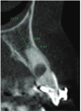
12.35
6.88
6.44
Figure 3: Initial computed tomography.
[36]. Although the HA is resistant to physiological resorption [37], its osteoconductive capacity remains uncertain [38,39].
On the other hand, the�-tricalcium phosphate is absorbed
slowly and it is considered an osteoconductive material [40].
hus, the HA maintains the gap while the �-tricalcium
phosphate is absorbed to promote bone regeneration simul-taneously [41].
In 2004, Boix et al. [42] evaluated the eicacy of this
material in peri-implant defects in dogs and concluded that the biomaterial generated signiicant increase in bone regeneration surrounding the dental implant.
4. Discussion
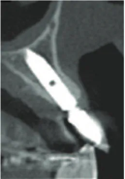
Figure 4: Peri-implant defect.
Figure 5: Peri-implant defect illed with biomaterial.
Several animal [6,7,30,44,45] and human [34,39,46–
54] studies were conducted to evaluate the reconstruction
of the peri-implant gap with biomaterials through clinical follow-up, histology, imaging, and immunohistochemistry. However, few of the literature reviews about biomaterials for peri-implant defects were found [55].
he ideal bone grat should present limited source, lack of morbidity in the donator site, no risk to disease transmission, eicient bone repair, immediate stability, versatility, easy manipulation, appropriate lifetime, and accessible cost [56].
he autogenous bone is considered the irst option for bone reconstruction in implantology since it presents char-acteristics of the ideal grat. However, this approach requires longer surgical procedure and may not obtain enough bone volume [57]. So, alternative treatments have been suggest for peri-implant reconstruction.
Jensen et al. [33] compared the performance of
autogenous bone, �-tricalcium phosphate, and anorganic
bovine bone by histological and histomorphometric analyses in pigs. he authors observed greater eicacy for the autogenous bone in comparison to the other grats.
On the other hand, Hockers et al. [18] compared grating with autogenous bone and demineralized bovine bone for reconstruction of peri-implant defects in dogs and found similar integration for both materials. Similarly, Santis et al.
[58] concluded that the autogenous bone and demineralized
bovine bone provided a high level of bone regeneration and satisfactory bone/implant contact for osseointegration.
In 2009, Beni´c et al. [59] performed a human study to
assess the success rate of peri-implant defects reconstruction with autogenous bone, demineralized bovine bone, and
Figure 6: Esthetic restoration.
Figure 7: Final computed tomography.
association of both materials. No diference was observed between the groups ater a 5-year follow-up.
Han et al. [60] compared bone regeneration in
peri-implant defects of dogs according to the following groups: (I) no illing, (II) autogenous bone, (III) Bio-Oss collagen, (IV) Bio-Oss, (V) no illing and collagen membrane, (VI) autoge-nous bone and collagen membrane, (VII) Bio-Oss collagen and collagen membrane, and (VIII) Bio-Oss and collagen membrane. he authors concluded that reconstruction of peri-implant defect with bone substitutes associated with membrane or not increases the percentage of bone/implant contact.
Guerra et al. [61] conducted a study on rabbits to compare grating with bovine bone, bovine bone associated with platelet-rich plasma, bovine bone protected by membrane, and blood clot. A higher percentage of bone/implant contact with bovine bone protected by collagen membrane was observed.
evaluated repair with blood clot, autogenous bone, Bio-Oss, and Bone-Ceramic in dogs and observed lower stability with Bio-Oss ater 2 months.
Wang and Lang [63] evaluated the more recent studies
in animal and human about this topic and they concluded that implants placed into the fresh extraction sockets do not prevent the resorption of the alveolar bone. In the research that was conducted bone regeneration with implant post-extractive implants would notice minor alveolar bone resorption. Moreover, other bone substitutes were tested: magnesium-enriched hydroxyapatite, human demineralized bone matrix, and deproteinized bovine bone mineral have been shown to be efective in ridge preservation. Applying the guided bone regeneration principle using bone substitutes together with a collagen membrane has shown clear efects on preserving alveolar ridge height as well as ridge width. Sot tissue grats or primary closure did not show a beneicial efect on preserving the alveolar bone.
5. Conclusions
Considering this literature review, the ixation of implants immediately ater tooth extraction is a reliable alternative to reduce the treatment length for patient’s rehabilitation. In general, this treatment requires the use of a biomaterial to increase bone/implant contact and enhance osseointegra-tion.
References
[1] B. M. B. Brkovic, H. S. Prasad, M. D. Rohrer et al., “Beta-tricalcium phosphate/type I collagen cones with or without a barrier membrane in human extraction socket healing: clini-cal, histologic, histomorphometric, and immunohistochemical
evaluation,”Clinical Oral Investigations, vol. 16, no. 2, pp. 581–
590, 2012.
[2] W. L. Tan, T. L. T. Wong, M. C. M. Wong, and N. P. Lang, “A systematic review of post-extractional alveolar hard and sot
tissue dimensional changes in humans,”Clinical Oral Implants
Research, vol. 23, no. 5, pp. 1–21, 2012.
[3] L. Schropp, A. Wenzel, L. Kostopoulos, and T. Karring, “Bone healing and sot tissue contour changes following single-tooth extraction: a clinical and radiographic 12-month prospective
study,” International Journal of Periodontics and Restorative
Dentistry, vol. 23, no. 4, pp. 313–323, 2003.
[4] B. Shi, Y. Zhou, Y. N. Wang, and X. R. Cheng, “Alveolar ridge preservation prior to implant placement with surgical-grade calcium sulfate and platelet-rich plasma: a pilot study in a
canine model,”he International Journal of Oral & Maxillofacial
Implants, vol. 22, no. 4, pp. 656–665, 2007.
[5] W. Schulte and G. Heimke, “he T¨ubinger immediate implant,” Die Quintessenz, vol. 27, no. 6, pp. 17–23, 1976.
[6] D. Botticelli, T. Berglundh, D. Buser, and J. Lindhe, “he jumping distance revisited: an experimental study in the dog,” Clinical Oral Implants Research, vol. 14, no. 1, pp. 35–42, 2003. [7] D. Botticelli, A. Renzi, J. Lindhe, and T. Berglundh, “Implants in
fresh extraction sockets: a prospective 5-year follow-up clinical
study,”Clinical Oral Implants Research, vol. 19, no. 12, pp. 1226–
1232, 2008.
[8] R. K. Schenk and H. R. Willenegger, “Histology of primary bone healing: modiications and limits of recovery of gaps in relation
to extent of the defect,”Unfallheilkunde, vol. 80, no. 5, pp. 155–
160, 1977.
[9] L. Carlsson, T. Rostlund, B. Albrektsson, and T. Albrektsson, “Implant ixation improved by close it. Cylindrical
implant-bone interface studied in rabbits,”Acta Orthopaedica
Scandi-navica, vol. 59, no. 3, pp. 272–275, 1988.
[10] K. Akimoto, W. Becker, R. Persson, D. A. Baker, M. D. Rohrer, and R. B. O’Neal, “Evaluation of titanium implants placed into
simulated extraction sockets: a study in dogs,”he International
Journal of Oral & Maxillofacial Implants, vol. 14, no. 3, pp. 351– 360, 1999.
[11] P. D. Costantino and C. D. Friedman, “Synthetic bone grat
substitutes,”Otolaryngologic Clinics of North America, vol. 27,
no. 5, pp. 1037–1074, 1994.
[12] T. J. Cypher and J. P. Grossman, “Biological principles of bone
grat healing,”Journal of Foot and Ankle Surgery, vol. 35, no. 5,
pp. 413–417, 1996.
[13] L. F. Coradazzi, I. R. Garcia Jr., and T. M. Manfrin, “Evaluation of autogenous bone grats, particulate or collected during osteotomy with implant burs: histologic and
histomorphome-tric analysis in rabbits,” he International Journal of Oral &
Maxillofacial Implants, vol. 22, no. 2, pp. 201–207, 2007. [14] A. Al-Sulaimani, S. A. Mokeem, and S. Anil, “Peri-implant
defect augmentation with autogenous bone: a study in beagle
dogs,”Journal of Oral Implantology, vol. 39, pp. 30–36, 2013.
[15] P. M. Trejo, R. Weltman, and R. Cafesse, “Treatment of intraosseous defects with bioabsorbable barriers alone or in combination with decalciied freeze-dried bone allograt: a
randomized clinical trial,”Journal of Periodontology, vol. 71, no.
12, pp. 1852–1861, 2000.
[16] M. B. H¨urzeler, C. R. Qui˜nones, A. Kirsch et al., “Maxillary sinus augmentation using diferent grating materials and dental implants in monkeys. Part I. Evaluation of anorganic
bovine-derived bone matrix,”Clinical Oral Implants Research, vol. 8, no.
6, pp. 476–486, 1997.
[17] C. H. F. H¨ammerle, G. C. Chiantella, T. Karring, and N. P. Lang, “he efect of a deproteinized bovine bone mineral on bone
regeneration around titanium dental implants,”Clinical Oral
Implants Research, vol. 9, no. 3, pp. 151–162, 1998.
[18] T. Hockers, D. Abensur, P. Valentini, R. Legrand, and C. H. F. Hammerle, “he combined use of bioresorbable membranes and xenograts or autograts in the treatment of bone defects
around implants: a study in beagle dogs,”Clinical Oral Implants
Research, vol. 10, no. 6, pp. 487–498, 1999.
[19] M. Caneva, D. Botticelli, E. Stellini, S. L. S. Souza, L. A. Salata, and N. P. Lang, “Magnesium-enriched hydroxyapatite at immediate implants: a histomorphometric study in dogs,” Clinical Oral Implants Research, vol. 22, no. 5, pp. 512–517, 2011. [20] M. Caneva, D. Botticelli, F. Pantani, G. M. Bafone, I. G. Rangel Jr., and N. P. Lang, “Deproteinized bovine bone mineral in marginal defects at implants installed immediately into
extraction sockets: an experimental study in dogs,”Clinical Oral
Implants Research, vol. 23, no. 1, pp. 106–112, 2012.
[21] A. Barone, M. Ricci, J. L. Calvo-Guirado, and U. Covani, “Bone remodelling ater regenerative procedures around implants placed in fresh extraction sockets: an experimental study in
Beagle dogs,”Clinical Oral Implants Research, vol. 22, no. 10, pp.
1131–1137, 2011.
placement and the treatment of bone defects with Bio-Oss in an
animal model.,”Clinical Implant Dentistry and Related Research,
vol. 14, no. 5, pp. 690–695, 2012.
[23] C. Caiero, S. Annibali, E. Gherlone et al., “Immediate transmu-cosal implant placement in molar extraction sites: a 12-month
prospective multicenter cohort study,”Clinical Oral Implants
Research, vol. 19, no. 5, pp. 476–482, 2008.
[24] S. Matarasso, G. E. Salvi, V. Iorio Siciliano, C. Caiero, A. Blasi, and N. P. Lang, “Dimensional ridge alterations following immediate implant placement in molar extraction sites: a
six-month prospective cohort study with surgical re-entry,”Clinical
Oral Implants Research, vol. 20, no. 10, pp. 1092–1098, 2009. [25] V. I. Siciliano, G. E. Salvi, S. Matarasso, C. Caiero, A. Blasi,
and N. P. Lang, “Sot tissues healing at immediate transmucosal implants placed into molar extraction sites with buccal self-contained dehiscences. A 12-month controlled clinical trial,” Clinical Oral Implants Research, vol. 20, no. 5, pp. 482–488, 2009. [26] Z. Artzi, A. Kozlovsky, C. E. Nemcovsky, and M. Weinreb, “he amount of newly formed bone in sinus grating procedures depends on tissue depth as well as the type and residual amount
of the grated material,”Journal of Clinical Periodontology, vol.
32, no. 2, pp. 193–199, 2005.
[27] M. Ara´ujo, E. Linder, J. Wennstr¨om, and J. Lindhe, “he inluence of Bio-Oss collagen on healing of an extraction socket:
an experimental study in the dog,” International Journal of
Periodontics and Restorative Dentistry, vol. 28, no. 2, pp. 123– 135, 2008.
[28] M. Ara´ujo, E. Linder, and J. Lindhe, “Efect of a xenograt on early bone formation in extraction sockets: an experimental
study in dog,”Clinical Oral Implants Research, vol. 20, no. 1, pp.
1–6, 2009.
[29] R. W. K. Wong and A. B. M. Rabie, “Efect of Bio-Oss collagen
and collagen matrix on bone formation,” Open Biomedical
Engineering Journal, vol. 4, pp. 71–76, 2010.
[30] M. G. Ara´ujo, E. Linder, and J. Lindhe, “Bio-Oss Collagen in the buccal gap at immediate implants: a 6-month study in the dog,” Clinical Oral Implants Research, vol. 22, no. 1, pp. 1–8, 2011. [31] R. Fujita, A. Yokoyama, Y. Nodasaka, T. Kohgo, and T. Kawasaki,
“Ultrastructure of ceramic-bone interface using hydroxyapatite
and�-tricalcium phosphate ceramics and replacement
mecha-nism of�-tricalcium phosphae in bone,”Tissue and Cell, vol. 35,
no. 6, pp. 427–440, 2003.
[32] F. Schwarz, M. Herten, D. Ferrari et al., “Guided bone regenera-tion at dehiscence-type defects using biphasic hydroxyapatite + beta tricalcium phosphate (Bone Ceramic) or a collagen-coated natural bone mineral (BioOss Collagen): an
immuno-histochemical study in dogs,”International Journal of Oral and
Maxillofacial Surgery, vol. 36, no. 12, pp. 1198–1206, 2007. [33] S. S. Jensen, N. Broggini, E. Hjørting-Hansen, R. Schenk, and
D. Buser, “Bone healing and grat resorption of autograt,
anor-ganic bovine bone and�-tricalcium phosphate. A histologic
and histomorphometric study in the mandibles of minipigs,” Clinical Oral Implants Research, vol. 17, no. 3, pp. 237–243, 2006. [34] I. R. Zerbo, A. L. J. J. Bronckers, G. L. de Lange, G. J. van Beek, and E. H. Burger, “Histology of human alveolar bone regeneration with a porous tricalcium phosphate. A report of
two cases,”Clinical Oral Implants Research, vol. 12, no. 4, pp.
379–384, 2001.
[35] E. T. Daif, “Efect of a multiporous beta- tricalicum phosphate
on bone density around dental,”Journal of Oral Implantology,
vol. 39, no. 3, pp. 339–344, 2013.
[36] N. Mardas, V. Chadha, and N. Donos, “Alveolar ridge preserva-tion with guided bone regenerapreserva-tion and a synthetic bone sub-stitute or a bovine-derived xenograt: a randomized, controlled
clinical trial,”Clinical Oral Implants Research, vol. 21, no. 7, pp.
688–698, 2010.
[37] S. Govindaraj, P. D. Costantino, and C. D. Friedman, “Current
use of bone substitutes in maxillofacial surgery,”Facial Plastic
Surgery, vol. 15, no. 1, pp. 73–81, 1999.
[38] O. R. Beirne, T. A. Curtis, and J. S. Greenspan, “Mandibular
augmentation with hydroxyapatite,”he Journal of Prosthetic
Dentistry, vol. 55, no. 3, pp. 362–367, 1986.
[39] S. S. Stahl and S. J. Froum, “Histologic and clinical responses to porous hydroxylapatite implants in human periodontal
defects. hree to twelve months postimplantation,”Journal of
Periodontology, vol. 58, no. 10, pp. 689–695, 1987.
[40] A. S. Breitbart, D. A. Stafenberg, C. H. M. horne et al., “Tricalcium phosphate and osteogenin: a bioactive onlay bone
grat substitute,”Plastic and Reconstructive Surgery, vol. 96, no.
3, pp. 699–708, 1995.
[41] N. Mardas, F. D’Aiuto, L. Mezzomo, M. Arzoumanidi, and N. Donos, “Radiographic alveolar bone changes following ridge
preservation with two diferent biomaterials,” Clinical Oral
Implants Research, vol. 22, no. 4, pp. 416–423, 2011.
[42] D. Boix, O. Gauthier, J. Guicheux et al., “Alveolar bone regen-eration for immediate implant placement using an injectable
bone substitute: an experimental study in dogs,” Journal of
Periodontology, vol. 75, no. 5, pp. 663–671, 2004.
[43] C. E. Misch and F. Dietsh, “Bone-grating materials in implant
dentistry,”Implant Dentistry, vol. 2, no. 3, pp. 158–167, 1993.
[44] D. Botticelli, T. Berglundh, and J. Lindhe, “Resolution of bone defects of varying dimension and coniguration in the marginal portion of the peri-implant bone: an experimental study in the
dog,”Journal of Clinical Periodontology, vol. 31, no. 4, pp. 309–
317, 2004.
[45] D. Botticelli, L. G. Persson, J. Lindhe, and T. Berglundh, “Bone tissue formation adjacent to implants placed in fresh extraction
sockets: an experimental study in dogs,”Clinical Oral Implants
Research, vol. 17, no. 4, pp. 351–358, 2006.
[46] D. A. Gelb, “Immediate implant surgery: three-year
retro-spective evaluation of 50 consecutive cases,”he International
Journal of Oral & Maxillofacial Implants, vol. 8, no. 4, pp. 388– 399, 1993.
[47] U. Br¨agger, C. H. F. H¨ammerle, and N. P. Lang, “Immediate transmucosal implants using the principle of guided tissue regeneration (II). A cross-sectional study comparing the clinical outcome 1 year ater immediate to standard implant placement,” Clinical Oral Implants Research, vol. 7, no. 3, pp. 268–276, 1996. [48] D. Schwartz-Arad and G. Chaushu, “Placement of implants into fresh extraction sites: 4 to 7 years retrospective evaluation of 95
immediate implants,”Journal of Periodontology, vol. 68, no. 11,
pp. 1110–1116, 1997.
[49] U. Grunder, G. Polizzi, R. Goen´e et al., “A 3-year prospective multicenter follow-up report on the immediate and
delayed-immediate placement of implants,”he International Journal of
Oral & Maxillofacial Implants, vol. 14, no. 2, pp. 210–216, 1999. [50] U. Lekholm, K. Wannfors, S. Isaksson, and B. Adielsson, “Oral
implants in combination with bone grats: a 3-year retrospective
multicenter study using the Br˚anemark implant system,”
[51] Z. Artzi, H. Tal, and D. Dayan, “Porous bovine bone mineral in healing of human extraction sockets. Part 1.
Histomorphomet-ric evaluations at 9 Months,”Journal of Periodontology, vol. 71,
no. 6, pp. 1015–1023, 2000.
[52] S. Froum, S.-C. Cho, E. Rosenberg, M. Rohrer, and D. Tarnow, “Histological comparison of healing extraction sockets implanted with bioactive glass or demineralized freeze-dried
bone allograt: a pilot study,”Journal of Periodontology, vol. 73,
no. 1, pp. 94–102, 2002.
[53] D. Carmagnola, P. Adriaens, and T. Berglundh, “Healing of
human extraction sockets illed with Bio-Oss,” Clinical Oral
Implants Research, vol. 14, no. 2, pp. 137–143, 2003.
[54] M. R. Norton, E. W. Odell, I. D. hompson, and R. J. Cook, “Eicacy of bovine bone mineral for alveolar augmentation: a
human histologic study,”Clinical Oral Implants Research, vol.
14, no. 6, pp. 775–783, 2003.
[55] J. Ortega-Mart´ınez, T. P´erez-Pascual, S. Mareque-Bueno, F. Hern´andez-Alfaro, and E. Ferr´es-Padr´o, “Immediate implants
following tooth extraction. A systematic review,”Medicina Oral,
Patologia Oral y Cirugia Bucal, vol. 17, no. 2, pp. e251–e261, 2012. [56] B. L. Eppley, W. S. Pietrzak, and M. W. Blanton, “Allograt and alloplastic bone substitutes: a review of science and technology
for the craniomaxillofacial surgeon,” Journal of Craniofacial
Surgery, vol. 16, no. 6, pp. 981–989, 2005.
[57] D. R. McAllister, M. J. Joyce, B. J. Mann, and C. T. Vangsness Jr., “Allograt update: the current status of tissue regulation,
pro-curement, processing, and sterilization,”he American Journal
of Sports Medicine, vol. 35, no. 12, pp. 2148–2158, 2007. [58] E. de Santis, D. Botticelli, F. Pantani, F. P. Pereira, M. Beolchini,
and N. P. Lang, “Bone regeneration at implants placed into
extraction sockets of maxillary incisors in dogs,”Clinical Oral
Implants Research, vol. 22, no. 4, pp. 430–437, 2011.
[59] G. I. Beni´c, R. E. Jung, D. W. Siegenthaler, and C. H. F. H¨ammerle, “Clinical and radiographic comparison of implants
in regenerated or native bone: 5-year results,” Clinical Oral
Implants Research, vol. 20, no. 5, pp. 507–513, 2009.
[60] J.-Y. Han, S.-I. Shin, Y. Herr, Y.-H. Kwon, and J.-H. Chung, “he efects of bone grating material and a collagen membrane in the ridge splitting technique: an experimental study in dogs,” Clinical Oral Implants Research, vol. 22, no. 12, pp. 1391–1398, 2011.
[61] I. Guerra, F. Morais Branco, M. Vasconcelos, A. Afonso, H. Figueiral, and R. Zita, “Evaluation of implant osseointegration with diferent regeneration techniques in the treatment of bone defects around implants: an experimental study in a rabbit
model,”Clinical Oral Implants Research, vol. 22, no. 3, pp. 314–
322, 2011.
[62] A. A. Antunes, P. Oliveira Neto, E. de Santis, M. Caneva, D. Botticelli, and L. A. Salata, “Comparisons between Bio-Oss and Straumann Bone Ceramic in immediate and staged
implant placement in dogs mandible bone defects,”Clinical Oral
Implants Research, vol. 24, pp. 135–142, 2013.
[63] R. E. Wang and N. P. Lang, “Ridge preservation ater tooth
extraction,”Clinical Oral Implants Research, vol. 23, no. 6, pp.
Submit your manuscripts at
http://www.hindawi.com
Scientifica
Hindawi Publishing Corporationhttp://www.hindawi.com Volume 2014
Hindawi Publishing Corporation
http://www.hindawi.com Volume 2014
Hindawi Publishing Corporation
http://www.hindawi.com Volume 2014
Hindawi Publishing Corporation
http://www.hindawi.com Volume 2014
Ceramics
Journal ofHindawi Publishing Corporation
http://www.hindawi.com Volume 2014
Nanoparticles
Journal ofHindawi Publishing Corporation
http://www.hindawi.com Volume 2014
Hindawi Publishing Corporation
http://www.hindawi.com Volume 2014 International Journal of
Biomaterials
Hindawi Publishing Corporation
http://www.hindawi.com Volume 2014
Nanoscience
Journal ofTextiles
Hindawi Publishing Corporation
http://www.hindawi.com Volume 2014
Journal of
Hindawi Publishing Corporation
http://www.hindawi.com Volume 2014
Crystallography
Journal ofHindawi Publishing Corporation
http://www.hindawi.com Volume 2014
The Scientiic
World Journal
Hindawi Publishing Corporationhttp://www.hindawi.com Volume 2014
Hindawi Publishing Corporation
http://www.hindawi.com Volume 2014
Coatings
Journal ofAdvances in
Materials Science and Engineering Hindawi Publishing Corporation
http://www.hindawi.com Volume 2014
Hindawi Publishing Corporation
http://www.hindawi.com Volume 2014
Hindawi Publishing Corporation
http://www.hindawi.com Volume 2014
Metallurgy
Journal ofHindawi Publishing Corporation
http://www.hindawi.com Volume 2014
BioMed
Research International
Materials
Journal ofHindawi Publishing Corporation
http://www.hindawi.com Volume 2014
N
a
no
ma
te
ria
ls
Hindawi Publishing Corporation
http://www.hindawi.com Volume 2014
Journal of