Living in Poor Neighbourhoods in Urban Latin American
Lı´via Ribeiro Mendonc¸a1, Rafael Valente Veiga1, Vitor Camilo Cavalcante Dattoli1, Camila Alexandrina Figueiredo1, Rosemeire Fiaccone2, Jackson Santos2,3, A´ lvaro Augusto Cruz4, Laura Cunha Rodrigues5, Philip John Cooper6, Lain Carlos Pontes-de-Carvalho7, Maurı´cio Lima Barreto3,
Neuza Maria Alcantara-Neves1*
1Instituto de Cieˆncias da Sau´de, Universidade Federal da Bahia, Salvador, Bahia, Brazil,2Instituto de Matema´tica, Universidade Federal da Bahia, Salvador, Bahia, Brazil, 3Instituto de Sau´de Coletiva, Universidade Federal da Bahia, Salvador, Bahia, Brazil,4ProAR – Nu´cleo de Exceleˆncia em Asma, Universidade Federal da Bahia, Salvador, Bahia, Brazil,5London School of Hygiene and Tropical Medicine, University of London, London, United Kingdom,6Molecular and Biochemical Parasitology, Liverpool School of Tropical Medicine, Liverpool, United Kingdom,7Centro de Pesquisas Gonc¸alo Moniz, Fundac¸a˜o Oswaldo Cruz, Salvador, Bahia, Brazil
Abstract
Background:Toxocara canisandT. catiare parasites of dogs and cats, respectively, that infect humans and cause human toxocariasis. Infection may cause asthma-like symptoms but is often asymptomatic and is associated with a marked eosinophilia. Previous epidemiological studies indicate thatT. canisinfection may be associated with the development of atopy and asthma.
Objectives:To investigate possible associations betweenToxocaraspp. seropositivity and atopy and childhood wheezing in a population of children living in non-affluent areas of a large Latin American city.
Methods: The study was conducted in the city of Salvador, Brazil. Data on wheezing symptoms were collected by questionnaire, and atopy was measured by the presence of aeroallergen-specific IgE (sIgE). Skin prick test (SPT), total IgE and peripheral eosinophilia were measured.Toxocaraseropositivity was determined by the presence of anti-ToxocaraIgG antibodies, and intestinal helminth infections were determined by stool microscopy.
Findings:Children aged 4 to 11 years were studied, of whom 47% were seropositive for anti-ToxocaraIgG; eosinophilia
.4% occurred in 74.2% and.10% in 25.4%; 59.6% had elevated levels of total IgE; 36.8% had sIgE$0.70 kU/L and 30.4% had SPT for at least one aeroallergen; 22.4% had current wheezing symptoms. Anti-ToxocaraIgG was positively associated with elevated eosinophils counts, total IgE and the presence of specific IgE to aeroallergens but was inversely associated with skin prick test reactivity.
Conclusion:The prevalence ofToxocaraseropositivity was high in the studied population of children living in conditions of poverty in urban Brazil. Toxocara infection, although associated with total IgE, sIgE and eosinophilia, may prevent the development of skin hypersensitivity to aeroallergens, possibly through increased polyclonal IgE and the induction of a modified Th2 immune reaction.
Citation:Mendonc¸a LR, Veiga RV, Dattoli VCC, Figueiredo CA, Fiaccone R, et al. (2012)ToxocaraSeropositivity, Atopy and Wheezing in Children Living in Poor Neighbourhoods in Urban Latin American. PLoS Negl Trop Dis 6(11): e1886. doi:10.1371/journal.pntd.0001886
Editor:Simon Brooker, London School of Hygiene & Tropical Medicine, United Kingdom
ReceivedOctober 12, 2011;AcceptedSeptember 7, 2012;PublishedNovember 1, 2012
Copyright:ß2012 Mendonc¸a et al. This is an open-access article distributed under the terms of the Creative Commons Attribution License, which permits
unrestricted use, distribution, and reproduction in any medium, provided the original author and source are credited.
Funding:This study was conducted through the SCAALA (Social Change, Asthma and Allergy in Latin America) initiative, funded by the Wellcome Trust, Grant No. 072405/Z/03/Z. The funders had no role in study design, data collection and analysis, decision to publish, or preparation of the manuscript.
Competing Interests:The authors have declared that no competing interests exist.
* E-mail: neuzalcantara@gmail.com
Introduction
There is evidence that the prevalence of allergic diseases has increased worldwide in recent decades, especially among popula-tions living in large cities and living a Western lifestyle [1]. A better understanding of the causes and risk factors associated with this epidemic is important to identify novel preventive strategies against these diseases [2]. Epidemiological studies conducted in various geographic locations have shown that helminth infections are, under different circumstances, associated with a reduced or increased prevalence of atopy and allergic diseases [3,4,5,6].
of atopy and asthma symptoms [14]. Chan and collaborators [15] have previously shown that toxocariasis may increase predisposition to the development of allergic diseases, especially in children. It has also been demonstrated that toxocariasis is associated with elevated levels of specific IgE against aeroallergens (sIgE), serum total IgE, eosinophil counts [16], increased skin sensitivity to aeroallergens [17], atopic asthma in children [18,19] and decreased lung function [20]. However, not all data support these associations: Zacharasiewicz and collabora-tors [21] were unable to show an association betweenToxocaraspp. seropositivity and allergen skin test reactivity, and they and others [18,21,22] did not observe an association betweenToxocarainfection and asthma.
The diagnosis of human toxocariasis is problematic because obtaining the excretory-secretory products ofToxocaralarvae required for serologic assays is highly labour-intensive and time-consuming. Most serologic studies of human and animal toxocariasis use the excretory-secretory antigens ofT. canislarvae (TcESLA) becauseT. canisfemales are easier to obtain from puppies. Due to the considerable antigenic cross-reactivity between theToxocaralarvae of both species, the detection of antibodies using the T. canis antigen does not discriminate between the two infections [23].
Because of the conflicting findings in the literature on the effects of Toxocara infection on atopy and asthma, we investigated this association in children living in poor urban neighbourhoods in Latin America where there is a high seroprevalence of specific IgG toToxocaraspp [24]. This study was carried out in the context of other chronic helminth infections of childhood that have also been associated with atopy and asthma [6]. After controlling for potential confounding factors, including intestinal helminths, we found that children who were seropositive for anti-ToxocaraIgG had more eosinophilia and elevated levels of total and allergen specific IgE, which is consistent with the findings of previous studies [16,17,18,19]. However, we also reported, for the first time in the literature, thatToxocaraseropositivity was associated with a reduced prevalence of skin prick test (SPT) reactivity to common aeroallergens and that it may play an important role as an effect modifier in the association between sIgE and SPT.
Methods
Study population
This study was performed in the city of Salvador in Northeast Brazil, which has a population of 2,800,000. The study was
performed with a cohort of 1,445 children aged 4 to 11 years who lived in non-affluent neighbourhoods and were chosen to represent areas of the city without sanitation. The cohort was chosen as part of a study conducted between 1997 and 2001 to assess the impact of a sanitation program on the occurrence of diarrhoea [25]. The children were resurveyed in 2005 to collect data on risk factors for wheezing [26]. The legal guardian of each child filled out an ISAAC Phase II-based questionnaire. Other social, demographic and environmental data were collected using validated questionnaires. Informed consent was obtained from the parents or guardians of the children, and ethical approval was granted by the Instituto de Sau´de Coletiva da Universidade Federal da Bahia and the National Commission on Ethics in Research (CONEP), Brazil.
Definitions of atopy and wheezing
Because the prevalence of sIgE for each of the studied allergens was greater than the SPT and the frequencies of SPT positivity among those without sIgE was very low [fungi (0.5%) dog epithelium (1.1%) and cat epithelium (0.9%)], atopy was defined as the presence of at least one positive test of the serum for anti-aeroallergen IgE$0.70 kU/L (anti- Dermatophagoides pteronyssinus, Blomia tropicalis, Blattella germanica and Periplaneta americana), irrespective of SPT results.
Children were classified as currently wheezing if parents reported wheezing in the previous 12 months and the children had at least one of the following: (i) diagnosis of asthma ever, (ii) wheezing with exercise in the last 12 months, (iii)$4 episodes of wheezing in the last 12 months or (iv) waking up at night because of wheezing in the last 12 months. These questions were included to increase the specificity for current wheezing as a marker for asthma disease. All other children were classified as non-wheeziers. Atopic and non-atopic wheezings were defined as symptoms of wheezing in the presence or absence, respectively, of serum IgE $0.70 kU/L for any of the tested aeroallergens.
Parasitological analysis
Two stool samples were collected from each child two days apart and analysed using the gravitational sedimentation [27] and Kato-Katz techniques [28] to detect eggs of Ascaris lumbricoides, Trichuris trichiura, hookworms and Schistosoma mansoni. Because hookworms and Enterobius vermicularis eggs were rarely observed (0.2% and 1.4%, respectively), these infections were excluded from the analyses. NoS. mansonieggs were observed in the stool samples.
Collection of blood and skin prick test (SPT) exams The children were evaluated in a mobile clinic in each of the study neighbourhoods, where they were evaluated by a medical team (doctor, nurse and laboratory technician), blood was collected (into EDTA-treated tubes), and skin prick testing for seven relevant aeroallergens was performed. At this time, the results of the stool examinations were provided to the parents, and appropriate treatment for parasite infections was given. Blood was taken to obtain differential blood cell counts (using an automated counter; Counter Electronics, Hialeah, FL, USA) and to measure total IgE, allergen-specific IgE and IgG toToxocaraspp. in plasma. SPTs were performed on the right forearm of each child using extracts (ALK-Abello, Sa˜o Paulo, Brazil) of Dermatophagoides pteronyssinus, Blomia tropicalis,Blatella germanica, Periplaneta americana, fungi, and cat and dog dander. Saline and 10 mg/mL histamine solution were used as negative and positive controls, respectively. Reactions were read after 15 minutes, and a mean wheal size of at least 3 mm greater than the negative control was considered positive.
Author Summary
Measurement of total IgE and specific IgE to aeroallergens and toA. lumbricoides
The measurement of total IgE was performed as described previously [29]. Briefly, high binding microassay plates (Costar, Cambridge, ME, USA) were coated with 4mg/mL of an anti-human IgE antibody (Pharmingen, San Diego, CA, USA) overnight at 4uC. Plates were blocked overnight at 4uC with PBS containing 10% foetal bovine serum (FBS) and 0.05% Tween 20. Samples were diluted 1:10 in PBS containing 2.5% FBS and 0.05% Tween 20 and incubated overnight at 4uC. Plates were incubated sequentially with biotinylated anti-human IgE (Sigma Aldrich, San Louis, MO, USA), streptavidin/peroxidase (Pharmigen, San Jose, CA, USA) and substrate (a mixture of hydrogen peroxide and o-phenylene-diamine; Sigma Aldrich, St Louis, MO, USA). Between all steps, the plates were washed three times with PBS containing 0.05% Tween 20 (PBS-T) and once with PBS. All incubations were for one hour at room temperature, except for the streptavidin-peroxidase and substrate steps, which were 30 minutes. A pool of sera from parasite-infected subjects was used as the positive control. Umbilical cord serum from a newborn of a non-atopic and non-parasitised mother was used as the negative control. The cut-off for elevated levels of total IgE was defined as 0.2mg/ml, which represented the median plus the half the interquartile range for 54 negative control sera (from children with 3 consecutive stool samples that were negative for parasites, allergen-specific IgE levels of,0.35 kU/L, and,2% peripheral blood eosinophilia) [29].
Measurement of the levels of specific IgE to B. tropicalis, D. pteronyssinus, P. americana, B. germanica and A. lumbricoides was performed using the ImmunoCAP assay (Phadia Diagnostics AB, Uppsala, Sweden). These four specific mite and cockroach allergens were chosen to measure atopy based on the findings of skin prick test against a panel of seven relevant aeroallergens, which showed these to be the most relevant allergens in our study population. We used two cut-off points for aeroallergen-specific concentrations ($0.35 kU/L and $0.70 kU/L) to investigate their association with Toxocaraseropositivity; however, only the higher cut-off point was used to define atopy.
Excretory/secretory products ofT. canislarvae
Excretory/secretory products ofT. canislarvae (TcESLA) were obtained as described previously [30] with appropriate modifica-tions [31]. Briefly, puppies from parasite-infected bitches were treated with piperazine (100 mg/kg) and mineral oil. The uteri of adult T. canis females were dissected, the eggs removed and incubated in 2% formalin until embryonation. The egg mem-branes were disrupted using glass beads, and the released larvae were purified using a 15-mm pore polystyrene membrane filter. The larvae were cultured in RPMI medium (Sigma Chemical Co., St. Louis, USA) at 37uC in a CO2 incubator, and the culture supernatants containing TcESLA were cryopreserved at 70uC in the presence of 1 mM phenylmethylsulfonyl fluoride (PMSF; Sigma Chemical Co., St. Louis, USA) until use. TcESLA was concentrated using Amicon filters (Millipore Corporate, Billerica, MO, USA) with pores permeable to molecules of 3000 kDa and subsequently dialysed against phosphate buffered saline (PBS), pH 7.4. The protein content of the samples was determined using the Lowry technique (1951)[32], and the antigen was aliquoted and stored at 70uC, until further use.
Absorption of sera withA. lumbricoidesandT. trichiura
extracts
To eliminate cross-reactive antigens shared by the ascarid wormsA. lumbricoidesandToxocara spp., human sera were absorbed
with somatic antigens fromA. lumbricoidesbefore the measurement of anti-Toxocara IgG. A. lumbricoides antigen was prepared from adult worms obtained from children infected and treated with albendazole and 5 mg bisacodyl (Dulcolax). The worms were washed in saline and crushed in an electric grinder (Bead-Beart, Biospec, USA) in the presence of PBS that contained protease inhibitors [1 mM phenylmethylsulfonyl fluoride (PMSF), 2 mM ethylenediamine tetra-acetic acid (EDTA), 2 mM tosyl phenyla-lanyl chloromethyl ketone (TPCK), and 50mM p-tosyl-L-lysine chloromethyl ketone (TLCK) (Sigma Chemical Co., St. Louis, USA)]. The suspension was centrifuged, and the soluble fraction stored at270uC after determining the protein concentration using the Lowry method [32]. For absorption of sera, 100mL of each serum was incubated with 250mL of a solution containing: 4.0 mg/mL A. lumbricoides antigen, 100mL polyethylene glycol (Sigma Chemical Co., St. Louis, USA) and 50mL PBS. After incubation for 30 minutes at room temperature under agitation, the material was centrifuged for 10 minutes. The supernatant was collected, re-absorbed with A. lumbricoides antigen and kept at 270uC until assayed. The second absorption was performed because some cross-reactive antibodies remained in the sera after the first absorption. Because 10.7% of the children were infected withT. trichiura, a sample of the studied sera was also absorbed with this parasite extract and compared to the same sera absorbed with A. lumbricoides alone or with both parasites. Because absorption with A. lumbricoides alone or with both parasites provided comparable titers of anti-Toxocara IgG, the remaining sera were absorbed withA. lumbricoidesantigen alone.
Detection of serum anti-ToxocaraIgG antibodies The detection of anti-ToxocaraIgG antibodies was carried out as previously described by de Savigny with modifications [33]. Briefly, 96-well plate wells were incubated overnight at 4uC with 3.2mg/mL of TcESLA in pH 9.6 carbonate/bicarbonate buffer. The plates were blocked with 0.15 M phosphate-buffered saline, pH 7.4 (PBS), containing 10% FBS (Sigma Aldrich, St Louis, MO, USA). Sera that had been pre-absorbed withA. lumbricoidesextract and diluted at 1:1000 in PBS containing 0.05% Tween 20 and 2.5% FBS (PBS-T-FBS) were added to the plates. After incubation, a solution of biotinylated anti-human IgG (BD, Pharmigen, San Jose, CA, USA) was added, followed by incubations with streptavidin-peroxidase (BD, Pharmigen, San Jose, CA, USA), and substrate (Sigma Aldrich, St Louis, MO, USA). Washings, incubations and reading were performed as described above for the measurement of total serum IgE. The reaction was blocked with 2 N sulphuric acid and read using a spectrophotometer at 490 nm (Biotek EL-800, CA, USA). The cut-off for the assay was obtained using the mean plus three standard deviations of the anti-Toxocara IgG assay from the negative control sera, which were obtained from 20 children without contact with dogs and cats. Because this assay does not discriminate infection byT. canisorT. cati, we used the results of this assay as marker of past or present infection by bothToxocara species [23].
Statistical analysis
considered: body mass index (BMI), maternal educational level, parental asthma, parental smoking, household connection to the municipal sewage system, living on a paved street, frequency of garbage collection, number of siblings, the presence of cat(s) and/ or dog(s) in the house, the presence of mould or dampness on the walls of the house (by inspection), the presence of cockroach and rodents at home, attendance at day-care centre and period of attendance, and presence ofA. lumbricoides and T. trichiurainfections in stool samples. These variables were selected because they were associated with seropositivity to Toxocaraor atopy or asthma in univariate analyses (Table 1) or because they had been identified as confounders in a previous analysis using data from these children [34]. To build multivariate logistic regression models, we used a procedure in which step-wise forward selection of variables was performed. Significant variables from the univariate analysis were included, and each non-significant variable was included sequentially; if a variable became significant, it was kept in the model, but if it remained non-significant, it was discarded. The interaction ofToxocaraseropositivity with the association between SPT and sIgE was analysed by univariate regression analysis, and the statistical significance of the interaction was provided by the Breslow-Day’s test for odds ratio homogeneity.
To analyse the association ofToxocaraseropositivity with wheeze phenotypes (atopic x non-atopic), multivariate logistic regression analyses were performed as described previously [35]. Thus, non-atopic wheeziers were compared with non-non-atopic non-wheeziers (to estimate the risk of wheezing associated with toxocariasis among non-atopic children), while atopic wheeziers were com-pared separately with two groups to demonstrate the importance of choosing the appropriated reference group and the differences generated when different comparison groups are chosen: group 1 -non-atopic and non-wheeziers (to estimate risk of wheezing associated with toxocariasis among atopic children uncontrolled by the effect of atopy); and group 2 - atopic and non-wheeziers (to estimate risk of wheezing associated with toxocariasis among atopic children controlled by the effect of atopy). We used multinomial logistic regression because it treats the categories of the polytomy (atopic wheeziers, atopic wheeziers, atopic non-wheeziers, and non-atopic non wheeziers) in a non-arbitrary order and also addresses several sets of log-odds that correspond to different dichotomies.
Results
Of the 1,445 children enrolled in the study, complete data were obtained from 1,148, all of which were included in the analysis. No statistically significant differences were observed in the prevalence of the outcomes when excluded children (n = 297) were compared with those included in the analysis (data not shown).
Table 1 shows the distribution of study variables and outcomes among the study children and the associations with Toxocara seropositivity, as analysed by univariate analysis. Seroprevalence of Toxocara IgG increased with age and was greater among children withA. lumbricoidesand T.trichiurainfections and among those without a household connection to the sewage system. Toxocaraseropositivity was positively associated with eosinophilia and with high levels of total and specific IgE ($0.35 and $0.70 kU/L) and was negatively associated with skin prick test (SPT) reactivity.
Tables 2 and 3 show the multivariate logistic analysis of the association between Toxocara seropositivity with the study out-comes. Models were adjusted for the following confounders: gender, age, maternal schooling, parental asthma, presence of mould, sewage access and infections with A. lumbricoidesand T.
trichiura. Positive associations were observed between theToxocara seropositivity and total IgE and eosinophilia (at cut-offs of.4% and.10%) (Table 2). The presence of sIgE, defined using cut-offs of$0.35 and$0.70 kU/L for at least one tested allergen, was also positively and significantly associated withToxocaraseropositivity. A statistically significant inverse association was observed between Toxocaraseropositivity and SPT (Table 3). When anti-ToxocaraIgG was stratified by optical density (to represent levels of anti-Toxocara IgG), dose-response associations were observed, such that greater optical densities were associated with a greater prevalence of all study outcomes (Tables 2 and 3), with the exception of SPT, in which higher optical densities were associated with a reduced prevalence of SPT.
The effect ofToxocaraseropositivity on the association between sIgE and SPT positivity was analysed. We found that the association of sIgE with SPT increased with an increase in the sIgE levels and that this association was weaker in Toxocara seropositive children (Table 4).
We evaluated the associations between Toxocara seropositivity and the levels of anti-Toxocara IgG antibodies with wheezing phenotypes. The results are shown in Table 5. A positive and weak association (statistically non-significant, but borderline) was found between high levels of anti-ToxocaraIgG and non-atopic asthma. Although a positive association between anti-Toxocara IgG seropositivity and atopic wheezing was found when non-atopic non-asthmatic children were used as reference group, this association disappeared when we used the appropriate reference group (atopic non-asthmatic children), as recommended previously by Barreto and colleagues [35].
Discussion
Human infections with Toxocaraspp. are generally difficult to diagnose because the parasites are inaccessible and most infections are asymptomatic. Thus, the prevalence of toxocariasis is grossly underestimated, posing a significant challenge to investigators interested in evaluating the public health impact of this infection [36]. Previous estimates of Toxocara seroprevalence in Latin America are highly variable, ranging from 4% to 52% in Brazil [37]. The latter prevalence was reported among adults in a village in the Amazon region [38]. Prevalences of 38% and 32% have been reported in children from Argentina [39] and Peru [40], respectively. A previous study in Salvador, where the present study was performed, estimated a prevalence of 46% among blood donors with eosinophilia but no evidence of intestinal helminth infection [24], which was similar to the high seroprevalence of 47% that was observed in the present study among children living in poor urban neighbourhoods and who were not selected by eosinophilia or helminth-infection status.
common in toxocariasis [41]. The association between Toxocara seropositivity and intestinal helminth infections could also be explained by immunological cross-reactivity, although we believe
that any false positive serologic reactions associated with intestinal helminths would have been minimised by the extensive absorption of sera withA. lumbricoidesantigens carried out before measurement
Table 1.Frequencies of the studied variables and their associations with anti-ToxocaraIgG seroposivity in 1,148 children.
Variables N % Anti-ToxocaraIgG seropositivity n = 540 (47.0%)
n(%) Crude OR (95% CI)
Gender
Female 532 46.3 243(45.7) 1
Male 616 53.7 297(48.2) 1.11(0.88; 1.40)
Age (years)
#5 298 26.0 133(44.6) 1
6–7 465 40.5 203(43.7) 0.96(0.72; 1.29)
$8 385 33.5 204(53.0) 1.40(1.03; 1.89)
Maternal Schooling
1st grade or less 251 21.9 149(59.4) 1
Incomplete 2nd grade 554 48.3 280(50.5) 0.70(0.52; 0.95)
Complete 2nd grade or more 343 29.9 111(32.4) 0.33(0.23; 0.46)
Parental Asthma
No 994 86.6 465(46.8) 1
Yes 154 13.4 75(48.7) 1.08(0.77; 1.52)
Mold at household
No 345 30.1 167(48.4) 1
Yes 803 69.9 373(46.5) 0.92(0.72; 1.19)
Connection to sewage system
No 194 16.9 109(56.2) 1
Yes 954 83.1 431(45.2) 0.64(0.47; 0.88)
Ascaris and/or Trichuris
No 914 79.6 375(41.0) 1
Yes 234 20.4 165(70.5) 3.44(2.52; 4.69)
Eosinophilia
,4% 296 25.8 80 (27) 1
$4% 852 74.2 460 (54) 3.17(2.35; 4.27)
,10% 856 74.6 345 (40.3) 1
$10% 292 25.4 195 (66.8) 2.98(2.23; 3.97)
Total IgE
,0.2mg/ml 464 40.4 186 (40.1) 1
$0.2mg/ml 684 59.6 354 (51.8) 1.60(1.26; 2.04)
*Specific IgE reactivity
,0.35 kU/L 591 51.5 252 (42.6) 1
$0.35 kU/L 557 48.5 288 (51.7) 1.44(1.14; 1.82)
,0.70 kU/L 726 63.2 327 (45) 1
$0.70 kU/L 422 36.8 213 (50.5) 1.24(0.98; 1.58)
*Skin prick test reactivity
No 797 69.4 394 (49.4) 1
Yes 351 30.6 146 (41.6) 0.73(0.56; 0.94)
**Wheeze plus asthma symptoms
No 890 77.5 409 (46) 1
Yes 258 22.5 131 (50.8) 1.21(0.92; 1.60)
*For at least one of the tested allergens.
**Wheeze plus (i) diagnosis of asthma ever; (ii) wheezing with exercise in the last 12 months; (iii)$4 episodes of wheezing in the last 12 months; (iv) waking up at night because of wheezing in the last 12 months. Boldface numbers show those that are statistically significant at p,0.05.
ofToxocaraantibodies, as well as by the high serum dilution used in this assay.
The present study investigated the relationship betweenToxocara seropositivity and several markers of allergic-type responses and allergic disease, including eosinophilia, total IgE, markers of atopy, and atopic and non-atopic wheezing. Typically, helminth infec-tions, such as byToxocaraspp., stimulate Th2 immune responses, a type of immune response that is considered to be central to the development of atopy and allergy. Experimental infections of mice with T. canishave been associated with increased inflammatory activity, intense migration of eosinophils to the lungs and increased plasma levels of pro-inflammatory cytokines such as IL-6 and IFN-c and eosinophil-associated chemokines such as eotaxin and RANTES [42]. Our findings demonstrate thatToxocara seropos-itivity was associated with high levels of total IgE and eosinophilia, even after adjustment for co-infections with intestinal helminths, confirming thatToxocarainfection is a strong inducer of IgE and eosinophilia. Previous studies have demonstrated that eosinophilia is present in up to 87% of individuals with toxocariasis [40,43].
Toxocaraseropositivity was also associated with the presence of specific IgE to mite and cockroach allergens. There is evidence from several studies of extensive cross-reactivity between mites and helminth parasites: 1) Johansson and collaborators (2001) [44]
reported cross-reactivity of IgE antibodies between a fish nematode (Anisakis simplex) and mites (Acarus siro, Lepidoglyphus destructor,Tyrophagus putrescentiaeand D. pteronyssinus), 2) Ponte and collaborators (2011) [45] reported a high frequency of cross-reactive IgE antibodies betweenB. tropicalisandA. lumbricoides(an ascarid worm that is closely related to Toxocara spp.), and 3) Acevedo and collaborators (2009) [46] reported the presence of multiple antigens that were cross-reactive betweenA. lumbricoides and B. tropicalis, including tropomyosin and glutathione-S-trans-ferase. Despite using a more stringent cut-off for positivity for allergen-specific IgE ($0.70 kU/L rather than that usually recommended for the definition of atopy, $0.35 kU/L) in the present study to minimise the problem of cross-reactivity of low-affinity IgE, we observed a positive association between sIgE and Toxocara seropositivity. This observation could be explained by cross-reactivity between arthropod allergens andToxocaraantigens as described above [45,46] by inducing production of allergen-specific IgE by plasma cells through polyclonal signals associated with helminth infections such as IL-4 or by the fact that children who develop strong Th2 responses to Toxocara may be more ‘atopic’ in the sense that they are more likely to develop IgE responses to environmental allergens.
Although we observed a positive association between Toxocara seropositivity and the presence of sIgE,Toxocaraseropositivity was
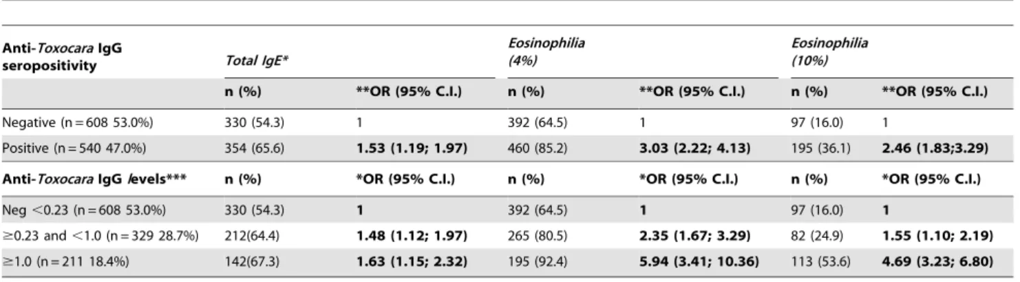
Table 2.Associations between anti-ToxocaraIgG seropositivity and total IgE and eosinophilia of$4% and$10% in 1,148 children.
Anti-ToxocaraIgG
seropositivity Total IgE*
Eosinophilia (4%)
Eosinophilia (10%)
n (%) **OR (95% C.I.) n (%) **OR (95% C.I.) n (%) **OR (95% C.I.)
Negative (n = 608 53.0%) 330 (54.3) 1 392 (64.5) 1 97 (16.0) 1
Positive (n = 540 47.0%) 354 (65.6) 1.53 (1.19; 1.97) 460 (85.2) 3.03 (2.22; 4.13) 195 (36.1) 2.46 (1.83;3.29)
Anti-ToxocaraIgGlevels*** n (%) *OR (95% C.I.) n (%) *OR (95% C.I.) n (%) *OR (95% C.I.)
Neg,0.23 (n = 608 53.0%) 330 (54.3) 1 392 (64.5) 1 97 (16.0) 1
$0.23 and,1.0 (n = 329 28.7%) 212(64.4) 1.48 (1.12; 1.97) 265 (80.5) 2.35 (1.67; 3.29) 82 (24.9) 1.55 (1.10; 2.19)
$1.0 (n = 211 18.4%) 142(67.3) 1.63 (1.15; 2.32) 195 (92.4) 5.94 (3.41; 10.36) 113 (53.6) 4.69 (3.23; 6.80)
*Positivity for total IgE defined by a cut-off of 0.2mg/mL;
**OR adjusted for gender, age, maternal schooling, parental asthma, mold, sewage access, infections withA.lumbricoidesandT. trichuris. ***Shown are strata of optical densities to represent antibody levels Boldface numbers show those that are statistically significant at P,0.05. doi:10.1371/journal.pntd.0001886.t002
Table 3.Associations between anti-ToxocaraIgG seropositivity and specific IgE (defined by ($0.35 and$0.70 kU/L) and skin prick test (SPT) reactivity in 1,148 children.
Anti-ToxocaraIgG
seropisitivity #IgE ($0.35 kU/L) #IgE ($0.70 kU/L) #SPT
n (%) *OR (95%CI) n (%) *OR (95% CI) n (%) *OR (95%CI)
Negative (n = 608 53.0%) 269 (44.2) 1 209 (34.4) 1 205 (33.7) 1
Positive (n = 540 47.0%) 288 (53.3) 1.51 (1.18; 1.94) 213 (39.4) 1.34 (1.03;1.73) 146 (27.0) 0.74 (0.57; 0.97)
**Anti-ToxocaraIgGlevels n (%) *OR (95%CI) n (%) *OR (95% CI) n (%) *OR (95%CI)
Negative,0.23 (n = 608; 53.0%) 269 (44.2) 1 209 (34.4) 1 205 (33.7) 1
$0.23 to,1.0 (n = 329; 28.7%) 162 (49.2) 1.27 (0.96; 1.67) 122 (37.1) 1.17 (0.87; 1.57) 98 (29.8) 0.81 (0.60; 1.10)
$1.0 (n = 211; 18.4%) 126 (59.7) 2.08 (1.48; 2.94) 91 (43.1) 1.71 (1.20; 2.42) 48 (22.7) 0.61 (0.42; 0.90)
#
For at least one of the tested allergens;
*OR adjusted by gender, maternal schooling, parental asthma, mold, sewage system, infection byA.lumbricoidesandT. Trichuris;
inversely associated with SPT to the same aeroallergens, and it had a strong modulator effect on the association between sIgE and SPT. The absence of allergen-specific skin reactivity, despite high sIgE values in the same individuals, has several possible explanations, including the following: 1) ‘mast cell saturation’ -the presence of high levels of parasite-induced polyclonal IgE ‘saturates’ high-affinity FceR1 receptors on mast cells, thus reducing the probability that allergen cross-linking of specific IgE will lead to cell activation [47]; 2) IgG blocking antibodies, particularly those of the IgG4 class, may bind allergen epitopes, thus preventing access of such epitopes to specific IgE antibodies bound to the mast cell [47]; 3) cross-reactive carbohydrate determinants - cross-reactive IgE antibodies, which are reactive to common carbohydrates shared by mites and helminths such as phosphorylcholine-modified glycans or glycans containing Galb1-4(Fuca1-3)GlcNAc- (Lewis X, LeX), have low affinity to the allergen epitopes, and weak binding may reduce the chance of cross-linking of the IgE bound to FceR1 receptors on the mast cell
surface [48] (this phenomenon has been described for plants and pollen allergens [49]); and 4) the downmodulation of SPT has been attributed to the so-called ‘‘modified Th2 response’’ [3,50,51], in which helminth infection induces regulatory popu-lations of T cells that produce immune regulatory cytokines such as IL-10, which may increase the threshold for mast cell activation [52]. Therefore, the negative association between anti-Toxocara antibody seropositivity and SPT found in this work could be due to at least two different phenomena. First, the infection byToxocara could modulate the immune system so that the ability of sIgE to mediate SPT is reduced (e.g., by competition withT. canis-elicited polyclonal/cross-reactive IgE). This phenomenon is consistent with the findings that the whole anti-Toxocaraantibody seropositive sub-group had higher sIgE levels than the anti-Toxocaraantibody seronegative sub-group (Table 3). Second, immunological phe-nomena, such as IL-10 production, that is induced by theToxocara infection could break the association between sIgE and SPT. In this case, one would expect a weaker association between sIgE
Table 4.Effect ofToxocara spp. seropositivity in the association of sIgE with SPT reactivity in the 1,148 studied children.
Toxocaraseronegative Toxocaraseropositive *p-value
#
sIgE (kU/L) #SPT #SPT
n(%)/N Crude OR (95% CI) n(%)/N Crude OR (95% CI)
Negative 36(9.0)/399 1 32(9.8)/327 1
Positive 169(80.9)/209 42.60 (26.21; 69.25) 114(53.5)/213 10.62 (6.75; 16.70) ,0,001
#
sIgE levels n (%) *OR (95%CI) n (%) *OR (95% CI) n (%)
,0.70 36(9.0)/399 1 32(9.8)/327 1
0.70 to,3.5 42(55.3)/76 12.46 (6.58; 23.59) 44(35.2)/125 5.01 (2.91; 8.63) 0,0316
$3.5 127(95.5)/133 213.43 (46.55; 978.58) 70(79.5)/88 35.85 (15.38; 83.59) 0.0239
#
For at least one tested allergen;
*Breslow-Day test for odds ratio homogeneity; Boldface numbers show those that are statistically significant at P,0.05. doi:10.1371/journal.pntd.0001886.t004
Table 5.Polytomous logistic regression analysis comparing the associations between anti-ToxocaraIgG seropositivity and non-atopic wheeze and non-atopic wheeze phenotypes in 1,148 children.
Anti-ToxocaraIgG seropositivity (N = 1,148)
Non-atopic wheeze plus asthma symptoms#
Atopic wheeze plus asthma symptoms#
Reference group atopic, non-wheeziers
Reference group atopic,
non-wheeziers Reference group atopic non-wheeziers
N = 706 N = 706 N = 422
n (%)/N ** OR (95% CI) n (%)/N ** OR (95% CI) n (%)/N **(95% CI)
Negative 69(17.3)/399 1 58(14.9)/388 1 58(27.8)/209 1
Positive 70(21.4)/327 1.19 (0.80; 1.77) 61(19.2)/318 1.57 (1.03; 2.39) 61(28.6)/213 1.16 (0.74; 1.82)
**Anti -ToxocaraIgG
levels n (%)/N * OR (95% CI) n (%)/N * OR (95% CI) n (%)/N * (95% CI)
Negative,0.23 69(17.3)/399 1 58(14.9)/388 1 58(27.8)/209 1
$0.23 and,1.0 35(16.9)/207 0.94 0.59; 1.50) 34(16.5)/206 1.29 (0.80; 2.10) 34(27.9)/122 1.15 (0.69; 1.94)
$1.0 35(29.2)/120 1.63 (0.98; 2.75) 27(24.1)/112 2.19 (1.25; 3.84) 27(29.7)/91 1.20 (0.66; 2.17)
#
Wheeze plus: (i) diagnosis of asthma ever; (ii) wheezing with exercise in the last 12 months; (iii)$4 episodes of wheezing in the last 12 months; (iv) waking up at night because of wheezing in the last 12 months.
*OR adjusted by gender, maternal schooling, parental asthma, mold, sewage system, infection byA.lumbricoidesandT. Trichuris;
levels and SPT reactivity, as found in anti-Toxocara antibody seropositive children (Table 4). WhileToxocaraseropositivity was strongly associated with sIgE in our study population, it was not associated with atopic wheezing. In contrast, previous studies have observed associations betweenT. canisseropositivity and increased skin sensitivity to allergens [20] and atopic asthma [7]. However, such studies were performed in different populations from different geographic regions and included adults.
We found a weak positive and statistically non-significant but borderline association between Toxocara seropositivity and non-atopic wheezing among children with the highest levels of anti-ToxocaraIgG. However, a limitation of our study was the lack of power to show a statistically significant association of this finding. This finding may be explained by lung infestations withToxocara larvae, which are known to cause asthma-like symptoms. Further limitations of our study were the cross-sectional study design, which did not allow us to distinguish exposure (presumed to be Toxocarainfections) from our study outcomes (allergic and atopic markers and wheezing). We identifiedToxocarainfection using the presence of specific IgG antibodies - the presence of antibodies does not distinguish present from past infections. Similarly, we cannot preclude confounding by other helminth infections. For example, we did not measure pinworm infections, which are universal and require specific detection methods. There is extensive cross-reactivity between different helminth infections, but we tried to reduce false-positive reactions by pre-absorption of sera with A. lumbricoidesantigens and using sera with the highest dilution described in the literature. This step was an important strength of our study. Other strengths were the use of a large sample of children, two markers for atopy (sIgE and SPT) and distinct control groups that allowed us to distinguish more clearly the effects ofToxocaraseropositivity on atopy and wheezing.
The apparent protective effects of chronic helminth infections against atopy and the clinical manifestation of allergy and autoimmune diseases are counterbalanced by the adverse effects of these infections on childhood growth and nutrition and possible adverse effects on the immune response to vaccines [53]. Given that the prevalence of intestinal helminths and schistosomiasis has declined dramatically in Latin American countries such as Brazil over recent years,Toxocarainfection is now likely to assume greater importance as a neglected public health problem and the most common endemic helminth infection in these countries.
The data from the present study revealed that almost half the children aged between 4 and 11 years living in poor neighbour-hoods in a large city in the tropical region of Brazil have evidence of infection withToxocara. Although this infection was associated with enhanced inflammatory markers (eosinophilia, total IgE) and an increased prevalence of sIgE, it appeared to be protective against immediate hypersensitivity reactions in the skin induced by common aeroallergens.
Acknowledgments
The authors would like to thank the families of the children who participated in this study and the individuals who contributed directly or indirectly to this work, including laboratory technicians, field workers, and students.
Author Contributions
Conceived and designed the experiments: NMAN AAC PJC MLB. Performed the experiments: LRM VCCD RVV. Analyzed the data: RVV RF JS LCR. Contributed reagents/materials/analysis tools: LCPC CAF. Wrote the paper: LRM CAF NMAN. Revised the paper: AAC PJC LCPC LCR MLB NMAN.
References
1. von Mutius E, Weiland SK, Fritzsch C, Duhme H, Keil U (1998) Increasing prevalence of hay fever and atopy among children in Leipzig, East Germany. Lancet 351: 862–866.
2. Falcone FH, Pritchard DI (2005) Parasite role reversal: worms on trial. Trends Parasitol 21: 157–160.
3. Yazdanbakhsh M, van den Biggelaar A, Maizels RM (2001) Th2 responses without atopy: immunoregulation in chronic helminth infections and reduced allergic disease. Trends Immunol 22: 372–377.
4. Fallon P, Mangan N (2007) Suppression of TH2-type allergic reactions by helminth infection. Nat Rev Immunol 7: 220–230.
5. Cooper PJ (2009) Interactions between helminth parasites and allergy. Curr Opin Allergy Clin Immunol 9: 29–37.
6. Alcantara-Neves NM, Veiga RV, Dattoli VC, Fiaccone RL, Esquivel R, et al. (2012) The effect of single and multiple infections on atopy and wheezing in children. J Allergy Clin Immunol 129: 359–367.
7. Cooper PJ (2008) Toxocara canis infection: an important and neglected environmental risk factor for asthma? Clin Exp Allergy 38: 551–553. 8. Rodrigues LC, Newcombe PJ, Cunha SS, Alcantara-Neves NM, Genser B, et al.
(2008) Early infection with Trichuris trichiura and allergen skin test reactivity in later childhood. Clin Exp Allergy 38: 1769–1777.
9. Moncayo A, Vaca M, Oviedo G, Erazo S, Quinzo I, et al. (2010) Risk factors for atopic and non-atopic asthma in a rural area of Ecuador. Thorax 65: 409–416. 10. Araujo MI, Lopes AA, Medeiros M, Cruz AA, Sousa-Atta L, et al. (2000) Inverse association between skin response to aeroallergens and Schistosoma mansoni infection. Int Arch Allergy Immunol 123: 145–148.
11. van den Biggelaar AH, Lopuhaa C, van Ree R, van der Zee JS, Jans J, et al. (2001) The prevalence of parasite infestation and house dust mite sensitization in Gabonese schoolchildren. Int Arch Allergy Immunol 126: 231–238. 12. Cooper PJ, Chico ME, Rodrigues LC, Ordonez M, Strachan D, et al. (2003)
Reduced risk of atopy among school-age children infected with geohelminth parasites in a rural area of the tropics. J Allergy Clin Immunol 111: 995–1000. 13. Dagoye D, Bekele Z, Woldemichael K, Nida H, Yimam M, et al. (2003) Wheezing, allergy, and parasite infection in children in urban and rural Ethiopia. Am J Respir Crit Care Med 167: 1369–1373.
14. Despommier D (2003) Toxocariasis: clinical aspects, epidemiology, medical ecology, and molecular aspects. Clin Microbiol Rev 16: 265–272.
15. Chan PW, Anuar AK, Fong MY, Debruyne JA, Ibrahim J (2001) Toxocara seroprevalence and childhood asthma among Malaysian children. Pediatr Int 43: 350–353.
16. Buijs J, Borsboom G, Renting M, Hilgersom WJ, van Wieringen JC, et al. (1997) Relationship between allergic manifestations and Toxocara seropositivity: a cross-sectional study among elementary school children. Eur Respir J 10: 1467– 1475.
17. Gonzalez-Quintela A, Gude F, Campos J, Garea MT, Romero PA, et al. (2006) Toxocara infection seroprevalence and its relationship with atopic features in a general adult population. Int Arch Allergy Immunol 139: 317–324. 18. Kustimur S, Dogruman Al F, Oguzulgen K, Bakir H, Maral I, et al. (2007)
Toxocara seroprevalence in adults with bronchial asthma. Trans R Soc Trop Med Hyg 101: 270–274.
19. Kuk S, Ozel E, Ogˇuztu¨rk H, Kirkil G, Kaplan M (2006) Seroprevalence of Toxocara antibodies in patients with adult asthma. South Med J 99: 719–722. 20. Walsh MG (2011) Toxocara infection and diminished lung function in a nationally representative sample from the United States population. Int J Parasitol 41: 243–247.
21. Zacharasiewicz A, Auer H, Brath H, Stohlhofer B, Frank W, et al. (2000) [Toxocara and bronchial hyperreactivity–results of a seroprevalence study]. Wien Klin Wochenschr 112: 922–926.
22. Sharghi N, Schantz PM, Caramico L, Ballas K, Teague BA, et al. (2001) Environmental exposure to Toxocara as a possible risk factor for asthma: a clinic-based case-control study. Clin Infect Dis 32: E111–116.
23. Kennedy MW, Maizels RM, Meghji M, Young L, Qureshi F, et al. (1987) Species-specific and common epitopes on the secreted and surface antigens of Toxocara cati and Toxocara canis infective larvae. Parasite Immunol 9: 407–420. 24. Dattoli VC, Freire SM, Mendonca LR, Santos PC, Meyer R, et al. (2011)
Toxocara canis infection is associated with eosinophilia and total IgE in blood donors from a large Brazilian centre. Trop Med Int Health 16: 514–517. 25. Barreto ML, Genser B, Strina A, Teixeira MG, Assis AM, et al. (2007) Effect of
city-wide sanitation programme on reduction in rate of childhood diarrhoea in northeast Brazil: assessment by two cohort studies. Lancet 370: 1622–1628. 26. Barreto ML, Cunha SS, Alcaˆntara-Neves N, Carvalho LP, Cruz AA, et al.
(2006) Risk factors and immunological pathways for asthma and other allergic diseases in children: background and methodology of a longitudinal study in a large urban center in Northeastern Brazil (Salvador-SCAALA study). BMC Pulm Med 6: 15.
28. Katz N, Chaves A, Pellegrino J (1972) A simple device for quantitative stool thick-smear technique in Schistosomiasis mansoni. Rev Inst Med Trop Sao Paulo 14: 397–400.
29. Figueiredo C, Barreto M, Rodrigues L, Cooper P, Silva N, et al. (2010) Chronic intestinal helminth infections are associated with immune hyporesponsiveness and induction of a regulatory network. Infect Immun 78: 3160–3167. 30. de Savigny D (1975) In vitro maintenance of Toxocara canis larvae and a simple
method for the production of Toxocara ES antigen for the uses in serodiagnostic test for visceral larva migrans. Journal of Parasitology 61: 781–782. 31. Alcaˆntara-Neves NM, dos Santos AB, Mendonc¸a LR, Figueiredo CA,
Pontes-de-Carvalho L (2008) An improved method to obtain antigen-excreting Toxocara canis larvae. Exp Parasitol 119: 349–351.
32. Lowry OH, Rosebrough NJ, Farr AL, Randall RJ (1951) Protein measurement with the Folin-Phenol reagents. J Biol Chem 193: 265–275.
33. De Savigny DH, Tizard IR (1975) Serodiagnosis of Toxocara larva migrans visceral. Candian Journal of Public Health 66: 52–56.
34. Matos SM, Jesus SR, Saldiva SR, Prado MS, D’Innocenzo S, et al. (2011) Overweight, asthma symptoms, atopy and pulmonary function in children of 4– 12 years of age: findings from the SCAALA cohort in Salvador, Bahia, Brazil. Public Health Nutr 14: 1270–1278.
35. Barreto ML, Cunha SS, Fiaccone R, Esquivel R, Amorim LD, et al. (2010) Poverty, dirt, infections and non-atopic wheezing in children from a Brazilian urban center. Respir Res 11: 167.
36. Hotez PJ, Wilkins PP (2009) Toxocariasis: America’s most common neglected infection of poverty and a helminthiasis of global importance? PLoS Negl Trop Dis 3: e400.
37. Chieffi PP, Santos SV, Queiroz ML, Lescano SA (2009) Human toxocariasis: contribution by Brazilian researchers. Rev Inst Med Trop Sao Paulo 51: 301– 308.
38. Damian MM, Martins M, Sardinha JF, Souza LO, Chaves A, et al. (2007) [Frequency of the antibody anti-Toxocara canis in a community along the Uatuma river, State of Amazonas]. Rev Soc Bras Med Trop 40: 661–664. 39. Alonso JM, Bojanich MV, Chamorro M, Gorodner JO (2000) Toxocara
seroprevalence in children from a subtropical city in Argentina. Rev Inst Med Trop Sao Paulo 42: 235–237.
40. Espinoza YA, Huapaya PH, Roldan WH, Jimenez S, Arce Z, et al. (2008) Clinical and serological evidence of Toxocara infection in school children from Morrope district, Lambayeque, Peru. Rev Inst Med Trop Sao Paulo 50: 101–105. 41. Regis SC, Mendonca LR, Silva Ndos S, Dattoli VC, Alcantara-Neves NM, et al.
(2011) Seroprevalence and risk factors for canine toxocariasis by detection of
specific IgG as a marker of infection in dogs from Salvador, Brazil. Acta Trop 120: 46–51.
42. Pecinali NR, Gomes RN, Amendoeira FC, Bastos AC, Martins MJ, et al. (2005) Influence of murine Toxocara canis infection on plasma and bronchoalveolar lavage fluid eosinophil numbers and its correlation with cytokine levels. Vet Parasitol 134: 121–130.
43. Martin UO, Machuca PB, Demonte MA, Contini L (2008) [Analysis of children with a presumptive diagnosis of toxocariasis in Santa Fe, Argentina]. Medicina (B Aires) 68: 353–357.
44. Johansson E, Aponno M, Lundberg M, van Hage-Hamsten M (2001) Allergenic cross-reactivity between the nematode Anisakis simplex and the dust mites Acarus siro, Lepidoglyphus destructor, Tyrophagus putrescentiae, and Derma-tophagoides pteronyssinus. Allergy 56: 660–666.
45. Ponte JC, Junqueira SB, Veiga RV, Barreto ML, Pontes-de-Carvalho LC, et al. (2011) A study on the immunological basis of the dissociation between type I-hypersensitivity skin reactions to Blomia tropicalis antigens and serum anti-B. tropicalis IgE antibodies. BMC Immunol 12: 34.
46. Acevedo N, Sanchez J, Erler A, Mercado D, Briza P, et al. (2009) IgE cross-reactivity between Ascaris and domestic mite allergens: the role of tropomyosin and the nematode polyprotein ABA-1. Allergy 64: 1635–1643.
47. Yazdanbakhsh M, Kremsner PG, van Ree R (2002) Allergy, parasites, and the hygiene hypothesis. Science 296: 490–494.
48. Kuijk LM, van Die I (2010) Worms to the rescue: can worm glycans protect from autoimmune diseases? IUBMB Life 62: 303–312.
49. Mari A, Iacovacci P, Afferni C, Barletta B, Tinghino R, et al. (1999) Specific IgE to cross-reactive carbohydrate determinants strongly affect the in vitro diagnosis of allergic diseases. J Allergy Clin Immunol 103: 1005–1011.
50. Maizels RM, Pearce EJ, Artis D, Yazdanbakhsh M, Wynn TA (2009) Regulation of pathogenesis and immunity in helminth infections. J Exp Med 206: 2059–2066.
51. Figueiredo CA, Barreto ML, Rodrigues LC, Cooper PJ, Silva NB, et al. (2010) Chronic intestinal helminth infections are associated with immune hyporespon-siveness and induction of a regulatory network. Infect Immun 78: 3160–3167. 52. Platts-Mills T, Vaughan J, Squillace S, Woodfolk J, Sporik R (2001)
Sensitisation, asthma, and a modified Th2 response in children exposed to cat allergen: a population-based cross-sectional study. Lancet 357: 752–756. 53. van Riet E, Hartgers FC, Yazdanbakhsh M (2007) Chronic helminth infections