INTRODUCTION
Address to: Dra. Sara Menezes de Oliveira. Depto. de Patologia e Medicina Legal/
UFC. Rua Monsenhor Furtado s/n, Rodolfo Teófi lo, 60441-750 Fortaleza, CE, Brasil.
Phone: 55 85 9933-3347 e-mail: [email protected] Received 15 October 2014 Accepted 5 December 2014
Association between allergic responses and
Schistosoma mansoni
infection in residents
in a low-endemic setting in Brazil
Sara Menezes de Oliveira
[1],
Fernando Schemelzer de Moraes Bezerra
[1],[2],[3],
Teiliane Rodrigues Carneiro
[1],[4],
Marta Cristhiany Cunha Pinheiro
[2],[3]and José Ajax Nogueira Queiroz
[1][1]. Departamento de Patologia e Medicina Legal, Universidade Federal do Ceará, Fortaleza, CE. [2]. Departamento de Análises Clínicas e Toxicológicas, Universidade Federal do Ceará, Fortaleza, CE. [3]. Departamento de Saúde Comunitária, Universidade Federal do Ceará, Fortaleza, CE. [4]. Departamento de Fisiologia e Farmacologia, Universidade Federal do Ceará, Fortaleza, CE.
ABSTRACT
Introduction: Schistosomiasis is endemic in 76 countries and territories. Several studies have found an inverse correlation
between parasitic disease and the development of allergies. The purpose of the present study was to determine whether infection with Schistosoma mansoni in subjects with a low parasite load is protective against allergy. The fi nal sample consisted of 39 S. mansoni-positive and 52 S. mansoni-negative residents of a small community in northeastern Brazil. Methods: All subjects
were submitted to the Kato-Katz test, anti-S. mansoni IgG measurement, the prick test for aeroallergens, eosinophil counts and
serum IgE measurement. Results: Subjects who reacted to one or more antigens in the prick test were considered allergic. Only
7 S. mansoni-positive subjects (17.9%) reacted to one or more antigens, whereas 20 S. mansoni-negative subjects (38.5%) tested
positive for allergy. Conclusions: Our fi ndings suggest that, in areas of low endemicity, infection with S. mansoni signifi cantly
reduces the risk of the development of allergy in subjects with a low parasite load.
Keywords: Schistosomiasis. Schistosoma mansoni. Allergy.
Schistosomiasis is endemic in 76 countries and territories. An estimated 200-250 million people are currently infected with schistosomiasis, and another 600-780 million people are
at risk of infection1,2.
This disease is caused when granulomatous infl ammatory lesions form around host tissues infected by Schistosoma mansoni eggs3. Eggs deposited in the liver stimulate cluster
of differentiation 4+ (CD4+) T cells, which in turn activate
macrophages and induce late hypersensitivity reactions leading
to the formation of granulomas4. Initially, the immune response
consists of an acute Th1 cell response directed at the adult worm; then, when eggs are deposited, the response is gradually shifted to a Th2 response. Failure to develop an effective Th2
response results in Th1- and Th17-mediated exacerbation of granulomatous infl ammation5.
During the acute stage, the immune response to helminth
infection is mediated by Th1 cells. This type of response generates heightened production of interferon-γ (IFN-γ)6 and tumor necrosis factor-α (TNF-α), which activate macrophages and induce the immunoglobulin G (IgG)-mediated opsonization and phagocytosis of antigens7. In the chronic stage, the immune response is mostly mediated by Th2 cells, leading to elevated interleukin 4 (IL-4) and interleukin 5 (IL-5) levels and interleukin
10 (IL-10) secretion and the subsequent reduction of IFN-γ8.
Several researchers have reported an inverse correlation
between parasitic disease and the development of allergic diseases, leading to the hypothesis that parasites may have a protective (immunomodulatory) effect against atopy9. For instance, a study conducted in Equador reported an inverse correlation between helminth infection and allergy skin test
results in children10.
Recent studies have also shown that asthma and allergic
diseases are less prevalent in rural settings11, possibly due to
METHODS
demonstrated between helminth infection and allergic diseases Many epidemiologists investigating the increasing prevalence of allergic diseases subscribe to the hygiene hypothesis14, which asserts that the lack of exposure to infectious agents and parasites, improvements in hygiene and the use of vaccines and antibiotics (especially in developed countries) can compromise the ability of the immune system to respond adequately to certain challenges, conceivably due to an imbalance between the Th1 and Th2 cell-mediated immune responses15.
The lower frequency of positive allergic tests and decreased asthma severity in populations infected with helminths may be due to several factors, such as enhanced polyclonal IgE production, reduced levels of allergen-specifi c IgE, high concentrations of antigen-specifi c IgG4, the activation of regulatory cells and the production of regulatory cytokines. For instance, IL-10 can inhibit the release of histamine and other mast cell mediators. Because infection with S. mansoni is associated with increased
IL-10 production, this may be the main mechanism by which
allergic response is suppressed in infected individuals16.
In general, studies evaluating the relationship between helminth infection, sensitivity to allergy skin tests and symptoms of allergic diseases have shown that the allergic response is reduced following infection with Schistosoma sp. Likewise,
cross-sectional studies indicate that subjects infected with
S. mansoni and Schistosoma haematobium are less frequently positive for aeroallergens on skin testing10. It should be
noted that these studies were conducted in regions where schistosomiasis is considered highly endemic. The purpose of the present study was to determine whether infection with
S. mansoni in subjects with a low parasite load is also protective against allergic diseases.
Study area and population
The study was carried out in Planalto do Cajueiro, a locality in the municipality of Maranguape (State of Ceará, Northeastern Brazil). The prevalence of schistosomiasis in the region rose from 8.53% in 2006 to 13.76% in 2007.
Located 30km from Fortaleza (the State capital), Maranguape covers an area of 591km2 and has 113,561 inhabitants17. All members of the community (n=903) were invited to participate in the study, but only 357 agreed to undergo parasite examination, and 250 were submitted to serological tests. Subjects fulfi lling the following criteria were excluded from the study: I) not a permanent resident of Planalto do Cajueiro, II) using anti-allergic drugs, such as corticoids or immunosuppressants, III) a history of immunodepression, IV) pregnant, V) a history of anaphylactic shock, VI) needle or skin test phobia, or VII) under 2 years of age.
Thus, 91 participants were included in the analysis.
To identify subjects with and without allergy, all participants (both Sm+ and Sm‒) were submitted to the prick test (PT). Thus,
four groups were established: Schistosoma mansoni-positive and
allergic (Sm+PT+); Schistosoma mansoni-positive and not allergic
(Sm+PT‒); Schistosoma mansoni-negative and allergic (Sm‒PT+)
and Schistosoma mansoni-negative and not allergic (Sm‒PT‒).
Diagnostic methods
Egg detection (Kato-Katz technique): Egg detection was
performed according to the Kato-Katz technique18, using a Helm-Test® kit. To increase the sensitivity of the test, three slides
of each fecal sample (rather than one slide, as recommended by the manufacturer) were used for the detection of eggs from
S. mansoni and other helminths.
Anti-Schistosoma mansoni IgG antibodies: Using adult worm
antigen in conjunction with a protocol slightly adapted from Colley
et al.19, an ELISA was performed to test for anti-Schistosoma mansoni IgG antibodies. The optical density was measured with
an automatic ELISA reader (BioTeck®) using a 490nm fi lter. Reactions with an optical density above 0.283 were considered positive. This cut-off value was based on the average optical density plus two standard deviations of 35 control serum
samples collected from S. mansoni-negative individuals from a
region where schistosomiasis is not endemic.
Prick test: the preferred site for the prick test is the inner
forearm. Following antisepsis with 70% alcohol, a 25µL aliquot of each allergenic extract was introduced into the skin surface with a standard disposable pricker according to a predefi ned grid20.
The allergens included three species of dust mites
(Dermatophagoides pteronissimus, Dermatophagoides farinae and Blomia tropicalis), cockroaches (mix) and fungi (mix).
The reaction was measured after 20 min of exposure.
Hemogram: a complete hemogram was performed with
an automated hematology analyzer (Sysmex KX-21N). To quantify eosinophils, a smear of the sample was submitted to rapid panoptic staining for a differential leukocyte count under
the microscope.
Serum IgE concentrations: the total serum IgE concentration
was measured with a commercially available kit (Monobind IgE AccuLite™ CLIA), following the manufacturerʼs instructions. The reading was performed by ELISA, at 450nm.
Statistical analysis
RESULTS
DISCUSSION
Ethical considerations
The study protocol had been previously approved by the Research Ethics Committee of the School of Medicine of the Federal University of Ceará [Universidade Federal do Ceará
(UFC)] on March 4, 2010 (fi led under #329/09).
A sample of 250 residents from a community with an 11.2% prevalence of schistosomiasis was examined. Thirty-nine of these subjects were S. mansoni-positive by Kato-Katz and
ELISA. According to the results of ELISA for anti-S mansoni
IgG antibodies, 118 samples (47.2%) were positive and 132 (52.8%) were negative. A positive result by Kato-Katz or ELISA indicates current infection and/or an antibody immune response to the parasite, respectively. The 91 selected participants (39 Sm+
and 52 Sm‒) were submitted to the prick test, and those reacting to one or more antigens were considered allergic. Only 7 Sm+
subjects (17.9%) reacted to one or more allergens. In contrast, 20 Sm‒ subjects (38.5%) tested positive for allergy (Figure 1). When analyzed with Fisherʼs exact test, the fi ndings shown
in Figure 1 yielded the following results: p=0.0394, relative
risk=0.5185, IC95%=0.2619-1.027 (confi dence interval at 95%) and odds ratio=0.350, IC95%=0.130-0.9426. The fact that 18 of the 91 participants presented co-infection with other helminths or protozoa is not likely to have affected the results, considering the diversity of species and the negligible incidence of each.
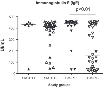
Based on the results of the Kato-Katz and prick tests, the following groups were defi ned: Sm+PT+ (n=7), Sm+PT‒
(n=32), Sm‒PT+ (n=20) and Sm‒PT‒ (n=32). The median IgE
concentrations were 434.0 UI/mL (38.5-452.5) for the Sm+PT+
group, 416.0 UI/mL (42.5-457.5) for the Sm+PT‒ group, 442.8
UI/mL (34.5-463.0) for the Sm‒PT+ group, and 154.8 UI/mL
(14.0-461.5) for the Sm‒PT‒ group. When submitted to the Mann-Whitney test, signifi cant differences were found between the SM+PT- and SM-PT- groups (p=0.0094) (Figure 2).
82.1%
61.5% 17.9%
38.5%
0.0% 20.0% 40.0% 60.0% 80.0% 100.0% 120.0%
S. mansoni + S. mansoni
-allergic not -allergic
FIGURE 1 - Relationship between the diagnosis of allergy and infection with Schistosoma mansoni.
Immunoglobulin E (IgE)
SM+PT+ SM+PT- SM-PT+ SM-PT-0
100 200 300 400 500
p<0.01
UI
/m
L
Study groups
FIGURE 2 - Median IgE concentrations in the four study groups. SM+PT+: Schistosoma mansoni-positive and allergic;
SM+PT‒:Schistosoma mansoni-positive and not allergic; SM‒PT+:
Schistosoma mansoni-negative and allergic; SMm‒PT‒: Schistosoma mansoni-negative and not allergic.
The median eosinophil counts were 21,504 cells/µL (12,702-39,865) for the Sm+PT+ group, 18,630 cells/µL (2,522-47,244)
for the Sm+PT‒ group, 464 cells/µL (52-1,200) for the Sm‒PT+
group and 147 cells/µL (0-1,044) for the Sm‒PT‒ group. When submitted to the Kruskal-Wallis test followed by Dunnʼs multiple comparison test, signifi cant differences were found between the Sm+PT+ and Sm‒PT+ (p<0.01), Sm+PT+ and Sm‒
PT‒ (p<0.01), Sm+PT‒ and Sm‒PT+ (p<0.001), and Sm+PT‒ and
Sm‒PT‒ groups (p<0.001) (Figure 3).
Since 1976, when interventions to control schistosomiasis began, the epidemiological profi le of the disease has changed in many regions of Brazil. In particular, control measures have had widely documented benefi cial impacts on disease prevalence, severity and morbidity. However, these infections are still transmitted throughout Ceará state, causing mostly
mild infections21.
In tropical regions, chronic helminth infection and allergic
diseases are known to be inversely correlated22. Helminth infection is an environmental factor that may skew the T cell
response to reduce the risk of allergic disease23, and S. mansoni
SM+PT+ SM+PT- SM-PT+ SM-PT-0
10,000 20,000
a,b
a,b
ce
ll
s/
µ
Study groups
FIGURE 3 - Median eosinophil counts in the four study groups. SM+PT+: Schistosoma mansoni-positive and allergic;
SM+PT‒:Schistosoma mansoni-positive and not allergic; SM‒PT+:
Schistosoma mansoni-negative and allergic; SM‒PT‒: Schistosoma mansoni-negative and not allergic.
To evaluate the influence of S. mansoni infection on the
allergic response, we submitted our subjects to the prick test using environmental allergens. According to the literature, in regions where schistosomiasis is highly endemic and the parasite load is high, reactivity to skin allergy tests tends to be reduced. In our study, only 7 Sm+ subjects (17.9%) reacted to one or more of the
allergens tested, whereas 20 Sm‒ subjects (38.5%) tested positive for one or more allergens. These results support the hypothesis
that helminth infection has a protective effect on the development
of allergic diseases. Similar fi ndings were reported by Catapani
et al.26 in a study on allergic diseases in a Brazilian region where
schistosomiasis is endemic, as these authors also found a lower
prevalence of asthma among infected individuals. This result
may be explained by the ability of chronic helminth infection to induce immunomodulatory responses associated with increased IL-10 or transforming growth factor-β (TGF-β) production by several regulatory cell types, thereby suppressing the mechanisms responsible for the development of allergy27. Based on the prick test results in our study, subjects with and without schistosomiasis were further divided into allergic and non-allergic categories, thereby defi ning four groups (Sm+PT+, Sm+PT‒, Sm‒PT+ and Sm‒PT‒). Considering the important role IgE plays in both allergic responses and helminth infections, the total serum IgE concentration was measured. As expected, these levels were higher in S. mansoni
-positive and/or prick test--positive subjects than in their negative
counterparts28. Intragroup differences in optical density may be
due to low responsiveness or variation in genetic characteristics of certain individuals and/or unknown allergies.
Our results are supported by those of Cooper et al.29, who found a positive correlation between the inhibition of skin reactivity to aeroallergens in subjects infected with helminths and increased total IgE levels. Likewise, Moraes et al.30 reported that total serum IgE levels were higher in patients with atopy than in patients without this condition.
in the presence of antibodies and/or the complement system,
eosinophils mediate the destruction of schistosomula in vitro34.
When the Kruskal-Wallis test, followed by Dunnʼs multiple comparison test, was used to analyze the differences in eosinophil counts between the four groups, signifi cantly higher numbers were observed for S. mansoni-positive subjects. However, there
was no signifi cant association between high eosinophil counts and prick test reactivity, likely because eosinophilia is a common
reaction to infections and allergies alike35.
However, eosinophil counts differed signifi cantly between
the Sm+PT‒ (18,630 cells/µL, 2,522-47,244) and Sm‒PT+
(464 cells/µL, 52-1,200) groups, possibly because large numbers of these cells are recruited during the immune response
to S. mansoni.
Given the heterogeneity of exposure and the diversity of parasite species, helminth infection is not invariably protective against
aeroallergens36. However, the results of the present study suggest
that, in areas of low endemicity, low-level S. mansoni infection
signifi cantly reduces the risk of the development of allergy.
ACKNOWLEDGMENTS
The authors declare that there is no confl ict of interest. CONFLICT OF INTEREST
FINANCIAL SUPPORT
REFERENCES
We would like to thank the staff at the Laboratory of Research in Parasitology and Mollusk Biology, the staff at the Dr. José Maria Leitão Laboratory of Clinical Analysis, and the staff at the Laboratory of Hemoglobinopathy and Genetic
Blood Diseases [Laboratório de Hemoglobinopatias e Doenças Hematológicas Genéticas (LHDHG)]. We are also grateful to
Prof. Dr. Filipe Inácios and Dr. Elza from the Universidade de Évora (Portugal) and to Dr. Judith Arruda (allergologist) for their valuable assistance.
Coordenação de Aperfeiçoamento de Pessoal de Nível Superior (CAPES) and Conselho Nacional de Desenvolvimento
Científi co e Tecnológico (CNPq).
2. World Health Organization (WHO). Schistosomiasis. Fact Sheet 115 [Internet]. Geneva: WHO; 2014 February. [Cited 2014 April]. Available at: http://www.who.int/mediacentre/factsheets/fs115/en/. 3. Cheever AW, Hoffmann KF, Wynn TA. Immunopathology of
Schistosomiasis mansoni in mice and men. Immunol Today 2000; 21:465-466.
4. Hussein MR, Abou-Dief EE, El-Hady HA, Mahmoud SS, Salah EM. Quantitative comparison of infected Schistosomiasis mansoni and Haematobium: animal model analysis of the granuloma cell population. J Egypt Soc Parasitol 2005; 35:467-476.
5. Stadecker MJ, Asahi H, Finger E, Hernandez HJ, Rutitzky L, Sun J. The immunobiology of Th1 polarization in high-pathology schistosomiasis. Immunol Rev 2004; 201:168-179.
6. Correa-Oliveira R, Malaquias LC, Falcão PL, Viana IR, Bahia-Oliveira LM, Silveira AM, et al. Cytokines as determinants of resistance and pathology in human S. mansoni infection. Braz J Med Biol Res 1998; 31:171-177.
7. De Jesus AR, Silva A, Santana LB, Magalhaes A, De Jesus AA, Almeida RP, et al Clinical and immunologic evalution of 31 patients with acute schistosomiasis mansoni. J Infect Dis 2002; 185:98-105. 8. Malaquias LCC, Falcão PL, Silveira MAS, Gazzinelli G, Prata A,
Coffmann RL, et al. Cytokine regulation of human response to Schistosoma mansoni: analysis of the role of IL-4, IL-5 and IL-10 on peripheral blood mononuclear cell responses. J Immunol Res 1997; 46:393-398.
9. Van den Biggelaar AH, Van Ree R, Rodrigues LC, Lell B, Deelder AM, Kremsner P, et al. Decreased Atopy in Children infected with Schistosoma haematobium: a role for parasite-induced interleukin-10. Lancet Infect Dis 2000; 356:1723-1727.
10. Medeiros M, Figueiredo JP, Almeida MC, Matos MA, Araújo MI, Cruz AA, et al. Schistosoma mansoni infection is associated with a reduced course of asthma. J Allergy Clin Immunol 2003; 111: 947-951.
11. Von Mutius E. Asthma and allergies in rural areas of Europe. Proc Am Thorac Soc 2007; 4:212-216.
12. Cooper PJ, Rodrigues LC, Cruz AA, Barreto ML. Asthma in Latin America: a public health challenge and research opportunity. Allergy 2009: 64:5-17.
13. Pacífi co LGG, Marinho FAV, Fonseca CT, Barsante MM, Pinho V, Sales-Junior PA, et al. Schistosoma mansoni Antigens Modulate Experimental Allergic Asthma in a Murine Model: a Major Role for CD4+ CD25+ Foxp3+ T Cells Independent of Interleukin-10. Infect Immun 2008; 77:98-107.
14. Strachan DP. Hay fever, hygiene and household size. Br J Hosp Med (Lond) 1989; 299:1259-1260.
15. Yazdanbakhsh M, Kremsner P, Van Ree R. Allergy, parasites, and the hygiene hypothesis. Science 2002; 296:490-494.
16. Almeida MCF, Lima GS, Cardoso LS, Souza RP, Campos RA, Cruz AA, et al. The Effect of Antihelminthic Treatment on Subjects with Asthma from an Endemic Area of Schistosomiasis: A Randomized, Double-Blinded, and Placebo-Controlled Trial. J Parasitol Res 2012; 2012:1-11.
17. Instituto Brasileiro de Geografi a e Estatística (IBGE). Censo Demográfi co 2010 [Internet]. Rio de Janeiro: IBGE; 2010 [Cited 2014 September 20]. Available at: http://censo2010.ibge.gov.br/. 18. Katz N, Chaves A, Pellegrino JP. A simple device for quantitative
stool thick-smear technique in schistosomiasis mansoni. Rev Inst Med Trop Sao Paulo 1972; 14:397-400.
19. Colley DG, Hieny SE, Bartholomew RK, Cook JA. Immune Response During Human Schistosomiasis Mansoni. III. Regulatory Effect Of Patient Sera On Human Lymphocyte Blastogenic Responses To Schistosomal Antigen Preparations. Am J Trop Med Hyg 1977; 26:917.
20. Heinzerling L, Mari A, Bergmann K, Bresciani M, Burbach G, Darsow U, et al. The skin prick test - European standards. Clin Transl Allergy 2013; 3:3.
21. Carneiro TR, Peralta RHS, Pinheiro MCC, Oliveira SM, Peralta JM, Bezerra FSM. A conventional polymerase chain reaction-based method for the diagnosis of human schistosomiasis in stool samples from individuals in a low-endemicity area. Mem Inst Oswaldo Cruz 2013; 108:1037-1044.
22. Maizels RM. Infections and allergy-helminths, hygiene and host immune regulation. Curr Opin Immunol 2005; 17:656-661. 23. Leonardi-Bee J, Pritchard D, Britton J. Asthma and current
intestinal infection: Systematic Review and Meta-Analysis. Am J Respir Crit Care Med 2006; 174:514-523.
24. Layland LE, Straubinger K, Ritter M, Loffredo-Verde E, Garn H, et al. Schistosoma mansoni-Mediated Suppression of Allergic Airway Infl ammation Requires Patency and Foxp3+ Treg Cells.
PLoS Negl Trop Dis 2013; 7:e2379.
25. Abbas AK, Lichtman AH. Imunologia Celular e Molecular. 5th ed.
Rio de Janeiro: Elsevier; 2005.
26. Catapani WR, Pinto PLS, Amaral-Neto V, Mendes E. Prevalence of alergic diseases in patients with schistosomiasis mansoni. J Allergy Clin Immunol 1997; 100:142.
27. Wiria AE, Djuardi Y, Supali T, Sartono E, Yazdanbakhsh M. Helminth infection in populations undergoing epidemiological transition: a friend or foe? Semin Immunopathol 2012; 34:889-901. 28. Burton OT, Oettgen HC. Beyond immediate hypersensitivity:
evolving roles for IgE antibodies in immune homeostasis and allergic diseases. Immunol Rev (Boston) 2011; 242:128-143. 29. Cooper PJ, Chico ME, Bland M, Griffi n GE, Nutman TB. Allergic
symptoms, atopy, and geohelminth infections in a rural area of Ecuador. Am J Respir Crit Care Med 2003; 168:313-317.
30. Moraes LS, Barros MD, Takano AO, Assami NM. Risk factors, clinical and laboratory aspects of asthma in children. J Pediatr 2001; 77:447-454.
31. Thorne KJI, Mazza G. Eosinophilia, activated eosinophils and human schistosomiasis. J Cell Sci 1991; 98:265-270.
32. Butterworth AE, Wassom DL, Gleich GJ, Loegering DA, David JR. Damage to schistosomula of Schistosoma mansoni induced directly by eosinophil major basic protein. J Immunol 1979; 122:221-229. 33. Cox F. Immune effector mechanisms in parasitic infections.
Parasitol Today 1998; 14:504.
34. David JR, Butterworth AE, Vadas MA. Mechanism of the interaction mediating killing of Schistosoma mansoni by human Eosinophils. Am J Trop Med Hyg 1980; 29:842-848.
35. Moira AP, Sousa-Figueiredo JC, Jones FM, Fitzsimmons CM, Betson M, Kabatereine NB, et al. Schistosoma mansoni infection in preschool-aged children: development of immunoglobulin E and immunoglobulin G4 responses to parasite allergen-like Protein. Am J Infect Dis 2013; 207:362-366.
36. McSorley HJ, O’Gorman MT, Blair N, Sutherland TE, Filbey KJ, Maizels RM. Suppression of type 2 immunity and allergic airway infl ammation by secreted products of the helminth Heligmosomoides