Mechanism for the Effect of Fractionated Low-Dose
Ionizing Radiation Exposure
Mark P. Little*, Anna Gola, Ioanna Tzoulaki
Department of Epidemiology and Public Health, Faculty of Medicine, Imperial College London, London, United Kingdom
Abstract
Atherosclerosis is the main cause of coronary heart disease and stroke, the two major causes of death in developed society. There is emerging evidence of excess risk of cardiovascular disease at low radiation doses in various occupationally exposed groups receiving small daily radiation doses. Assuming that they are causal, the mechanisms for effects of chronic fractionated radiation exposures on cardiovascular disease are unclear. We outline a spatial reaction-diffusion model for atherosclerosis and perform stability analysis, based wherever possible on human data. We show that a predicted consequence of multiple small radiation doses is to cause mean chemo-attractant (MCP-1) concentration to increase linearly with cumulative dose. The main driver for the increase in MCP-1 is monocyte death, and consequent reduction in MCP-1 degradation. The radiation-induced risks predicted by the model are quantitatively consistent with those observed in a number of occupationally-exposed groups. The changes in equilibrium MCP-1 concentrations with low density lipoprotein cholesterol concentration are also consistent with experimental and epidemiologic data. This proposed mechanism would be experimentally testable. If true, it also has substantive implications for radiological protection, which at present does not take cardiovascular disease into account. The Japanese A-bomb survivor data implies that cardiovascular disease and cancer mortality contribute similarly to radiogenic risk. The major uncertainty in assessing the low-dose risk of cardiovascular disease is the shape of the dose response relationship, which is unclear in the Japanese data. The analysis of the present paper suggests that linear extrapolation would be appropriate for this endpoint.
Citation:Little MP, Gola A, Tzoulaki I (2009) A Model of Cardiovascular Disease Giving a Plausible Mechanism for the Effect of Fractionated Low-Dose Ionizing Radiation Exposure. PLoS Comput Biol 5(10): e1000539. doi:10.1371/journal.pcbi.1000539
Editor:Richard B. Richardson, McMaster University, Canada
ReceivedJuly 7, 2009;AcceptedSeptember 21, 2009;PublishedOctober 23, 2009
Copyright:ß2009 Little et al. This is an open-access article distributed under the terms of the Creative Commons Attribution License, which permits unrestricted use, distribution, and reproduction in any medium, provided the original author and source are credited.
Funding:The project was funded in part by the European Commission under contract FP6-036465 (NOTE). The funders had no role in study design, data collection and analysis, decision to publish, or preparation of the manuscript.
Competing Interests:The authors have declared that no competing interests exist. * E-mail: mark.little@imperial.ac.uk
Introduction
Atherosclerosis is the main cause of coronary heart disease and stroke, the two major causes of death in developed society [1]. Though previously initiation of atherosclerosis was attributed mainly to lipid accumulation within the arterial walls, it is now widely accepted that inflammation plays a vital role in the initiation and progression of the disease [2–5].
For some time cardiovascular effects of high dose radiotherapy (RT) have been known [6,7]. A variety of effects are observed, presumed to result from inactivation of large numbers of cells and associated functional impairment of the affected tissue. Among such effects are direct damage to the structures of the heart – including marked diffuse fibrotic damage, especially of the pericardium and myocardium, pericardial adhesions, microvascu-lar damage and stenosis of the valves 2 and to the coronary arteries; these sorts of damage occur both in patients receiving RT and in experimental animals [6]. There is emerging evidence of excess risk of cardiovascular disease at much lower radiation doses and occurring a long time after radiation exposure in the Japanese atomic bomb survivor Life Span Study (LSS) cohort [8,9] and in various occupationally-exposed groups [10–14] although not in all (e.g., [15]). Assuming that they are causal, the likely mechanisms
for such effects of low dose and/or chronic radiation exposures on cardiovascular disease are not clear [16,17]. It is of interest that elevated levels of the pro-inflammatory cytokines IL-6, CRP, TNF-aand INF-c, but also increased levels of the (generally) anti-inflammatory cytokine IL-10, have been observed in the Japanese atomic bomb survivors [18,19]. There was also dose-related elevation in erythrocyte sedimentation rate and in levels of IgG, IgA and total immunoglobulins in this cohort, all markers of systemic inflammation [19].
of the model will be performed. We shall be particularly concerned with mechanisms for effects of cholesterol and fractionated low dose radiation exposure in this inflammation model, and outline a case for radiation-induced monocyte cell death as a candidate pathway.
Models
Spatial atherosclerosis model
In this section we shall consider a spatial atherosclerosis model based on a simplification of the biology outlined in Text S1 section A.1. The model is entirely concerned with processes in the intima (the tissue immediately adjacent to the endothelial cell lining of the arteries),VI, where the disease process is thought to be initiated, with boundary conditions determined in part by species concentrations in the lumen, VL. Specifically, the model is concerned with atherosclerosis in the large arteries, for example the coronary, carotid and other cerebral arteries, lesions in which account for the largest part of cardiovascular morbidity and mortality [4]. In Text S1 section A.2 we outline a rather fuller version of this spatial model, incorporating more of the biological detail of section A.1 of Text S1. The main point of the section is the stability analysis that we perform in the final part, this being the reason for the simplifications. The processes are a combination of stage 2 and stage 3 processes outlined in section A.1 of Text S1. The set of reaction-diffusion equations is as follows:
dE0
dt ~t(E0ss{E0){
X Nmax ,L{1
i~0
iLiE0Li ð1Þ
dE1 dt ~
X Nmax ,L{1
i~0
iLiE0Li{dEE1 ð2Þ
LC Lt ~
rCEE1zrCMMzrCTTzDC+2C{dCMMC{dCTTC{dCmmC
ð3Þ
LP
Lt~rPTTzDP+
2P ð4Þ
Lm
Ltz+:½mxm(C)+C~m½m{rMPzDm+
2m ð5Þ
LM
Lt z+:½MxM(C)+C~rMmP{dM(g=M)MzDM+
2M ð6Þ
Lg
Ltz+:½gxM(C)+C
~rin(g=M)ML0{dM(g=M)gz+:½(g=M)DM+M
ð7Þ
LT Ltz+
:½Tx
T(C)+C~{dTTzDT+2T ð8Þ
LN
Lt ~dM(g=M)½gzdMMMzdTdTTT ð9Þ
where E0,E1 are the undamaged and damaged EC
concentra-tions, C is the chemo-attractant (monocyte chemo-attractant protein 1 (MCP-1)) concentration, P is the proliferation factor (macrophage colony-stimulating factor (M-CSF)), m is the monocyte concentration,M is the macrophage concentration,g is the bound lipid concentration and N is the necrotic core concentration.Liis the LDL concentration with oxidation statei (the number of vitamin E molecules unoxidized - 1) (soL1is the
LDL with all vitamin E oxidized, although itself unoxidised andL0
is the fully oxidised LDL concentration).xm(C),xM(C),xT(C)are the chemotactic factors (assumed constant) associated with monocytes, macrophages and T-lymphocytes, respectively; the mechanism for chemotaxis (as given by the terms involving these coefficients) is similar to that of Keller and Segel [23,24]. DC,DP,Dm,DM,DT are the rates of diffusion of the associated species. MCP-1 (also known as CCL2) is known to recruit monocytes, T-lymphocytes and dendritic cells to sites of tissue injury. Table S1 gives further details of candidate molecules for some of the model variables.
While many of the components of these equations are standard (further details are given in Text S1 section A), a few deserve further explanation. In equation (3) we assume that chemo-attractant is degraded (via the term{dCMMC{dCTTC{dCmmC) at a rate proportional to the concentration of macrophages, T-cells and monocytes; McKayet al.[20] do not assume such degradation. We assume this because chemo-attractant molecules are assumed to adhere to cell-surface markers on these cell species (the mechanism by which they are assumed to attract); a similar assumption was made by Ibragimovet al.[22]. In equation (7) we assume that the bound lipid concentration,g, is increased (internalised within macrophages) at a rate determined by the concentration of macrophages,M, and the concentration of fully oxidized LDL, L0 (the rinML0 term in
equation (7)), but that this bound lipid is released when the macrophages die (the{dM(g=M)M(g=M)~{dM(g=M)gterm in equation (7)), a function of macrophage concentration and bound lipid concentration, as given in McKay et al. [20]. As discussed in Text S1 section A (equations (A.23)–(A.24) and preceding), we shall assume that rinðg=MÞ~rin,highz½rin,0{rin,highexp½{R3g=M
Author Summary
and dMðg=MÞ~dM,0zR2g=M. The macrophage flux, as per
equation (6), is given byJM~xM(C)M+C{DM+M. Therefore the bound lipid flux (all carried by macrophage diffusion and chemo-taxis) isJg~
g
M½xM(C)M+C{DM+M~gxM(C)+C{ g MDM+M. This leads to a chemotaxis termgxM(C)+Csimilar to that assumed by McKayet al.[20]. We obtain a diffusion term that is entirely due to macrophage diffusion, +:½(g=M)DM+M~DMð(1=M)+g:+M{
(g=M2)+M:+Mz(g=M)+2MÞ, in contrast with McKayet al.who assume a standard diffusion term in the lipid,Dg+2g; we fail to see how bound lipid can diffuse apart from macrophages - by definition it
is bound within macrophages. [+:~
L
Lxz L
Lyz L
Lzis the divergence
(div) operator,+~
L
Lx, L
Ly, L
Lz
is the gradient (grad) operator and
+2~
L2
Lx2z
L2
Ly2z
L2
Lz2 is the Laplacean operator.] Further details
are given in Text S1 section A.
Boundary conditions
Let P be the outward unit normal on the boundary (LVI|LVL), where LVL is the boundary between intima and lumen, andLVIis the boundary between intima and media. Then we have:
LC
LP~0 onLVI|LVL ð10Þ
LP
LP~0 onLVI|LVL ð11Þ
Lm LP~
{bm(C,mL)~{(1=Dm)Jm(C,mL) onLVL
0 otherwise
ð12Þ
LM
LP~0 onLVI|LVL ð13Þ
Lg
LP~0 onLVI|LVL ð14Þ
LT LP~
{bT(C,TL)~{(1=DT)JT(C,TL) onLVL
0 otherwise
ð15Þ
LN
LP~0 onLVI|LVL ð16Þ
[mL,TL are the monocyte and T-lymphocyte concentrations on the immediate lumenal side of the EC layer.] These boundary conditions are similar to those assumed by Ibragimovet al. [22]. Note that in (12) and (15) we convert from the monocyte and T-lymphocyte flux (Jm(C,mL),JT(C,TL) respectively) to the rate of change of concentration per unit distance (bm(C,mL),bT(C,TL) respectively) via the inverse of the respective diffusion constants. We assume the following parametric form of the boundary monocyte and T-lymphocyte flux in (12) and (15):
Jm(C,mL)~ Dmbm(C,mL)
~DmmL:max½bm,0zbm,1C,01CwCTmonC1 0 otherwise
ð17Þ
JT(C,TL)~ DTbT(C,TL)
~DTTL:max½bT,0zbT,1C,01CwCTTonC1 0 otherwise
ð18Þ
The fundamentally linear form of these is inspired by data in Takakuet al.[25] and Kloucheet al.[26]. The threshold levels of chemo-attractant,CTm,CTT, below which these fluxes are zero, is inspired by similar assumptions made by Ibragimovet al.[22]; as we discuss below, non-zero threshold levels are needed for there to be a stable solution.
Results
Equilibrium solution
We assume that the system is in spatial and temporal equilibrium at some timet0, and is subject to some perturbation
after that point. LetE0,eq,E1,eq,:::,Neqbe the equilibrium values of the various quantities, and letE0,D,E1,D,:::,ND be the differences
from these equilibrium values after perturbation – so that, for example, E0~E0,eqzE0,D, and similarly for the other species.
Therefore, forE0,eq,E1,eq,:::,Neqto be the equilibrium values we have by (1)–(9):
E0,eq~
tE0ss
tz P
Nmax ,L{1
i~0 iLiLi
ð19Þ
E1,eq~
E0,eq
dE X Nmax ,L{1
i~0
iLiLi ð20Þ
rCEE1,eqzrCMMeqzrCTTeq
~dCMMeqCeqzdCTTeqCeqzdCmmeqCeq
ð21Þ
rPTTeq~0 ð22Þ
mmeqPeq~rMmeqPeq ð23Þ
rMmeqPeq~dM(geq=Meq)Meq ð24Þ
rin(geq=Meq)MeqL0~dM(geq=Meq)geq ð25Þ
dTTeq~0 ð26Þ
dM(geq=Meq)½geqzdMMMeqzdTdTTTeq~0 ð27Þ
therefore assume this. Assuming the coefficients are non-trivial (rPT=0,dM(geq=Meq)=0,dMM=0,m=rM) these simplify to (19) and (20) and:
Teq~geq~Meq~meqPeq~0 ð28Þ
rCEE1,eq~dCmmeqCeq ð29Þ
Putting together (19), (20) and (29) we see that:
Ceq~
rCEtE0,ss P Nmax ,L{1
i~0 iLiLi
!
dCmmeqdE tz P Nmax ,L{1
i~0 iLiLi
! ð30Þ
This implies a non-linear relationship between LDL(Li)Nmax ,L {1
i~0
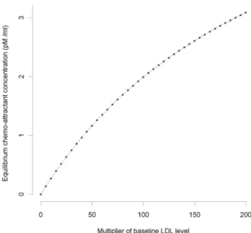
and the equilibrium chemo-attractant level Ceq. However, as is clear from Figures 1–2, only for very high levels of LDL, multiples in excess of 50 of the baseline levels (see Table S3), are there appreciable departures from linearity.
We shall often assume thatPeq~0. In all that follows we assume a limiting process, so thatlimgeq,Meq?0dM(geq=Meq)~dM(0)~dM,0, limgeq,Meq?0rin(geq=Meq)~rin(0)~rin,0. If we perform the obvious linearisations in equations (1)–(9) and ignore all second and higher order terms inE0,D,E1,D,:::,NDwe obtain:
dE0,D
dt ~{tE0,D{
X Nmax ,L{1
i~0
iLiE0,DLi ð31Þ
dE1,D
dt ~
X Nmax ,L{1
i~0
iLiE0,DLi{dEE1,D ð32Þ
LCD
Lt ~rCEE1,DzrCMMDzrCTTDzDC+
2C
D{dCMMDCeq
{dCTTDCeq{dCm½mDCeqzmeqCD
ð33Þ
LPD
Lt ~rPTTDzDP+
2P
D ð34Þ
LmD
Lt zmeqxm(C)+
2C
D
~meq½m{rMPDzPeq½m{rMmDzDm+2mD
ð35Þ
LMD
Lt ~rMmeqPDzrMPeqmD{dM(0)MDzDM+
2M
D ð36Þ
LgD
Lt ~rin(0)MDL0{dM(0)gD ð37Þ
LTD
Lt ~{dTTDzDT+
2T
D ð38Þ
LND
Lt ~dM(0)½gDzdMMMDzdTdTTTD ð39Þ
The boundary conditions (10)–(16), together with (17), (18) translate to:
Figure 1. MCP-1 concentration vs baseline LDL (small). Equilibrium chemo-attractant (MCP-1) concentration,Ceq, as a function
of small multiples of baseline LDL level (Table S3), using parameters given in Tables S2, S3.
doi:10.1371/journal.pcbi.1000539.g001
Figure 2. MCP-1 concentration vs baseline LDL (large). Equilibrium chemo-attractant (MCP-1) concentration,Ceq, as a function
of large multiples of baseline LDL level (Table S3), using parameters given in Tables S2, S3.
LCD
LP~ LPD
LP~ LmD
LP~ LMD
LP~ LgD
LP~ LTD
LP~ LND
LP~0 onLVI|LVLð40Þ
Green’s first identity (for general scalarC2functionsu,vand domain
V) states that:
ð
V
u+2vdx~{
ð
V
+u:+vdxz
ð
LV
uLv
LPds ð41Þ
Integrating (33)–(39) over the intima,VI, we have by (41) that:
d dt
ð
VI
CDdx~rCE ð
VI
E1,Ddxz½rCM{dCMCeq ð
VI MDdx
z½rCT{dCTCeq ð
VI
TDdx{dCm½Ceq ð
VI mDdx
zmeq ð
VI CDdx
ð42Þ
d dt
ð
VI
PDdx~rPT ð
VI
TDdx ð43Þ
d dt
ð
VI
mDdx~meq½m{rM ð
VI
PDdxzPeq½m{rM ð
VI
mDdx ð44Þ
d dt
ð
VI
MDdx~rMmeq ð
VI
PDdxzrMPeq ð
VI
mDdx{dM(0) ð
VI
MDdxð45Þ
d dt
ð
VI
gDdx~rin(0)
ð
VI
MDL0dx{dM(0) ð
VI
gDdx ð46Þ
d dt
ð
VI
TDdx~{dT ð
VI
TDdx ð47Þ
d dt
ð
VI
NDdx~dM(0)½ ð
VI
gDdxzdMM ð
VI
MDdxzdTdTT ð
VI
TDdxð48Þ
In Text S1 section B we outline solutions to (31)–(32), (42)–(48) in various cases. As shown there, iftw0anddEw0then:
limt??E0,D(t)~0 and limt??E1,D(t)~0 ð49Þ
From Table S2 it is clear that these conditions are likely to be always satisfied: t~4:6x10{6s{1w0 and dE~1x10{4s{1
w0. If Peq~0 then using the results of Text S1 section B ((B.11)–(B.16)):
ð
VI
CD(t)dx~
~
ð
VI
PD(0)dxz
rPT dT
ð
VI TD(0)dx 0 B @ 1 C A
½rCM{dCMCeqrM dM(0)dCm
{Ceq½m{rM t{
1
dCmmeq
2 6 6 6 4 3 7 7 7 5
{Ceq meq
ð
VI
mD(0)dx{
meq½m{rMrPT
Ð
VI TD(0)dx
dT2
0
B @
1
C
AzO( exp½{dt)
ð50Þ
ð
VI
PD(t)dx~
ð
VI
PD(0)dxz
rPT dT
ð
VI
TD(0)dxzO( exp½{dt) ð51Þ
ð
VI
mD(t)dx~
ð
VI mD(0)dx
zmeq½m{rM t Ð
VI
PD(0)dxz
rPT
dT Ð
VI TD(0)dx
!
{rPT dT2 Ð
VI TD(0)dx
8 > > > > < > > > > : 9 > > > > = > > > > ;
zO( exp½{dt)
ð52Þ
ð
VI
MD(t)dx~
rMmeq Ð
VI
PD(0)dxz
rPT
dT Ð
VI TD(0)dx
!
dM(0)
zO( exp½{dt)
ð53Þ
ð
VI
gD(t)dx~rin(0)L0 rMmeq
Ð
VI
PD(0)dxz
rPT dT
Ð
VI
TD(0)dx
!
dM(0)2
zO( exp½{dt)
ð54Þ
ð
VI
ND(t)dx~ ð
VI
ND(0)dxz ð
VI gD(0)dx
z
dTT{rin(0)L0rMmeqrPT dT2dM(0) {dMMrMmeqrPT
dT2
2 6 6 6 4 3 7 7 7 5 ð VI
TD(0)dxz rin(0)L0 dM(0)
zdMM
ð
VI
MD(0)dx ð
VI
PD(0)dxz
rPT
dT
ð
VI
TD(0)dx 0 B @ 1 C A
rin(0)L0rMmeq t{ 2 dM(0)
dM(0)
zdMMrMmeq t{ 1 dM(0)
2 6 6 6 6 6 4 3 7 7 7 7 7 5
zO( exp½{dt)
ð55Þ
for somedw0, so that in generalCD,PD,mD,MD,gD,ND do not
in particular that unlessm~rM andrin(0)L0zdM(0)dMM~0, or possibly (more likely) thatÐ
VI
PD(0)dx~
Ð
VI
TD(0)dx~0, then there
will be time trends in the averaged quantities for Ð
VI
CD(t)dx,
Ð
VI
mD(t)dxand
Ð
VI
ND(t)dx, so that in particular stability cannot be
re-attained.
In Figures 3–7 we plot the variation of the spatially-averaged chemo-attractant, C, derived assuming a non-zero equilibrium concentration of monocytes (Table S3) and using (B.1b’). The perturbation is assumed to take place via killing of monocytes in the intima, which in this case could be produced by ionizing radiation, and also via damage to endothelial cells, produced in the same way. We do not assume instantaneous changes in any of the other species, i.e.,TD(0)~MD(0)~PD(0)~0. The reason for this
is that by (28) Teq~Meq~Peq~0 (assuming as we do that
meqw0), so that radiation would not have any species to act on in
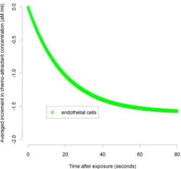
equilibrium. For the parameters used here (given in Tables S2 and S3), the overwhelming contribution is via monocyte killing: by 80 seconds the contribution from this term is 4.5610217M ml21
compared with a contribution of 21.6610218M ml21 via damage to endothelial cells. As can be seen, the change in chemo-attractant concentration occurs (for monocytes and in aggregate) relatively quickly, over a timescale of minutes, although the endothelial cell killing component varies more slowly, over a timescale of hours; after 24 hours this and all other averaged quantities are virtually constant. In contrast to the above general case, when only the monocyte population (of all the species) is perturbed, for a sufficiently long time after exposure (days or more) we have by (50), (52) that Ð
VI
CD(t)dx&{Cmeq
eq Ð
VI
mD(0)dxand
Ð
VI
mD(t)dx& Ð VI
mD(0)dx, and by (51), (53), (54) the averaged
change in all other species tends to zero. In this case we see that:
Figure 4. MCP-1 variation over 0–80 seconds after 10 mGy (endothelial).Spatial average (over intima) of increment in chemo-attractant (MCP-1) concentration after 10 mGy of acutely delivered radiation, using parameters given in Tables S2, S3. The component of changes in chemo-attractant (MCP-1) level due to endothelial cell killing 0–80 seconds after 10 mGy are shown.
doi:10.1371/journal.pcbi.1000539.g004
Figure 5. MCP-1 variation over 0–0.5 hours after 10 mGy (monocyte, total). Spatial average (over intima) of increment in chemo-attractant (MCP-1) concentration after 10 mGy of acutely delivered radiation, using parameters given in Tables S2, S3. The components of changes in chemo-attractant (MCP-1) level due to monocyte cell killing and total (monocyte+endothelial) cell killing 0–
0.5 hours after 10 mGy are shown. doi:10.1371/journal.pcbi.1000539.g005 Figure 3. MCP-1 variation over 0–80 seconds after 10 mGy
(monocyte, total). Spatial average (over intima) of increment in chemo-attractant (MCP-1) concentration after 10 mGy of acutely delivered radiation, using parameters given in Tables S2, S3. The components of changes in chemo-attractant (MCP-1) level due to monocyte cell killing and total (monocyte+endothelial) cell killing 0– 80 seconds after 10 mGy are shown.
Ceqz Ð
VI CD(t)dx
Ð
VI dx
2
6 4
3
7 5 meqz
Ð
VI mD(t)dx
Ð
VI dx
2
6 4
3
7 5
&Ceqmeq 1{
Ð
VI mD(t)dx
meq Ð
VI dx
2
6 4
3
7
5 1z
Ð
VI mD(t)dx
meq Ð
VI dx
2
6 4
3
7
5&Ceqmeq
ð56Þ
approximated to first order, so that by (30), at least in average, equilibrium can be re-established at these new values ofCandm. We conjecture that in fact equilibrium is re-established for all quantities in this case. It is easy to see that if there were to be further small perturbations in CD,PD,mD,MD,gD,TD,ND, at
intervals of days or more, the resulting changes in the spatially-averaged quantities would be approximately additive in the corresponding increments, as shown in Figure 7. Moreover, from (50) the excess chemo-attractant (MCP-1) in relation to the
monocyte perturbation is{Ceq meq Ð
VI
mD(0)dx. Therefore, so long as
the individual monocyte perturbations are small and temporally separated (by a day or more), the increment in chemo-attractant will not depend on anything other than the cumulative absorbed dose, as indicated in Figure 7.
In Figure 8 we plot the percent proportion of the population whose cumulative chemo-attractant (MCP-1) concentration ex-ceeds the threshold min½CTm,CTT; as we discuss below, this threshold is the critical point for system stability, exceedance of which makes development of cardiovascular disease much more likely. [The probability is derived assuming that the population distribution MCP-1 is Gaussian with mean and standard deviation (SD) determined by the adult female data of Cannonet al.[27]; the mean is augmented by the radiation-induced increment, given by (B.1b’).] For a range of threshold values between 0.25 and 1.00
Figure 8. Risk of MCP-1 exceeding threshold levelvs dose. Cumulative risk of exceeding of chemo-attractant (MCP-1) threshold (<cumulative risk of cardiovascular disease) min[CTm,CTT], as function of
radiation dose and threshold value (mean+multiple of population SD [27] (see Table S3)).
doi:10.1371/journal.pcbi.1000539.g008 Figure 6. MCP-1 variation over 0–0.5 hours after 10 mGy
(endothelial). Spatial average (over intima) of increment in chemo-attractant (MCP-1) concentration after 10 mGy of acutely delivered radiation, using parameters given in Tables S2, S3. The component of changes in chemo-attractant (MCP-1) level due to endothelial cell killing 0–0.5 hours after 10 mGy are shown.
doi:10.1371/journal.pcbi.1000539.g006
Figure 7. MCP-1 variation over time after 10 mGy/day.As for Figures 3–6, but assuming fractionated multiple radiation doses, 10 mGy/day.
times the SD in excess of the mean, we have baseline risks of exceeding the threshold (i.e., cardiovascular disease) of 16–40%. Most developed countries have cumulative cardiovascular disease mortality in the range 20–40% and the world mean is 30% [28], so that this range of values of the MCP-1 threshold, min½CTm,CTT, is plausible. For this range of threshold values, Figure 8 demonstrates that risks vary remarkably linearly with dose over the dose interval 0–4 Gy. As for Figure 7, the risk will not depend on anything other than the cumulative absorbed dose, as long as this is given in small daily increments.
Discussion
We have outlined a model for early stage atherosclerotic lesion formation, and performed a stability analysis for a simplified version of the model. While some components of the system (in particular the T-lymphocyte concentration, T) are stable, in the sense that after perturbations of the system the species concentrations return to their equilibrium value, various other species, in particular the proliferation factor concentration,P, the chemo-attractant concentration, C, the monocyte concentration,m, and the necrotic core,N, are generally not stable. In particular, the mean level of chemo-attractant increases continuously and rapidly after instantaneous perturbation by a radiation dose, over a timescale of minutes. However, as we note below, because of cellular repair processes, which are not taken into account in our model, there are reasons for assuming that perturbation
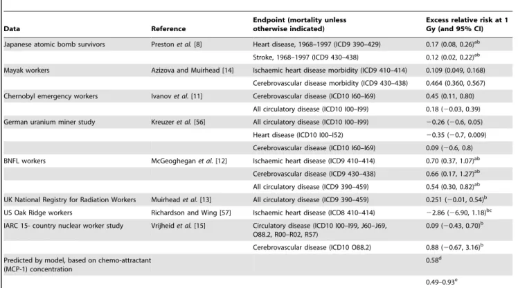
by radiation would not be instantaneous, so that this process might be extended over at least a period of hours after exposure. The main driver for the increase in chemo-attractant is the death of monocytes and the consequent reduction in monocyte-induced degradation in chemo-attractant concentration, the{dCmmCterm in (3). It is well known that radiation can cause cell death [29], and the degree of cell killing and damage that we assume is consistent with radio-biological expectation [30,31]. Although the change in chemo-attractant (MCP-1) concentration that we assume after 10 mGy is relatively modest, 4.5610217M ml21, a fractionated dose of 1 Gy would result in 4.5610215M ml21, comparable with the normal concentration of MCP-1 in adult plasma, 7.9610215M ml21[27]. The fact that the range of excess relative risks predicted by our model, 0.49–0.93 Gy21, is consistent with those in a number of occupational studies (Table 1) adds to the plausibility of this mechanism.
We have also shown that the model predicts that equilibrium level of chemo-attractant (MCP-1) increases more or less directly with levels of LDL, and in particular oxidized LDL, with slight non-linearity at very high levels of MCP-1. This is in accordance with experimental [32,33] and epidemiological observations [34]. Specifically, there is experimental evidence that addition of minimally-oxidised LDL results in a <22-fold increase in levels
of MCP-1 in ECs in anin vitroco-culture system [32]. In a group of baboons fed a high cholesterol, high fat diet, oxLDL in serum increased by about 19.6% (95% 228.9, 68.1) after 7 weeks, resulting in an increase in serum MCP-1 at that point of 66.7%
Table 1.Risks in various human cohorts, and predicted by model.
Data Reference
Endpoint (mortality unless otherwise indicated)
Excess relative risk at 1 Gy (and 95% CI) Japanese atomic bomb survivors Prestonet al.[8] Heart disease, 1968–1997 (ICD9 390–429) 0.17 (0.08, 0.26)ab
Stroke, 1968–1997 (ICD9 430–438) 0.12 (0.02, 0.22)ab
Mayak workers Azizova and Muirhead [14] Ischaemic heart disease morbidity (ICD9 410–414) 0.109 (0.049, 0.168)
Cerebrovascular disease morbidity (ICD9 430–438) 0.464 (0.360, 0.567)
Chernobyl emergency workers Ivanovet al.[11] Cerebrovascular disease (ICD10 I60–I69) 0.45 (0.11, 0.80)
All circulatory disease (ICD10 I00–I99) 0.18 (20.03, 0.39)
German uranium miner study Kreuzeret al.[56] All circulatory disease (ICD10 I00–I99) 20.26 (20.6, 0.05)
Heart disease (ICD10 I00–I52) 20.35 (20.7, 0.009)
Cerebrovascular disease (ICD10 I60–I69) 0.09 (20.6, 0.8)
BNFL workers McGeogheganet al.[12] Ischaemic heart disease (ICD9 410–414) 0.70 (0.37, 1.07)ab
Cerebrovascular disease (ICD9 430–438) 0.66 (0.17, 1.27)ab
All circulatory disease (ICD9 390–459) 0.54 (0.30, 0.82)ab
UK National Registry for Radiation Workers Muirheadet al.[13] All circulatory disease (ICD9 390–459) 0.251 (20.01, 0.54)b
US Oak Ridge workers Richardson and Wing [57] Ischaemic heart disease (ICD8 410–414) 22.86 (26.90, 1.18)bc
IARC 15- country nuclear worker study Vrijheidet al.[15] Circulatory disease (ICD10 I00–I99, J60–J69, O88.2, R00–R02, R57)
0.09 (20.43, 0.70)b
Cerebrovascular disease (ICD10 O88.2) 0.88 (20.67, 3.16)b
Predicted by model, based on chemo-attractant (MCP-1) concentration
0.58d
0.49–0.93e
Excess relative risks (per Gy) of cardiovascular disease in the Japanese atomic bomb survivors and in various occupationally exposed groups, compared with excess relative chemo-attractant (MCP-1) concentration at 1 Gy predicted by model.
a90% CI.
bExcess relative risk Sv21. cAssuming 10 year lag.
d1 Gy assumed given as 100 daily doses of 10 mGy, ERR evaluated by dividing excess MCP-1 concentration by baseline level from data of Cannonet al.[27]. eERR at 1 Gy of cumulative risk of exceeding threshold (
<cumulative cardiovascular risk), as given by Figure 8, for levels of MCP-1 threshold min[CTm,CTT] in the range [mean+0.25 SD, mean+1.00 SD], mean and SD taken from data of Cannonet al.[27].
(95% 54.2, 79.1) [33]. Both of these are consistent with the linear relationship (without constant term) predicted by our model (30). If radiation dose were to be given in a fractionated manner, with doses separated by a period of hours or more, the model predicts that chemo-attractant (e.g., MCP-1) would increase linearly with cumula-tive accumulated dose, with a corresponding decrease in the intimal monocyte concentration, as shown in Figure 7. This would carry on until the chemo-attractant concentration at the boundary,C, exceeds one or other of the thresholds CTm,CTT, beyond which point an equilibrium solution is no longer possible. At these points, there would be increased trans-intimal flux of monocytes and T-lymphocytes from the lumen, which would result (via (48)) in a continuous increase in necrotic lesion size, and therefore risk of atherosclerosis. The doses used here are moderate (10 mGy/day), such as might occur in occupational exposure settings, and would account for the observed radiation-associated excess risk that has been seen in various groups of nuclear workers [10–14]. The model implies that at least until the chemo-attractant thresholdmin½CTm,CTTis exceeded the system is stable, assuming that the conjecture we make after (56) is valid.
If the chemo-attractant thresholdmin½CTm,CTTis exceeded as a result of the perturbation term,CD, resulting in monocyte or
T-lymphocyte flux across the EC layer, then extra terms need adding to the right hand side of (44) and (47),
{DmmLbm,1
ð
LVL
CD1bm,0zbm,1½CeqzCDw01CeqzCDwCTmdx ð57Þ
and
{DTTLbT,1
ð
LVL
CD1bT,0zbT,1½CeqzCDw01CeqzCDwCTTdx ð58Þ
respectively. Apparently paradoxically, if Ceqvmin½CTm,CTT,
then we must have CDw0 for the terms inside the integrals to
contribute non-trivially, and so these terms will be negative and therefore tend to reduce the averaged levels of monocytes and T-lymphocytes in the system. By (42) this will tend to increase the chemo-attractant concentration still further. In other words, once this chemo-attractant threshold is crossed the system tends (on average) to become yet more unstable.
There are of course other agents that damage monocytes or ECs that would cause the chemo-attractant level to increase, so that although for an individual this threshold might never be passed, in a large population there would be a continuous (and approxi-mately linear) increase in cardiovascular risk with dose as shown in Figure 8. The same phenomenon would also occur at higher doses (e.g., at radiotherapeutic levels of dose), at a correspondingly higher level, although the relative magnitude of the perturbations would make the neglect of all but first order perturbations that we assumed in deriving (31)–(39) possibly invalid; there is abundant evidence of radiation-induced disease in groups exposed to certain forms of RT [6,7]. Critical to our model, and indeed the understanding of atherosclerosis, is whether there really are such thresholds in chemo-attractant levels for the trans-intimal monocyte and T-lymphocyte flux. We assume the presence of such thresholds for the purposes of our stability analysis, as we have to if there is to be a stable solution, but it is possible nevertheless that these thresholds are zero, in which case, assuming the model is correct, the atherosclerotic process must be inherently unstable. As indicated above, if this is model is correct and is to be consistent with the observed cumulative cardiovascular disease mortality in developed populations [28],
then the chemo-attractant (MCP-1) threshold must lie in the range [mean+0.256population SD, mean+1.006population SD] (the
mean and population SD being as in Cannonet al.[27]), in other words [1.0, 1.7]610214M ml21.
We implicitly assume that atherosclerosis is mainly responsible for the observed excess risk of cardiovascular morbidity or mortality following fractionated low-dose irradiation of the heart and major arteries. This assumption is supported by experimental data inApoE2/2mice [35]. However, some human symptoms are due to (myocardial) ischaemia which could be caused by either macrovascular (atherosclerotic) or microvascular damage. At higher (radiotherapy) doses, both human and animal data suggest that both types of lesion occur [6]. Although the generally high prevalence of atherosclerosis in humans suggests that this is the more probable cause of ischaemia following low-dose radiation, it is possible that microvascular disease also plays a role. It should be noted that we have been addressing mechanisms for induction of atherosclerosis following fractionated low-dose radiation to the large arteries (coronary, carotid etc). There is a large literature on fibrotic, pericardial, myocardial and other morbidity sequelae of high-dose irradiation of the heart and large arteries, both for humans and animals [6]. The pro-inflammatory mechanisms for these are reasonably well understood, and quite different from those hypothesized here [36]. That the true mechanisms for low-dose effects are likely to be very different is also suggested by the pronounced fractionation effect seen for high-dose exposure in relation to heart failure in rats [37,38], in contrast to the somewhat lower risks observed in the Japanese atomic bomb survivors compared with occupationally exposed groups (Table 1).
Indirect mechanisms for the action of radiation could also be postulated. At high doses it is clear that inflammatory markers are up-regulatedin vitroandin vivo, although at lower doses if anything the evidence points to down-regulation of inflammation [16]. In terms of the model this could be mediated by an increase in radical flux, which could, via lipid peroxidation, lead to EC damage. This in turn would lead to an increase in the chemo-attractant signal. Radiation is known to cause long-term variation in certain T-cell subpopulations (CD4+) in the Japanese atomic bomb survivors [39], and this mechanism could also be readily incorporated in the model. Long-term radiation-associated changes in cholesterol concentration have been observed in the Japanese atomic bomb survivors [40], presumably a result of some change in liver metabolism; these too could be easily incorporated in the model. It is of interest in this respect that there is a highly statistically significant trend with internal (plutoniuma-particle) dose to the liver for ischaemic heart disease and cerebrovascular disease in the latest analysis of the Mayak worker data [14]. Set against that, there is little evidence of excess risk of circulatory disease risk, specifically cardiac disease in groups exposed to the diagnostic contrast medium Thorotrast, which delivered a substantial a-particle liver dose [41,42].
cardiovascular risk, not doing so would imply a modest negative bias in modifications of the radiation response by age at exposure. Whilst the inflammatory process is recognized as an integral part of the atherosclerotic process [5] it does not explain the observation that the proliferation of vascular smooth muscle cells (VSMC) during atherosclerotic plaque development appears to be monoclonal [46]. Clonality suggests that plaque VSMCs must have undergone multiple rounds of division, and telomere loss studies argue that this is between 7–13 cumulative population doublings [47]. However, clonality itself is not synonymous with transformation of a single cell, and subsequent studies have shown that large patches in the normal vessel media are monoclonal [48,49]. Thus, clonality is more likely to be explained by the presence of developmental clones in the normal vessel wall, rather than a mutation. Finally, in contrast to tumours, plaque VSMCs show poor proliferation, enhanced apoptosis, and early senescence [50]. These features would not confer a proliferative or survival advantage to plaque VSMCs. Furthermore, plaque VSMC proliferation is now seen to be beneficial in atherosclerosis [51], so that the pathological consequences of a mutation promoting VSMC proliferation are unclear.
The limitations of the modelling performed here should be acknowledged. Even in the fuller model considered in Text S1 section A there is much biology not included – simplifications have been made for analytical simplicity. Although not strictly a defect in the model, we assume in our motivating example that a certain (dose-dependent) fraction of the monocytes are killed instantaneously by radiation exposure. The magnitude of this fraction is based on data from a human bone-marrow colony-forming assay (for cells under hypoxic conditions) of Gordon [30] (Table S3), performed 9 days after irradiation. It is known that cells take a variable length of time to die after irradiation, as a result of the repair and mis-repair processes they are thought to be subject to [52]. As such, a possibly more realistic scenario would have assumed this total cell damage exponentially distributed over time rather than occurring instanta-neously. However, it is unlikely that the variable delay in expression of monocyte mortality, which is likely to be 99.7% complete within three hours of irradiation [52], will make much difference to the predictions of our model, concerned as it largely is with the consequences of fractionated radiation doses separated by days or more. It would not be too difficult to modify the equations (5), (6) and (8) to incorporate the simple repair-misrepair model outlined in Brenneret al.[52], although for the purposes of the present paper we regard this as an unnecessary elaboration.
That said, the simpler model proposed here we trust captures what is known about the main features of interaction of oxidized LDL and various other molecular species (MCP-1, G-CSF, bound lipid) with the various cellular species (monocytes, macrophages, T-lymphocytes) that are known to be of significance for induction of atherosclerosis. The mathematics underlying these reaction and diffusion processes is reasonably standard. What is interesting and novel about the present paper is that using only experimentally derived parameters (taken wherever possible from human data) (Tables S2, S3) we have reproduced what is observed in other experimental and epidemiologic data (Figures 7–8, Table 1).
This proposed mechanism would in principle be experimentally testable. This would best be donein vitro, looking for changes in
MCP-1 levels, or other potential chemo-attractants, in a co-culture system similar to that developed by Takakuet al.[25]. This could be explored under a range of radiation exposure conditions (both localized and fractionated) and subsequent effects on, for example, adhesion properties could also be examined.In vivo experiments would be more complex (and expensive), but could also be performed, for example, using the ApoE2/2 knockout mouse model employed by Stewartet al.[35,53]. Even human data could be envisaged. In particular, if arterial tissue could be sampled from patients who have, a short time previously, received low-dose radiotherapy or high-dose diagnostic procedures (e.g., computer-ized tomography), together with suitable (age-matched) controls, one could determine whether intimal concentration of MCP-1 was significantly increased and the manner in which concentration changed with dose.
If the proposed mechanism were true, it also has substantive implications for radiological protection, which at present does not take cardiovascular disease into account [54]. Analysis of the Japanese atomic bomb survivor data implies that non-cancer disease mortality, in particular cardiovascular mortality, contrib-utes almost equally as cancer mortality to the radiogenic excess risk [8]. The major uncertainty in assessing the low-dose risk of cardiovascular disease is the shape of the dose response relationship, which is very unclear in the Japanese data [8,55]. The analysis of the present paper suggests that linear extrapolation would be generally appropriate for this endpoint.
Supporting Information
Text S1 Text S1 (sections A, B)
Found at: doi:10.1371/journal.pcbi.1000539.s001 (0.46 MB DOC)
Table S1 Candidate molecules for variables in the model.
Found at: doi:10.1371/journal.pcbi.1000539.s002 (0.04 MB DOC)
Table S2 Parameters and estimated values.
Found at: doi:10.1371/journal.pcbi.1000539.s003 (0.35 MB DOC)
Table S3 Values for perturbed species.
Found at: doi:10.1371/journal.pcbi.1000539.s004 (0.07 MB DOC)
Acknowledgments
The authors are grateful for the detailed and helpful comments of an associate editor and three referees. The Mayak worker analysis by Drs Azizova and Muirhead, which was cited in this paper, was conducted with support from the European Commission’s Euratom Nuclear Fission and Radiation Protection Programme as part of the SOUL project; more details of this analysis can be found in separate papers by the study investigators.
Author Contributions
Conceived and designed the experiments: MPL. Performed the experi-ments: MPL. Analyzed the data: MPL AG IT. Wrote the paper: MPL AG IT.
References
1. Murray C, Lopez A (1997) Alternative projections of mortality and disability by cause 1990–2020: Global Burden of Disease Study. Lancet 249: 1498–1505. 2. Ross R (1995) Cell biology of atherosclerosis. Annual Rev Physiol 57: 791–804. 3. Ross R (1999) Atherosclerosis is an inflammatory disease. Am Heart J 138:
S419–420.
4. Lusis A (2000) Atherosclerosis. Nature 407: 233–241.
5. Hansson GK (2005) Inflammation, atherosclerosis, and coronary artery disease. N Engl J Med 352: 1685–1695.
7. McGale P, Darby SC (2005) Low doses of ionizing radiation and circulatory diseases: a systematic review of the published epidemiological evidence. Radiat Res 163: 247–257, 711.
8. Preston DL, Shimizu Y, Pierce DA, Suyama A, Mabuchi K (2003) Studies of mortality of atomic bomb survivors. Report 13: solid cancer and noncancer disease mortality: 1950–1997. Radiat Res 160: 381–407.
9. Yamada M, Wong FL, Fujiwara S, Akahoshi M, Suzuki G (2004) Noncancer disease incidence in atomic bomb survivors, 1958–1998. Radiat Res 161: 622–632.
10. Howe GR, Zablotska LB, Fix JJ, Egel J, Buchanan J (2004) Analysis of the mortality experience amongst U.S. nuclear power industry workers after chronic low-dose exposure to ionizing radiation. Radiat Res 162: 517–526.
11. Ivanov VK, Maksioutov MA, Chekin SY, Petrov AV, Biryukov AP, et al. (2006) The risk of radiation-induced cerebrovascular disease in Chernobyl emergency workers. Health Phys 90: 199–207.
12. McGeoghegan D, Binks K, Gillies M, Jones S, Whaley S (2008) The non-cancer mortality experience of male workers at British Nuclear Fuels plc, 1946–2005. Int J Epidemiol 37: 506–518.
13. Muirhead CR, O’Hagan JA, Haylock RGE, Phillipson MA, Willcock T, et al. (2009) Mortality and cancer incidence following occupational radiation exposure: third analysis of the National Registry for Radiation Workers. Br J Cancer 100: 206–212.
14. Azizova TV, Muirhead CR (2009) Epidemiological evidence for circulatory diseases – occupational exposure. EU Scientific Seminar 2008. ‘‘Emerging evidence for radiation induced circulatory diseases’’. Proceedings of a scientific seminar held in Luxembourg on 25 November 2008. Radiat Prot 158: 33–46. (downloadable from http://ec.europa.eu/energy/nuclear/radiation_protection/ doc/publication/158.pdf).
15. Vrijheid M, Cardis E, Ashmore P, Auvinen A, Bae J-M, et al. (2007) Mortality from diseases other than cancer following low doses of ionizing radiation: results from the 15-Country Study of nuclear industry workers. Int J Epidemiol 36: 1126–1135.
16. Little MP, Tawn EJ, Tzoulaki I, Wakeford R, Hildebrandt G, et al. (2008) A systematic review of epidemiological associations between low and moderate doses of ionizing radiation and late cardiovascular effects, and their possible mechanisms. Radiat Res 169: 99–109.
17. Little MP, Tawn EJ, Tzoulaki I, Wakeford R, Hildebrandt G, et al. (2009) Review and meta-analysis of epidemiological associations between low/ moderate doses of ionizing radiation and circulatory disease risks, and their possible mechanisms. Radiat Environ Biophys;re-submitted.
18. Hayashi T, Kusunoki Y, Hakoda M, Morishita Y, Kubo Y, et al. (2003) Radiation dose-dependent increases in inflammatory response markers in A-bomb survivors. Int J Radiat Biol 79: 129–136.
19. Hayashi T, Morishita Y, Kubo Y, Kusunoki Y, Hayashi I, et al. (2005) Long-term effects of radiation dose on inflammatory markers in atomic bomb survivors. Am J Med 118: 83–86.
20. McKay C, McKee S, Mottram N, Mulholland T, Wilson S, et al. (2005) Towards a model of atherosclerosis. Strathclyde Mathematics Research Report (March 2005), No. 4.
21. Cobbold C, Sherratt J, Maxwell S (2002) Lipoprotein oxidation and its significance for atherosclerosis: a mathematical approach. Bull Math Biol 64: 65–95.
22. Ibragimov AI, McNeal CJ, Ritter LR, Walton JR (2005) A mathematical model of atherogenesis as an inflammatory response. Math Med Biol 22: 305–333. 23. Keller EF, Segel LA (1971) Model for chemotaxis. J Theoret Biol 30: 225–234. 24. Keller EF, Segel LA (1971) Traveling bands of chemotactic bacteria: a
theoretical analysis. J Theoret Biol 30: 235–248.
25. Takaku M, Wada Y, Jinnouchi K, Takeya M, Takahashi K, et al. (1999) An in vitro coculture model of transmigrant monocytes and foam cell formation. Arterioscler Thromb Vasc Biol 19: 2330–2339.
26. Klouche M, May AE, Hemmes M, Messner M, Kanse SM, et al. (1999) Enzymatically modified, nonoxidized LDL induces selective adhesion and transmigration of monocytes and T-lymphocytes through human endothelial cell monolayers. Arterioscler Thromb Vasc Biol 19: 784–793.
27. Cannon JG, Sabatier MJ, Marinik EL, Schwark EH, Haddow S, et al. (2009) Femoral artery diameter and arteriogenic cytokines in healthy women. Vascul Pharmacol 50: 104–109.
28. World Health Organisation Statistical Information System (WHOSIS) (2009) http://www.who.int/whosis/en/.
29. United Nations Scientific Committee on the Effects of Atomic Radiation (UNSCEAR) (1993) Sources and effects of ionizing radiation. UNSCEAR 1993 report to the General Assembly, with scientific annexes. New York: United Nations. 30. Gordon MY (1975) A comparison of the radiosensitivity and O.E.R. of human and mouse marrow progenitor cells cultured in agar in diffusion chambers. Int J Radiat Biol 28: 285–290.
31. Deschavanne PJ, Fertil B (1996) A review of human cell radiosensitivityin vitro. International Journal of Radiat Oncol Biol Phys 34: 251–266.
32. Cushing SD, Berliner JA, Valente AJ, Territo MC, Navab M, et al. (1990) Minimally modified low density lipoprotein induces monocyte chemotactic
protein 1 in human endothelial cells and smooth muscle cells. Proc Natl Acad Sci USA 87: 5134–5138.
33. Shi Q, Vandeberg JF, Jett C, Rice K, Leland MM, et al. (2005) Arterial endothelial dysfunction in baboons fed a high-cholesterol, high-fat diet. Am J Clin Nutr 82: 751–759.
34. Kowalski J, Okopien´ B, Madej A, Zielin´ski M, Belowski D, et al. (2003) Effects of atorvastatin, simvastatin, and fenofibrate therapy on monocyte chemoattractant protein-1 secretion in patients with hyperlipidemia. Eur J Clin Pharmacol 59: 189–193.
35. Hoving S, Heeneman S, Gijbels MJJ, te Poele JAM, Russell NS, et al. (2008) Single-dose and fractionated irradiation promote initiation and progression of atherosclerosis and induce an inflammatory plaque phenotype in ApoE2/2
mice. Int J Radiat Oncol Biol Phys 71: 848–857.
36. Schultz-Hector S, Trott K-R (2007) Radiation-induced cardiovascular diseases: is the epidemiologic evidence compatible with the radiobiologic data? Int J Radiat Oncol Biol Phys 67: 10–18.
37. Lauk S, Ru¨th S, Trott KR (1987) The effects of dose-fractionation on radiation-induced heart disease in rats. Radiother Oncol 8: 363–367.
38. Schultz-Hector S, Sund M, Thames HD (1992) Fractionation response and repair kinetics of radiation-induced heart failure in the rat. Radiother Oncol 23: 33–40.
39. Kusunoki Y, Kyoizumi S, Hirai Y, Suzuki T, Nakashima E, et al. (1998) Flow cytometry measurements of subsets of T, B and NK cells in peripheral blood lymphocytes of atomic bomb survivors. Radiat Res 150: 227–236.
40. Wong FL, Yamada M, Sasaki H, Kodama K, Hosoda Y (1999) Effects of radiation on the longitudinal trends of total serum cholesterol levels in the atomic bomb survivors: 1958–1986. Radiat Res 151: 736–746.
41. Travis LB, Land CE, Andersson M, Nyberg U, Goldman MB, et al. (2001) Mortality after cerebral angiography with or without radioactive Thorotrast: an international cohort of 3,143 two-year survivors. Radiat Res 156: 136–150. 42. dos Santos Silva I, Malveiro F, Jones ME, Swerdlow AJ (2003) Mortality after
radiological investigation with radioactive Thorotrast: a follow-up study of up to fifty years in Portugal. Radiat Res 159: 521–534.
43. Richardson RB (2008) Age-dependent changes in oxygen tension, radiation dose and sensitivity within normal and diseased coronary arteries - Part A: Dose from radon and thoron. Int J Radiat Biol 84: 838–848.
44. Richardson RB (2008) Age-dependent changes in oxygen tension, radiation dose and sensitivity within normal and diseased coronary arteries - Part B: Modeling oxygen diffusion into vessel walls. Int J Radiat Biol 84: 849–857.
45. Richardson RB (2008) Age-dependent changes in oxygen tension, radiation dose and sensitivity within normal and diseased coronary arteries - Part C: Oxygen effect and its implications on high- and low-LET dose. Int J Radiat Biol 84: 858–865.
46. Benditt EP, Benditt JM (1973) Evidence for a monoclonal origin of human atherosclerotic plaques. Proc Natl Acad Sci USA 70: 1753–1756.
47. Matthews C, Gorenne I, Scott S, Figg N, Kirkpatrick P, et al. (2006) Vascular smooth muscle cells undergo telomere-based senescence in human atheroscle-rosis: effects of telomerase and oxidative stress. Circ Res 99: 156–164. 48. Chung I-M, Schwartz SM, Murry CE (1998) Clonal architecture of normal and
atherosclerotic aorta - Implications for atherogenesis and vascular development. Am J Pathol 152: 913–923.
49. Schwartz SM, Murry CE (1998) Proliferation and the monoclonal origins of atherosclerotic lesions. Annu Rev Med 49: 437–460.
50. Zhang QJ, Goddard M, Shanahan C, Shapiro L, Bennett M (2002) Differential gene expression in vascular smooth muscle cells in primary atherosclerosis and in stent stenosis in humans. Arterioscler Thromb Vasc Biol 22: 2030–2036. 51. Braganza DM, Bennett MR (2001) New insights into atherosclerotic plaque
rupture. Postgrad Med J 77: 94–98.
52. Brenner DJ, Hlatky LR, Hahnfeldt PJ, Huang Y, Sachs RK (1998) The linear-quadratic model and most other common radiobiological models result in similar predictions of time-dose relationships. Radiat Res 150: 83–91. 53. Stewart FA, Heeneman S, te Poele J, Kruse J, Russell NS, et al. (2006) Ionizing
radiation accelerates the development of atherosclerotic lesions in ApoE2/2
mice and predisposes to an inflammatory plaque phenotype prone to hemorrhage. Am J Pathol 168: 649–658.
54. International Commission on Radiological Protection. The 2007 Recommen-dations of the International Commission on Radiological Protection. ICRP Publication 103. Annals ICRP 2007; 37 (2–4): i–ii+1–332.
55. Little MP (2004) Threshold and other departures from linear-quadratic curvature in the non-cancer mortality dose-response curve in the Japanese atomic bomb survivors. Radiat Environ Biophys 43: 67–75.
56. Kreuzer M, Kreisheimer M, Kandel M, Schnelzer M, Tschense A, et al. (2006) Mortality from cardiovascular diseases in the German uranium miners cohort study, 1946–1998. Radiat Environ Biophys 45: 159–166.