Original Article
A Survey on the Possibility of Utilizing
H2AX as a Biodosimeter in Radiation
Workers
Mostafa Mir 1, Yaghoub Yazdani 2, Jahanbakhsh Asadi 1, Alireza Khoshbin Khoshnazar 1
Abstract
Introduction
DNA damage is among the main consequences of radiation. Of many different classes of DNA damage, double-strand breaks are the most deleterious. Development of a sensitive biodosimetry method, which utilizes a detection material with a similar construction to the body, seems essential for monitoring radiation workers. In this study, histone H2AX protein was examined as a potential biodosimeter in radiation workers. Moreover, the presence of this protein after in vitro irradiation of blood samples was assessed simultaneously.
Materials and Methods
Blood samples from 46 radiation workers were analyzed in Golestan province, Iran. Meanwhile, two groups of blood samples (five blood samples in each group) were irradiated in vitro by doses of 1 to 0.2 Gy and 0.09 to 0.01 Gy from a 60Co source, respectively. H2AX level in lymphocytes was measured, using Western blot technique. ANOVA and Tukey’s tests were performed, using SPSS version 16. The significance level was considered to be 0.05.
Results
The results of Western blotting for the identification of H2AX protein in radiation workers were negative. However, H2AX level in lymphocytes of two in vitro irradiated groups showed a significant correlation with the radiation dose (P<0.0001).
Conclusion
The results showed that H2AX was a good indicator for acute or local exposure to ionizing radiation, while in chronically exposed individuals, including radiation workers, this protein was useless at least in autoradiography detection method. Regarding the presence of H2AX protein in blood samples, which were irradiated in vitro at low doses, it can be concluded that this protein has powerful repair mechanisms.
Keywords: Biodosimetry; H2AX; Radiation Worker
1- Department of Clinical Biochemistry, School of Medicine, Golestan University of Medical Sciences, Gorgan, Iran 2- Infectious Diseases Research Center and Laboratory Sciences Research Center, Golestan University of Medical
1. Introduction
Today, application of radiation is an undeniable necessity in human life [1]. Many investigators have noted the significant effects of radiation on health. As the review of the history of radiobiology indicates, the biological effects of high-dose radiation were known after the discovery of X-ray and radioactivity.
However, the effects of low-dose radiation on human health are still unclear and under investigation [2]. In fact, the threshold dose, i.e., the dose above which radiation leads to adverse effects on the body, has not been yet established [3]. Nevertheless, the maximum allowed dosage for radiation workers is determined to be 20 mSv per year. According to the guidelines by the International Commission on Radiation Protection (ICRP), to avoid the possible adverse effects of radiation such as cancer and genetic diseases, the annual received dose to general populations should not exceed 1 mSv [4]. In vivo studies on radiation workers, who are occupationally exposed to low levels of radiation, could provide a more accurate threshold level [5]. Dicentric chromosome count in the metaphase spread of stimulated lymphocytes is a common gold standard for biological dosimetry [6]. This gold standard is specific to ionizing radiation and remains unchanged for several months after receiving ionizing radiation [6]. However, at least a 52-hour interval is required between the time of blood sampling and dose estimation, which is not ideal [6, 7].
DNA double-strand break (DSB) is one of the main consequences of exposure to ionizing radiation. DSB is arguably the most dangerous DNA lesion that could induce genomic and cancerous changes in cells and lead to mortality at high levels [8]. Recently, an altered protein has been suggested as a potential indicator for exposure to ionizing radiation, which is significantly associated with radiation-induced DSB [9].
Histone H2AX, a variant of histone H2A, becomes phosphorylated on serine residue 139 in response to radiation exposure, so-called gamma-H2AX, which signals the repair of DSBs [10]. Protein detection seems to be a suitable method for initial triage, as responses to radiation begin within a few minutes and remain stable for several days after exposure [11]. The presence of gamma-H2AX protein can be detected by sensitive, quantitative, or semi-quantitative methods [11].
Various human specimens including lymphocytes, mouth cells, and skin can be used for the detection of gamma-H2AX after ionizing radiation [12]. Despite the abovementioned points, no comprehensive study has shown the efficacy of gamma-H2AX as a reliable biological dosimeter in radiation workers. Therefore, in this complementary study, we examined gamma-H2AX as a biological dosimeter in radiation workers, employed by hospitals in Golestan province, Iran. Lymphocytes from individuals, chronically exposed to low doses of ionizing radiation from CT scan, radiation therapy, nuclear medicine imaging, and diagnostic radiology, were gathered and analyzed.
2. Materials and Methods
2.1. Study subjects
The test subjects included 46 radiation workers (27 females and 19 males), within the age range of 23-52 years, who were occupationally exposed to chronic doses of radiation while performing their routine tasks at diagnostic radiology units, nuclear medicine departments, and CT scan facilities in Golestan province, Iran. Their work experience ranged from 10.5 months to 27 years.
demographic information, work experience, previous radiation therapy, prior chemotherapy, smoking history, medication use, and prior history of receiving radiation exceeding the standard limit. In case such information was not accessible, the subject was excluded from the study.
The peripheral blood of subjects was collected in tubes containing ethylenediaminetetraacetic acid (EDTA). The presence of H2AX in peripheral blood lymphocytes was assessed at the Clinical Biochemistry Laboratory of Golestan University of Medical Sciences. Lymphocytes were separated from other blood components by ficoll solution at 2500 rpm within 10 min. The cells were transferred to another tube and washed at 2500 rpm for 5 min three times in cold phosphate-buffered saline (PBS) solution.
2.2. In vitro irradiation
At this stage, blood samples were collected from a healthy subject, based on the mentioned questionnaire. The blood samples were distributed among several aliquots in plastic tubes. Three blood samples were irradiated at each dose at room temperature using a 60C0 source. The absorbed dose by blood samples was determined at 1, 0.8, 0.6, 0.4, 0.2, 0.09, 0.07, 0.05, 0.03, and 0.01 Gy with a rate of 67.55 cGy/min.
The calculated irradiation time ranged from 4.88 to 0.05 min. Lymphocytes were separated from other blood components by ficoll solution at 2500 rpm within 10 min. The cells were transferred to another tube and washed at 2500 rpm for 5 min three times in a cold PBS solution.
2.3. Extraction of cellular proteins
The lymphocytes in tubes were added to lysis buffer, containing 5% sodium dodecyl sulfate (SDS), 1 ml of Triton X-100, 100 mM of Tris-HCL (pH=7.4), 150 mM of NaCl, 5 mM of EDTA, 5% sodium deoxycholate, and 10% glycerol. The obtained solution was added to 10 μl/ml of phosphatase inhibitor cocktail
quantified by Pierce BCA Protein Assay Kit (Thermo, 23227), using bovine serum albumin (BSA) to form a standard curve.
2.4. Western blot analysis
Proteins are normally separated based on their
molecular weight in SDS-PAGE
electrophoresis. In our experiment, we used 15% gel, and then, 5x sample loading buffer, containing 10% SDS, 0.5% bromophenol blue, 60 mM of Tris-HCI (pH 6.8), 50% glycerol, and 14.4 Mm of 2-mercaptoethanol, was added to each tube and subsequently boiled for 5 min.
For blots, 100 µg of protein samples was loaded in each well. The sample proteins were separated on a 15% polyacrylamide gel and transferred to nitrocellulose membranes (Thermo) for 48 min at 110 V, using transfer buffer (15.6 Mm of Tris-base, 120 Mm of glycine, and 10% methanol, pH=8.4). The blots were incubated in 3% skim milk in PBS-T (0.05% PBS-Tween-20 at room temperature for 2-3 hrs on a rotating platform).
The blots were incubated overnight on a rotating platform at 4 °C in anti-phosphorylated histone H2A (Millipore, 05-636), with the dilution of 1:1000 in 3% skim milk/PBS-T. The blots were rinsed three times with BPS-T for 5, 19 and 15 min, respectively at room temperature on a rotating platform. They were then incubated with Goat Anti-Mouse IgG and horseradish peroxidase-conjugated secondary antibodies (Millipore, 12-349) at a dilution of 1:2000 in 3% milk/PBS-T for 1-2 hrs at room temperature on a rotating platform.
2.5. Statistical analysis
Statistical tests were performed using SPSS version 16.0. One-way analysis of variance (ANOVA) was used to determine any statistical differences in the level of H2AX, induced by gamma irradiation in lymphocytes. Also, Turkey’s test was performed for mutual comparison between the groups. In case the data were not normally distributed, Kolmogorov-Smirnov test was performed in each group. Spearman’s correlation test was also performed to evaluate the relationship between radiation dose and H2AX protein. P-value less than 0.05 were considered statistically significant.
3. Results
3.1. Western blot analysis of radiation workers and the control group
After determining the total protein concentration by BCA method, 100 μg of cellular protein from each sample was used for Western blot analysis (100 μg of protein was loaded in each gel well). The results of Western blotting for the identification of
H2AX protein in radiation workers and the control group showed no protein bands. In order to evaluate the sensitivity and accuracy of the method, we directly irradiated the blood samples by a 60Co source; the samples along with the specimens were added to additional wells. H2AX band corresponding the direct irradiation of blood samples (along with other specimens showing no protein band) is depicted in Figure 1. Moreover, we used beta-actin loading control as shown in Figure 2.
Figure 1. The results of Western blot analysis of cellular H2AX protein in radiation workers and the control group (on the left)
Figure 2. The results of Western blot analysis of cellular beta-actin protein
3.2. In vitro irradiation
To evaluate the relationship between radiation dose and formation of H2AX protein, ten different radiation doses were selected in two equal groups (receiving 10.2 Gy and 0.09 -0.01 Gy, respectively). At this stage, three samples were irradiated in vitro. The results of Western blot analysis of H2AX in human lymphocytes after in vitro irradiation are shown in Figures 3 and 4.
Figure 3. The results of Western blot analysis of H2AX protein, induced by radiation doses ranging from 1 to 0.2 Gy
Figure 4. The results of Western blot analysis of H2AX protein, induced by radiation doses ranging from 0.09 Gy to 0.01 Gy
3.3. Densitometry of Western blot bands
For the quantitative evaluation of in vitro irradiation, Western blot bands were scanned and analyzed using Image J software(National Institutes of Health (NIH) Version 1.49p 28 February 2014). Color intensity and the area under density profile were scored. We aimed to convert the profile areas and band intensities to numeric quantities for statistical analysis, making comparisons, and definitive conclusions.
significant meaning. In other words, these numbers do not describe the level of proteins in bands in milligram or any other unit. ANOVA test of densitometry profiles corresponding to 1-0.2 Gy and 0.09-0.01 Gy groups showed a significant difference (P<0.0001) in the density of protein bands. The obtained results are shown in tables 1 and 2.
Also, the results of Tukey’s test for the mutual comparison of groups showed that in the first group, all doses (0.8 Gy, 0.6 Gy, 0.4 Gy, and 0.2 Gy) were significantly different from 1 Gy (P<0.0001). Moreover, doses of 0.8, 0.6 and 0.4 were significantly different from 0.2 Gy.
Gy were not significantly different from one another.
In the second group, Tukey’s test showed that all doses (0.09 Gy, 0.07 Gy, 0.05Gy, and 0.03 Gy) were significantly different from 0.01 Gy (P<0.0001), while radiation doses of 0.09 Gy, 0.07 Gy, 0.05 Gy and 0.03 Gy were not significantly different. However, Spearman’s correlation test showed a significant association between radiation dose and
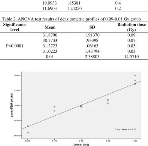
H2AX protein level in the first group (r=0.941, P<0.0001) and the second group to a lesser extent (r=0.595, P<0.0001). The data are shown in figures 1 and 2.
Table 1. ANOVA test results of densitometric profiles of 1-0.2 Gy group Significance level Mean SD Radiation dose (Gy)
P<0.0001
28.4130 1.38689 1
21.3927 .64996 0.8
19.7438 .25132 0.6
19.0933 .85381 0.4
11.6903 1.24250 0.2
Table 2. ANOVA test results of densitometric profiles of 0.09-0.01 Gy group Significance
level Mean SD
Radiation dose (Gy)
P<0.0001
31.8790 1.91370 0.09
30.7733 .93398 0.07
31.2723 .06165 0.05
31.0223 1.43794 0.03
0.01 2.38803 14.5710
Figures 2. The correlation between H2AX and radiation dose in the 0.09-0.01 Gy group, calculated by Spearman’s correlation test related to densitometry profiles
4. Discussion
Considering the increasing medical use of radiation in radiology, nuclear medicine, and CT scan, particular attention should be paid to the radiation dose. Radiation workers in therapeutic and diagnostic facilities, despite the appropriate use of personal protective devices and regulations, receive low exposure levels within a long period [13].
Cancerous and genetic effects of radiation are major health concerns after chronic exposure in occupationally exposed individuals, while their underlying mechanisms remain largely unclear [14, 15]. Generally, the results of previous human studies have showed that the received doses are within the annual permitted range Previous studies suggest that monitoring of radiation workers should not be solely based on physical dosimetry [13]. In fact, it is necessary to use biological indicators which could provide individual radiation damages [17]. Development of a simple, rapid and efficient method for biological dosimetry seems essential in cases of acute or chronic exposure [18].
A linear relationship has been documented between the number of DSBs and H2AX protein at different ionizing radiation doses [11]. This may be a potential source for the formation of chromosomal aberrations induced by radiation [19]. According to the results of this study, in radiation workers, who were occupationally exposed to chronic doses of
radiation, no band of H2AX protein was identified. This may be due to a robust and efficient repair system for DSBs at chronic radiation exposures [4]. In fact, at low doses of radiation received by workers, there is enough time for the repair of DSBs, in comparison with sudden received doses [3-20].
Contrarily, several studies have shown that chromosomal damages are more significant in radiation workers than the general population [21, 22]. On the other hand, DNA may be misreported instead of repaired in chronic radiation exposure, which may lead to chromosomal aberrations [12].
Main DNA damages induced by ionizing radiation include DSBs, single-strand breaks (SSBs) and DNA base damages [4-12] ; the ratio of SSB to DSB was estimated to be 20:1 [12], especially in low linear energy transfer radiation. However, SSBs could not lead to
H2AX foci formation [23]. In fact, the absence of H2AX protein band in radiation workers does not indicate genomic stability and SSBs may be present. On the other hand, DSB does not always lead to H2AX foci formation [24]. Rapid and complete disappearance of foci may also not be correlated with the repair of DSBs. Finally, the higher activity level of phosphatases may lead to the disappearance of H2AX in DSBs [19-25].
pathway for the repair of S and G2 phases of cell cycle; in this pathway, misrepair rarely occurs . Over 99% of cells in the human body (such as the sample in this study) are in G0 and G1 phases of cell cycle and the HR pathway in these cells is suppressed. Therefore, DSBs are repaired mostly in the NHEJ pathway, which can be prone to error [26].
Moreover, a significant correlation was found between irradiation dose and formation of
H2AX protein under in vitro conditions. Similar results have been obtained in other studies. Our results were in agreement with a study by Redon et al., who irradiated blood samples by doses of 5 to 0.2 Gy in ex vivo conditions; they found a linear response to this dose range. [1] Additionally, Havelek et al. aimed to determine whether phosphorylation of histone H2AX could be used as an indicator of received gamma radiation dose after whole- body irradiation of rats [17]; their results were also in agreement with our findings.
In this regard, Lefevre et al. examined the presence of H2AX foci in isolated peripheral blood lymphocytes in ex vivo gamma radiation (0.02-2 Gy). They concluded that H2AX can be a useful indicator, particularly for the
received radiation doses [7]. All these studies showed a significant relationship between radiation dose and H2AX protein level.
5. Conclusion
The results showed that H2AX measurement is effective for monitoring individuals with acute or local exposure (such as radiological accidents) and patients undergoing radiation therapy. However, H2AX showed no sensitivity to low chronic exposure in occupationally exposed individuals. Strong DNA repair mechanism, detection method of protein bands, and autoradiography may be the main underlying causes.
Acknowledgements
This study was approved by the Metabolic Disorders Research Center and funded by the Deputy of Research & Technology at Golestan University of Medical Sciences. We would like to thank all the participants and the staff at Clinical Biochemistry Laboratory, Laboratory of Medical Biotechnology and Radiation Oncology Center in Golestan Province.
References
1. Redon CE, Dickey JS, Bonner WM, Sedelnikova OA. γ-H2AX as a biomarker of DNA damage induced by
ionizing radiation in human peripheral blood lymphocytes and artificial skin. Adv Space Resv. 2009;43(8):1171-8.
2. Tucker JD. Low-dose ionizing radiation and chromosome translocations: a review of the major considerations for human biological dosimetry. Mutat Res. 2008;659(3):211-20.
3. Gricienė B, Slapšytė G. Assessment of chromosomal aberrations in workers chronically exposed to ionising radiation. Biologija. 2007;53(4).
4. Hall EJ, Giaccia AJ. Radiobiology for the Radiologist: Lippincott Williams & Wilkins; 2006.
5. Sudprasert W, Navasumrit P, Ruchirawat M. Effects of low-dose gamma radiation on DNA damage, chromosomal aberration and expression of repair genes in human blood cells. Int J Hyg Environ Health. 2006;209(6):503-11.
6. Wang Z, Hu H, Hu M, Zhang X, Wang Q, Qiao Y, et al. Ratio of γ-H2AX level in lymphocytes to that in
granulocytes detected using flow cytometry as a potential biodosimeter for radiation exposure. Radiat Environ Biophys. 2014;53(2):283-90.
7. Roch-Lefèvre S, Mandina T, Voisin P, Gaëtan G, Mesa JEG, Valente M, et al. Quantification of γ-H2AX foci in human lymphocytes: A method for biological dosimetry after ionizing radiation exposure. Radiat Res. 2010;174(2):185-94.
9. Rothkamm K, Krüger I, Thompson LH, Löbrich M. Pathways of DNA double-strand break repair during the mammalian cell cycle. Mol Cell Biol. 2003;23(16):5706-15.
10. Paull TT, Rogakou EP, Yamazaki V, Kirchgessner CU, Gellert M, Bonner WM. A critical role for histone H2AX in recruitment of repair factors to nuclear foci after DNA damage. Curr Biol. 2000;10(15):886-95. 11. Redon CE, Nakamura AJ, Gouliaeva K, Rahman A, Blakely WF, Bonner WM. The use of gamma-H2AX as a
biodosimeter for total-body radiation exposure in non-human primates. PloS one. 2010;5(11):e15544.
12. Rothkamm K, Horn S. gamma-H2AX as protein biomarker for radiation exposure. Ann Ist Super Sanita. 2009;45(3):265-71.
13. Garaj-Vrhovac V, Kopjar N, Poropat M. Evaluation of cytogenetic damage in nuclear medicine personnel occupationally exposed to low-level ionising radiation. Arh Hig Rada Toksikol. 2006 Mar;57(1):31-8.
14. Cengiz M, Gurkaynak M, Vural H, Aksoy N, Cengiz B, Yildiz F, et al. Tissue trace element change after total body irradiation. Nephron Exp Nephrol. 2003;94(1):e12-6.
15. Protasova O, Maksimova I, Cheprasov VY, Nikiforov A. Altered balance of macroelements and trace elements in blood serum, its ultrafiltrates, and hairs long after exposure to low doses of ionizing radiation. Biology Bulletin of the Russian Academy of Sciences. 2001;28(4):344-9.
16. Tavakkoli M, Moradalizade M, Ananisarab GhR HS. Evaluation of blood cell count in the radiology staff of Birjand Hospitals in 2011. Modern Care, Scientific Quarterly of Birjand Nursing and Midwifery Faculty. 2012;9(2):80-6.
17. Havelek R, Řezáčová M, Šinkorová Z, Zárybnická L, Tichý A, Vávrová J. Phosphorylation of histone H2AX
as an indicator of received dose of gamma radiation after whole-body irradiation of rats. Acta Veterinaria Brno. 2011;80(1):113-8.
18. Moroni M, Maeda D, Whitnall MH, Bonner WM, Redon CE. Evaluation of the Gamma-H2AX Assay for Radiation Biodosimetry in a Swine Model. Int J Mol Sci. 2013 Jul 8;14(7):14119-35.
19. Bouquet F, Muller C, Salles B. Report The Loss of γH2AX Signal is a Marker of DNA Double Strand Breaks
Repair Only at Low Levels of DNA Damage. Cell Cycle. 2006;5(10):1116-22.
20. Löbrich M, Rief N, Kühne M, Heckmann M, Fleckenstein J, Rübe C, et al. In vivo formation and repair of DNA double-strand breaks after computed tomography examinations. Proceedings of the National Academy of Sciences of the United States of America. 2005;102(25):8984-9.
21. Zakeri F, Assaei RG. Cytogenetic monitoring of personnel working in angiocardiography laboratories in Iran hospitals. Mutat Res. 2004 Aug 8;562(1-2):1-9.
22. Eken A, Aydın A, Erdem O, Akay C, Sanal HT, Soykut B, et al. Cytogenetic analysis of peripheral blood lymphocytes of hospital staff occupationally exposed to low doses of ionizing radiation. Toxicol Ind Health. 2010;26(5):273-80.
23. Horn S, Barnard S, Rothkamm K. Gamma-H2AX-based dose estimation for whole and partial body radiation exposure. PloS one. 2011;6(9):e25113.
24. Löbrich M, Shibata A, Beucher A, Fisher A, Ensminger M, Goodarzi AA, et al. γH2AX foci analysis for monitoring DNA double-strand break repair. Cell Cycle. 2010;9(4):662-9.
25. Redon CE, Nakamura AJ, Martin OA, Parekh PR, Weyemi US, Bonner WM. Recent developments in the use
of γ-H2AX as a quantitative DNA double-strand break biomarker. Aging (Albany NY). 2011 Feb;3(2):168-74.