Uremic Retention Solute Indoxyl Sulfate
Level Is Associated with Prolonged QTc
Interval in Early CKD Patients
Wei-Hua Tang1,2, Chao-Ping Wang3, Fu-Mei Chung3, Lynn L. H. Huang4, Teng-Hung Yu3, Wei-Chin Hung3, Li-Fen Lu5, Po-Yuan Chen6, Ching-Hsing Luo6, Kun-Tai Lee1,2,
Yau-Jiunn Lee7, Wen-Ter Lai1,2*
1Graduate Institute of Medicine, Collage of Medicine, Kaohsiung Medical University, Kaohsiung, Taiwan,
2Division of Cardiology, Department of Internal Medicine, Kaohsiung Medical University Hospital, Kaohsiung Medical University, Kaohsiung, Taiwan,3Division of Cardiology, Department of Internal Medicine E-Da Hospital, I-Shou University, Kaohsiung, Taiwan,4Institute of Biotechnology, National Cheng Kung University, Tainan, Taiwan,5Division of Cardiac Surgery, Department of Surgery, E-Da Hospital, I-Shou University, Kaohsiung, Taiwan,6Institute of Electric Engineering, National Cheng-Kung University, Tainan, Taiwan,7Lee’s Endocrinologic Clinic, Pingtung, Taiwan
Abstract
Total mortality and sudden cardiac death is highly prevalent in patients with chronic kidney disease (CKD). In CKD patients, the protein-bound uremic retention solute indoxyl sulfate (IS) is independently associated with cardiovascular disease. However, the underlying mechanisms of this association have yet to be elucidated. The relationship between IS and cardiac electrocardiographic parameters was investigated in a prospective observational study among early CKD patients. IS arrhythmogenic effect was evaluated by in vitro cardio-myocyte electrophysiological study and mathematical computer simulation. In a cohort of 100 early CKD patients, patients with corrected QT (QTc) prolongation had higher IS levels. Furthermore, serum IS level was independently associated with prolonged QTc interval. In vitro, the delay rectifier potassium current (IK) was found to be significantly decreased after the treatment of IS in a dose-dependent manner. The modulation of IS to the IK was through the regulation of the major potassium ion channel protein Kv 2.1 phosphorylation. In a com-puter simulation, the decrease of IK by IS could prolong the action potential duration (APD) and induce early afterdepolarization, which is known to be a trigger mechanism of lethal ventricular arrhythmias. In conclusion, serum IS level is independently associated with the prolonged QTc interval in early CKD patients. IS down-regulatedIKchannel protein
phos-phorylation and theIKcurrent activity that in turn increased the cardiomyocyte APD and
QTc interval in vitro and in the computer ORd model. These findings suggest that IS may play a role in the development of arrhythmogenesis in CKD patients.
a11111
OPEN ACCESS
Citation:Tang W-H, Wang C-P, Chung F-M, Huang LLH, Yu T-H, Hung W-C, et al. (2015) Uremic Retention Solute Indoxyl Sulfate Level Is Associated with Prolonged QTc Interval in Early CKD Patients. PLoS ONE 10(4): e0119545. doi:10.1371/journal. pone.0119545
Academic Editor:Leighton R James, University of Florida, UNITED STATES
Received:May 25, 2014
Accepted:January 14, 2015
Published:April 20, 2015
Copyright:© 2015 Tang et al. This is an open access article distributed under the terms of the
Creative Commons Attribution License, which permits unrestricted use, distribution, and reproduction in any medium, provided the original author and source are credited.
Data Availability Statement:All relevant data are within the paper.
Introduction
The importance of cardiovascular diseases (CVD) and cardiac arrhythmias in chronic kidney disease (CKD) is currently well established. Abnormal corrected QT (QTc) prolongation on the electrocardiogram, which is an independent risk factor for sudden cardiac death (SCD), is fre-quently found in patients with CKD and end-stage renal disease (ESRD) [1,2]. However, tradi-tional cardiovascular risk factors are insufficient to explain and accurately predict QTc
prolongation and SCD in this population. As the renal function deteriorates, levels of uremic re-tention toxins and proinflammatory cytokines increase [3]. Various inflammatory mediators, such as C-reactive protein (CRP), IL-6 and platelet-activating factor, have been associated with arrhythmias through the modulation of ion channel function [4,5]. In CKD patients, the protein bound uremic retention solutes, such as Indoxyl sulfate (IS), is associated with the elevation of inflammatory mediators [6,7], elevation of serum IL-6 [8], anti-oxidant modulation [6,7] and monocyte-mediated inflammation [9]. Our previous studies have also demonstrated that in-creased serum IS may be a possible risk factor in the pathogenesis of coronary atherosclerosis in CKD patients [10,11]. However, the association between IS levels and the cardiac arrhythmias risk indicator remains was still unknown. In the present study, we thus examined the relation-ship of serum IS levels with electrocardiographic parameters in a cohort of early CKD patients.
The delayed rectifier potassium current (IK) plays an important role in the repolarization of
cardiac action potentials and is one of the major currents that determines the action potentials duration and QT interval [12,13]. The potassium channel trafficking and regulation are in-volved in certain inherited or acquired cardiac channelopathies, such as long QT syndrome and heart failure [14]. For these reasons, the IS effect on cardiomyocyte electrophysiology was explored in vitro and in a mathematical computer simulation model in the present report.
Materials and Methods
Participants
The study investigated 100 consecutive CKD patients who underwent cardiac multi-slice CT or coronary angiography for exclusion of CAD due to typical and atypical chest pain, with in-termediate pretest indications for CAD, from June 2006 to June 2008 at the E-Da Hospital. Be-fore the examination, all of the each patients received a detailed interview covering their medical and personal histories. Patients with previous diagnoses of cerebrovascular diseases, heart failure, cardiomyopathy, coronary heart diseases, congenital heart disease, cardiac ar-rhythmias or taking medication that had a QT prolongation effect [15] were excluded from this study. Type 2 diabetes (T2DM) was defined as a past or current diagnosis of T2DM and/or the need for medical therapy. Hypertension was defined as a systolic blood pressure (SBP) 140 mmHg, a diastolic blood pressure (DBP)90 mmHg, or if the patient was under antihy-pertensive treatment. Dyslipidemia was defined according to the criteria of Adult Treatment Panel III, or if the patient was under lipid lowering treatment. Former and current smokers were analyzed as one group, and compared with those who had never smoked.
Estimated glomerular filtration rate (eGFR) had been calculated and followed up according to the extended Modification of Diet in Renal Disease Study formula 3–6 months before the study [16]. Written informed consent was obtained before the enrollment. This study was ap-proved by the Human Research Ethics Committee of I-Shou University E-Da Hospital.
Laboratory measurement
Peripheral blood samples were taken after fasting for 8 hours overnight and before the exami-nation. Complete blood counts and serum creatinine, sodium, potassium, calcium, uric acid,
9E%8B%E8%A8%88%E7%95%AB%E9%80%9A% E9%81%8E%E5%90%8D%E5%96%AE.pdf) and Chao-Ping Wang received the grant from I-Shou University E-DA Hospital with grant number EDAHP101020 (URL:http://www2.edah.org.tw/mrd/ plan/EDMRP.html). Neither the Kaohsiung Medical University Hospital nor I-Shou University E-Da Hospital had a role in study design, data collection and analysis, decision to publish, or preparation of the manuscript.
albumin, glucose and lipid profiles (included plasma triglycerides, total cholesterol, LDL-C, HDL-C) were determined as our previous reports [17,18].
To determine the blood total IS, A UPLC assay was used as our previously reported [10,17,18]. In brief, the blood for total IS determination was drawn, centrifuged, and stored at-80°C for subsequent assay immediately after blood sampling. The serum samples were depro-teinized by the addition of 3 parts methanol to 1 part serum for determination of IS. A UPLC assay, using detection at the 280 nm of the PDA detector, was performed at room temperature on an ACQULITY UPLC BEH phenyl column of 2.1 × 100 mm. Quantitative results were ob-tained and calculated as concentrations (μmol/L). The sensitivity of this assay was 1.061μmol/
L for IS.
ECG, QT and QTc interval measurement
Twelve-lead ECGs were collected at the baseline examination by standardized protocol. Stan-dard interval (heart rate, PR, QRS, QT intervals), amplitudes (R, S, and T waves and J and ST segment) were analyzed by standard protocol as described elsewhere [19]. The QT and QTc in-terval were calculated by the post-processing of ECG signals using the superimposed median beat method and Bazett's formula (QTc = QT/pRR). QRS interval with a bundle branch block pattern, duration longer than 120 ms, extremely rapid (>150 beats per minute) or extremely
slow heart rate (<40 beats per minute) ECG were excluded from this study [20,21]. The
defini-tion of prolonged QTc interval in our study was according to the 2009 AHA/ACCF/HRS rec-ommendations. The adjusted QT of 460 ms or longer in women and 450 ms or longer in men was considered a prolonged QT interval [22].
H9c2 cell culture and IS treatment
Embryonic rat heart-derived cardiac H9c2 cells (BCRC 60096, Bioresource Collection and Re-search Center, Taiwan) were cultured in DMEM supplemented with 10% fetal bovine serum under an atmosphere of 95% air 5% CO2 at 37°C. Experiments were carried out using mono-nucleated myoblasts culture for 2–5 days [23]. All culture media were controlled to pH 7.4 be-fore use. The cells were incubated 48 hours with IS (purchased from Sigma) at a concentration of 0.1μM, 1μM and 300μM, as previously described [24], before the experiment. In addition,
we did not investigate the effect of albumin and potassium in the present study for the follow-ing reasons. Meijers et al. revealed that the presence of albumin, even in abnormal high concen-tration, still did not significantly affect the protein-bound uremic toxin biological effect [25]. Further, in this study becausep-cresyl sulfate was synthesized as a potassium salt, cells exposed to culture medium supplemented with equimolar concentrations of potassium chloride were used as controls. We found that the addition of 1.0 mmol/L of potassium chloride did not alter the number of EMPs compared with growth medium alone [25].
Western blot analysis
Patch-clamp cell electrophysiological studies
The whole-cell potassium outward currents were recorded using an Axopatch 700A amplifier (Axon Instruments, Union City, CA, USA). The details of the methods has been described in previous reports [23,28,29]. Briefly, H9c2 cells were placed in a recording dish and perfused with a bath solution and the cells were voltage clamped. Step-pulse protocols and data acquisi-tion were performed using pCLAMP software (Axon Instruments). Membrane capacitance was calculated from the peak amplitudes and time constant decay of capacity transients elicited by 10 mV, hyperpolarizing voltage pulses from holding potential of -50 mV. All electrical re-cordings were performed at room temperature.
Mathematical computer model for cardiomyocyte action potential and
pseudo-ECG
The latest mathematical model of the O'Hara-Rudy dynamic human ventricular model (ORd model) was used in our experiment [30]. Cardiomyocyte action potential was mathematically constructed to include ionic currents, ionic pumps, ionic exchangers, and intracellular ionic regulation processes of Na+, K+and Ca2+.
A Markov model forIKis derived from previously published K channel models [31]. The
current traces figures and data ofIKin H9c2 cells with and without IS treatment was digitized
and formulated into a new Markov model computer equation and inserted into the ORd model to evaluate the IS effect on human cardiomyocyte action potential.
A pseudo-ECG was constructed and simulated by the transmural wedge model [32]. The numerical method of forward Euler with the Rush and Larsen method was used to compute pseudo-ECG with an integration time-step size (0.005ms) [33].
Statistical analysis
Data normality was analyzed using the Kolmogorov-Smirnov test. Continuous, normally dis-tributed variables are presented as mean ± SD, and non-normally disdis-tributed variables as medi-an (interquartile rmedi-ange). Statistical differences in variables were compared using the Wilcoxon rank-sum test. Categorical variables were recorded as frequencies and/or percentages, and inter-group comparisons were analyzed with the Fisher’s exact test. The general linear model-ing function analysis was used to control for potential confounders other than age and sex. Simple linear regression analysis was used to examine the association and independence be-tween serum IS and the values of other parameters. Using multiple logistic regression, variables were assessed for independent associations with the prolonged QTc interval. Multivariate ad-justed ORs are presented with 95% confidence interval (CI). All the tests were two-tailed, and a p value of<0.05 was considered statistically significant. All of the statistical analyses were
per-formed using SAS statistical software, version 8.2 (SAS Institute Inc., Cary, NC, USA).
Results
Clinical characteristics
The median serum IS level was 6.1μmol/L and the median of high sensitive CRP was
4.8 mg/L. The average heart rate was 73.0 ± 16.3 bpm, PR interval 161.5 ± 24.2ms, QRS duration 94.0 ± 17.3ms, QT interval 401.8 ± 44.6ms and QTc interval 436.7 ± 40.4ms.
Association between serum IS and patients
’
clinical characteristics
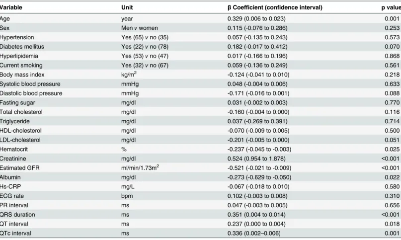
A univariate analysis was performed to test the association between the clinical and biochemi-cal variables with log-transformed serum IS levels (Table 2). Serum IS was found positively as-sociated with the age, creatinine, QRS duration, QT and QTc interval. In contrast, serum IS concentration was inversely associated to hematocrit, albumin, and eGFR.
Patient clinical laboratory data stratified by QTc status
The patients were divided into the normal QTc and prolonged QTc groups according to the criteria of AHA/ACCF/HRS [22] to investigate whether biological factors affect QTc (Table 3). Of the 100 patients in our study, 26 patients had prolonged QTc and had significantly higher serum IS levels but lower HDL levels compared to those of the normal QTc patients even after adjusting for age and sex.
Table 1. Patient demographics of 100 chronic kidney disease patients.
Age (years) 61 (54–69)
Men/Women (n, %) 56/44 (56/44)
Hypertension (% yes/no) 65/35
Diabetes mellitus (% yes/no) 22/78
Hyperlipidemia (% yes/no) 53/47
Current smoking (% yes/no) 32/68
Body mass index (kg/m2) 25.2±3.6
Systolic blood pressure (mmHg) 129±19
Diastolic blood pressure (mmHg) 75±11
Fasting sugar (mg/dl) 111.2±37.3
Total cholesterol (mg/dl) 181.0±41.6
Triglyceride (mg/dl) 154.7 (77.5–173.5)
HDL-cholesterol (mg/dl) 44.8±12.9
LDL-cholesterol (mg/dl) 106.1±37.2
Hematocrit (%) 40.3±4.6
Creatinine (mg/dl) 1.4±0.9
GFR-MDRDGFR-E (ml/min/1.73m2) 63.0±14.6
Albumin (mg/dl) 4.1±0.3
Indoxyl sulfate (μmol/L) 6.1 (0.9–6.1)
Hs-CRP (mg/L) 4.8 (0.8–4.0)
Electrocardiographic parameters
Rate (bpm) 73.0±16.3
PR interval (ms) 161.5±24.2
QRS duration (ms) 94.0±17.3
QT interval (ms) 401.8±44.6
QTc interval (ms) 436.7±40.4
Values expressed as number (percent), mean±SD, or median (25th to 75th percentile), as appropriate.
Bpm: beats per minute, HDL: density lipoprotein, LDL: low-density lipoprotein, Hs-CRP: high-sensitivity C-reactive protein.
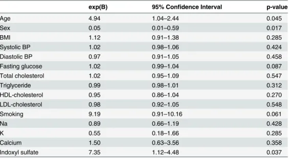
Association between IS and prolonged QTc interval
Multivariate logistic regression analysis was performed to estimate the effects of serum IS level together with several other parameters in the presence of prolonged QTc interval. The presence of prolonged QTc interval was associated with age, sex, and serum IS level (Table 4).
The effect of IS on H9c2 ventricular cardiomyocyte potassium outward
current
To evaluate the effect of IS on H9c2 cell, in the beginning, we tested the acute effect of IS on the delayed rectifier potassium current in cardiac H9c2 cells with an IS treatment of 16 hours over night. However, there was no significant change in the potassium current in the treated group compared to that of the controls. As a result, we prolong the duration of treatment to 48 hours and founded that the potassium current was significantly decreased in the IS treated group compared to that of the controls. To our knowledge, there are few reports investigating IS effect on cardiomyocytes, though Lekawanvijit et al. found that IS induced cardiomyocyte hypertro-phic change after 48 hours of IS treatment [24]. For this reason, we designed the condition of cell incubation for 48 hours.
The results of the patch-clamp cell electrophysiological study revealed that theIKwas
signif-icantly decreased after treatment with IS for 48 hours (Fig 1A). The average relationships be-tweenIKand membrane potential calculated from the measured peak current amplitudes
Table 2. Clinical and biochemical variables associated in univariate analysis with log indoxyl sulfate.
Variable Unit βCoefficient (confidence interval) p value
Age year 0.329 (0.006 to 0.023) 0.001
Sex Menvwomen 0.115 (-0.076 to 0.286) 0.253
Hypertension Yes (65)vno (35) 0.057 (-0.135 to 0.243) 0.573
Diabetes mellitus Yes (22)vno (78) 0.182 (-0.017 to 0.412) 0.070
Hyperlipidemia Yes (53)vno (47) 0.017 (-0.166 to 0.196) 0.868
Current smoking Yes (32)vno (67) 0.059 (-0.136 to 0.249) 0.561
Body mass index kg/m2 -0.124 (-0.041 to 0.010) 0.218
Systolic blood pressure mmHg 0.048 (-0.004 to 0.006) 0.633
Diastolic blood pressure mmHg -0.171 (-0.016 to 0.001) 0.088
Fasting sugar mg/dl 0.031 (-0.002 to 0.003) 0.770
Total cholesterol mg/dl -0.160 (-0.004 to 0.000) 0.116
Triglyceride mg/dl 0.037 (-0.269 to 0.391) 0.714
HDL-cholesterol mg/dl -0.070 (-0.009 to 0.005) 0.500
LDL-cholesterol mg/dl -0.201 (-0.005 to 0.000) 0.051
Hematocrit % -0.237 (-0.045 to -0.003) 0.025
Creatinine mg/dl 0.524 (0.954 to 1.878) <0.001
Estimated GFR ml/min/1.73m2 -0.521 (-0.021 to -0.009) <0.001
Albumin mg/dl -0.273 (-0.629 to -0.050) 0.022
Hs-CRP mg/L -0.067 (-0.018 to 0.010) 0.580
ECG rate bpm 0.102 (-0.003 to 0.008) 0.310
PR interval ms 0.047 (-0.003 to 0.005) 0.656
QRS duration ms 0.351 (0.004 to 0.014) <0.001
QT interval ms 0.237 (0.000 to 0.004) 0.018
QTc interval ms 0.336 (0.002–0.006) 0.001
HDL: high-density lipoprotein, LDL: low-density lipoprotein.
Table 3. Patient clinical laboratory data according to QTc classification.
QTc<450 ms in women /<460 ms in men QTc450 ms in women /460 ms in men p-value
No 74 26
Age (years) 59.0±9.0 66.4±11.4 0.002
Sex (male/female) 44/30 12/14 0.260
Current smoking (n, %) 23 (31.1) 9 (34.6) 0.805
BMI (kg/m2) 25.0±3.2 25.7±4.3 0.437
Systolic BP (mmHg) 128±14 130±31 0.620
Diastolic BP (mmHg) 76±10 73±13 0.341
Fasting glucose (mg/dl) 105.8±23.9 126.8±59.4 0.300
Total cholesterol (mg/dl) 184.3±36.5 170.8±54.0 0.071
Triglyceride (mg/dl) 103.5 (74.5–173.5) 112.0 (81.8–176.5) 0.506
HDL-cholesterol (mg/dl) 46.1±12.1 40.5±14.6 0.007
LDL-cholesterol (mg/dl) 107.6±31.5 101.3±51.8 0.216
NA (mEq/L) 139.9±2.5 139.0±4.0 0.190
K (mEq/L) 4.0±0.8 4.0±0.6 0.682
Calcium (mg/dl) 8.5±1.1 8.8±0.7 0.184
Hematocrit (%) 40.6±4.6 39.5±4.5 0.293
Creatinine (mg/dl) 1.1 (1.0–1.2) 1.1 (1.0–1.7) 0.157
Albumin (mg/dl) 4.1±0.3 4.0±0.4 0.218
Estimated GFR (ml/min/1.73m2) 64.6±12.2 57.8±20.1 0.343
Indoxyl sulfate (μmol/L) 2.8 (0.9–5.2) 6.1 (0.9–11.3) 0.019
Data are expressed as mean±SD, number (%), or median (interquartile range). BMI, body mass index; BP, blood pressure; HDL, high-density lipoprotein;
LDL, low-density lipoprotein.
doi:10.1371/journal.pone.0119545.t003
Table 4. Multiple logistic regression analysis with presence of prolonged QTc interval as the depen-dent variable.
exp(B) 95% Confidence Interval p-value
Age 4.94 1.04–2.44 0.045
Sex 0.05 0.01–0.59 0.017
BMI 1.12 0.91–1.38 0.285
Systolic BP 1.02 0.98–1.06 0.424
Diastolic BP 0.97 0.91–1.05 0.458
Fasting glucose 1.02 0.99–1.04 0.087
Total cholesterol 1.02 0.95–1.09 0.547
Triglyceride 0.99 0.98–1.01 0.312
HDL-cholesterol 0.95 0.86–1.04 0.270
LDL-cholesterol 0.98 0.92–1.05 0.548
Smoking 9.19 0.91–10.16 0.061
Na 0.89 0.66–1.19 0.428
K 0.55 0.18–1.66 0.285
Calcium 1.50 0.63–3.56 0.358
Indoxyl sulfate 7.35 1.12–4.48 0.037
BMI, body mass index; BP, blood pressure; HDL, high-density lipoprotein; LDL, low-density lipoprotein.
showed thatIKwas significantly decreased at the membrane potentials from 0 mV to 50 mV in
a dose-dependent manner (Fig 1B).
Kv2.1 Western Blot Analysis with and without IS treatment in H9c2
cardiomyocyte
Potassium ion channel protein Kv2.1 is the major subunit protein comprised of ion channels that generateIKin H9c2 cell [23]. Western Blot analysis revealed that there was an increase in
Kv2.1 level after the treatment of IS. However, there were no significant differences noted be-tween the groups (Fig 1C). In contrast, the expression of phosphorylated Kv2.1 was found to be significantly decreased in the IS-treated groups (Fig 1D).
Mathematical computer simulation of IS effect on human cardiomyocyte
electrophysiology
From the computer calculations and simulation results, the decrease ofIKcaused by the
in-crease of treated IS concentration will gradually prolong the constructed action potential dura-tion (APD) and pseudo ECG QT interval (Fig 2). In addition, early afterdepolarization (EAD) was noted in the higher suppression ofIKsimulation, which mimicked the high IS effect on the
Fig 1. The effect of IS on H9c2 cardiomyocyteIKand potassium channel protein Kv2.1 expression.(A)
The representative current traces for delayed rectifier potassium outward currents (Ik) in H9c2 cells with different indoxyl sulfate (IS) concentration treatment.Ikwere elicited by 300 ms depolarizing step pulses from -70 to 50 mV at a holding potential of -60 mV. (B) The average relationships betweenIk(pA/pF) and
membrane potential in the control, 0.1μM IS, 1μM IS and 300μM IS groups (n = 6 for each groups) comparing
the IS treated group with the control group,Ikwas significantly decreased at membrane potentials from 0 mV to 50 mV in a dose-dependent manner. (C and D) The expression of Kv2.1 and phosphorylated Kv2.1 by Western blot in the H9c2 cells treated with different concentration of IS (0.1μM, 1μM and 300μM). The
expressions of Kv2.1 were not significantly different among the control and IS-treated groups (C). However, the phosphorylated Kv2.1 was significantly decreased in the 1μM IS-and 300μM-IS treated groups (D).
(n = 6 for each groups)*:p<0.05 as compared with the control group.
cell electrophysiological studies (Fig 2, arrow). The constructed pseudo ECG also showed the ventricular arrhythmias like ECG when theIKwas severely suppressed (Fig 2, empty arrow).
Discussion
In the present study, we demonstrated that serum IS concentrations are correlated with age, he-matocrit, creatinine, estimated GFR, albumin, QRS duration, and QTc interval. The serum IS concentration was increased in QTc prolongation patients in contrast with the normal QTc controls; in addition, multiple logistic regression analysis also confirmed this independent as-sociation, even in a fully adjusted model. Furthermore, in cell electrophysiological study, IS de-creased theIKin rat ventricular cardiomyocyte through the regulation of the major potassium
ion channel protein Kv 2.1 phosphorylation. To our knowledge, this is the first report to ob-serve that the serum IS level is associated with QTc prolongation and that the possible mecha-nism of this phenomenon is through the down-regulation ofIKchannel protein
phosphorylation and theIKcurrent activity that in turn increases the APD and QTc interval.
Fig 2. Ventricular cardiomyocyte action potential (AP) and pseudo-ECG constructed by the O'Hara-Rudy dynamic human ventricular model.The suppression of inward rectifier potassium current (IK) mimics the effect of indoxyl sulfate toxicity to ventricular cardiomyocyte AP. The AP duration was gradually increased and the QT interval was also prolonged with the increment ofIKsuppression. The early afterdepolarization was noted in the higher suppression ofIKespecially in the mid-myocardial cardiomyocyte (arrow). The ventricular arrhythmias like ECG was also noted when theIKwas severely suppressed (empty arrow).
QT prolongation is usually found in patients with chronic renal diseases and multiple mech-anisms have been suggested, such as electrolyte imbalance, autonomic nerve dysfunction, rapid changes in electrolyte plasma concentrations during hemodialysis and cardiac hypertrophy [34]. However, there have been few reports demonstrating the relationship between early stages of CKD and QT interval. Cardiorenal syndrome (CRS), which indicates that in the heart and kidney, acute or chronic dysfunction of one organ may induce acute or chronic dysfunc-tion in the other, is caused by multiple factors, including non-dialyzable uremic toxins, such as IS [35].
Recently, IS has been reported to have the profibrotic and prohypertropic effects on cardio-myocytes [24], free radical production [7], endothelial microparticle release [36], vascular smooth muscle cell proliferation [37], and adherens junctions disruption of vascular endotheli-al cells [38]. Clinically, all of these pathogenetic states can contribute to vascular damage [39], progression of renal diseases [40], coronary artery disease [10], and even mortality [39] in CKD patients. In the present study, we found that there is a strong association between QTc in-terval and serum IS level in early CKD patients. Importantly, an increase in circulating IS levels can occur in the early stages (2 and 3) of CKD [41], and the combined prevalence of stages 2 and 3 accounts for 67% of all CKD stages [42].
The biological mechanisms involving IS level in the pathogenesis of QTc prolongation is not well understood. According to Ronco et al., IS is the strongest evidence-based uremic toxin involved in type 2 and type 4 CRS pathophysiology, which is mainly attributable to the profi-brotic effects [35]. Recently, IS has also been proven to have a direct effect on cardiac fibro-blasts and induced cardiac fibrosis in an animal study [24]. As cardiac fibrosis is known as one of the mechanisms of cardiac dysfunction, QT prolongation and cardiac arrhythmia [43], as well as high serum IS levels may be associated with QTc prolongation. In addition, distur-bances in gap junctional intercellular communication affect the electrical coupling between heart muscle cells and the underlie prolongation of QRS and QT intervals [44]. Several uremic retention toxins, such as homocysteine [45] andp-cresol [46] have been shown to be involved in the disassembly of connexin and the disruption of the adherens junction of cardiomyocyte and QT prolongation in an animal model [45]. Therefore, whether IS also involved in the mod-ulation of cardiomyocyte connexin needs further investigation.
IKis one of the core current determinants of the cardiomyocyte APD, with the increase of
APD prolonging the ECG QT interval [13]. The Kv2.1 protein is the major subunit protein comprising the ion channel in H9c2 cell, which generates theIK[23], and previous studies have
also shown the Kv2.1 channel activity is regulated by phosphorylation [47]. Here, we demon-strated that IS decreased the expression of phosphorylated Kv2.1 in the H9c2 cell, down-regulated the Kv2.1 channel activity and decreasedIK, which suggests that IS may play a role in
It is hard to demonstrate any channelopathies in human cardiomyocyte and their conse-quent effects on the human heart surface ECG. Recently, both experimental and theoretical models have been used in procedures studying the biological factors that induce arrhythmias [30]. In our computer simulation experiment, the prolonged APD, EAD phenomenon, pro-longed QT interval and ventricular arrhythmias-like ECG appearance were noted (Fig 2). The EAD induction and trigger activity were the major arrhythmogenesis in ventricular arrhyth-mias. The induction of EAD in the experiment suggests an IS arrhythmogenic effect and its possible role in the arrhythmias and SCD among CKD patients.
Among these non-dialyzable uremic toxins, many biological and pathological effects have been previously discussed [54,55]. However, there are lacks of reports showing the arrhythmo-genic effect of these non-dailyzable uremic toxins in human heart or animal models. IS is the representative molecule of uremic toxin and already known to be associated with the pathogen-esis of many uremic syndrome. Previous studies have shown that IS has profibrotic and prohy-pertropic effects on cardiomyocytes [24,56], which are also known to be related to free radical production and the elevation of inflammatory mediators, which are in turn proven to affect cardiac ion channel function [24,48,49,56]. It is therefore reasonable to select IS over other tox-ins for investigation into its arrhythmogenic effect. Further work is required to confirm these findings in other uremic toxins.
Some limitations of this study need to be considered. First, our study population was rela-tively small. Further, the cross-sectional design limits our ability to infer any causal relationship between total IS levels and QTc prolongation. Second, we did not determined the plasma mag-nesium and bicarbonate levels in our patients due to hospital standard cardiac multi-slice CT or coronary angiography examination preparation protocols; as such, it is unfortunate that we could not demonstrate whether these electrolytes also affect QTc prolongation. Third, in our study, the IS affectedIKin vitro at very low concentrations, even below the normal population
serum IS level [57]. Moreover, the potassium concentration in the added IS potassium salt (0.1uM to 0.3mM) was relatively low compared to the potassium concentration in the DMEM culture medium (5.3mM) used in the experiment. Hence, the additional potassium effect on the action potentials in the experiment could have been eliminated. As the cardiac electrical ac-tivity is a complex system, it is no doubt plausible that there exists another modulation system in vivo to contend with the IS biological effect, such as the equilibrium state of IS protein bind-ing capacity [24]. Finally, it is still unclear whether IS affects the expression of other cardiac ion channels, ion currents and cardiomyocyte electrophysiology. Further investigation
is warranted.
In conclusion, our study demonstrated that the QTc interval was prolonged in early CKD patients with a higher serum IS level. The arrhythmogenic effect of IS was shown through the inhibition ofIK. By the progress of renal disease and the interaction of CRS, the effect and role
of IS on the arrhythmogenesis among CKD patients might be enhanced in conjunction with the advance of renal function impairment. As a result, the arrhythmogenic effect of IS should be taken seriously.
Supporting Information
S1 Code. Mathematical computer model for cardiomyocyte action potential and pseudo-ECG.In our experiment, cardiomyocyte action potential was mathematically constructed by the latest mathematical model of the O’Hara-Rudy dynamic human ventricular model. The codes and equations we used in our experiment were downloaded and modified from the Open access and supplemental material journal PLoS computational Biology.
Acknowledgments
The authors would like to thank the E-Da Hospital and Kaohsiung Medical University Hospital of the Republic of China, Taiwan, for financially supporting this research under contracts of EDAHP103002 and KMUH101-1M06.
Author Contributions
Conceived and designed the experiments: WHT YJL WTL. Performed the experiments: WHT CPW WCH KTL PYC. Analyzed the data: FMC LLHH. Contributed reagents/materials/analy-sis tools: WHT LFL CHL. Wrote the paper: WHT FMC THY CPW.
References
1. Genovesi S, Rossi E, Nava M, Riva H, De Franceschi S, Fabbrini P, et al. A case series of chronic hae-modialysis patients: mortality, sudden death, and QT interval. Europace. 2013; 15: 1025–1033. doi:10. 1093/europace/eus412PMID:23284142
2. Straus SM, Kors JA, De Bruin ML, van der Hooft CS, Hofman A, Heeringa J, et al. Prolonged QTc inter-val and risk of sudden cardiac death in a population of older adults. J Am Coll Cardiol. 2006; 47: 362–367. PMID:16412861
3. Shamseddin MK, Parfrey PS. Sudden cardiac death in chronic kidney disease: epidemiology and pre-vention. Nat Rev Nephrol. 2011; 7: 145–154. doi:10.1038/nrneph.2010.191PMID:21283136
4. Hoffman BF, Guo SD, Feinmark SJ. Arrhythmias caused by platelet activating factor. J Cardiovasc Electrophysiol. 1996; 7: 120–133. PMID:8853022
5. Hoffman BF, Feinmark SJ, Guo SD. Electrophysiologic effects of interactions between activated canine neutrophils and cardiac myocytes. J Cardiovasc Electrophysiol. 1997; 8: 679–687. PMID:9209969
6. Aoyama I, Shimokata K, Niwa T. An oral adsorbent downregulates renal expression of genes that pro-mote interstitial inflammation and fibrosis in diabetic rats. Nephron. 2002; 92: 635–651. PMID: 12372949
7. Motojima M, Hosokawa A, Yamato H, Muraki T, Yoshioka T. Uremic toxins of organic anions up-regulate PAI-1 expression by induction of NF-kappaB and free radical in proximal tubular cells. Kidney Int. 2003; 63: 1671–1680. PMID:12675842
8. Lee CT, Kuo CC, Chen YM, Hsu CY, Lee WC, Tsai YC, et al. Factors associated with blood concentra-tions of indoxyl sulfate and p-cresol in patients undergoing peritoneal dialysis. Perit Dial Int. 2010; 30: 456–463. doi:10.3747/pdi.2009.00092PMID:20338972
9. Ito S, Higuchi Y, Yagi Y, Nishijima F, Yamato H, Ishii H, et al. Reduction of indoxyl sulfate by AST-120 attenuates monocyte inflammation related to chronic kidney disease. J Leukoc Biol. 2013; 93: 837–845. doi:10.1189/jlb.0112023PMID:23362306
10. Chiu CA, Lu LF, Yu TH, Hung WC, Chung FM, Tsai IT, et al. Increased levels of total P-Cresylsulphate and indoxyl sulphate are associated with coronary artery disease in patients with diabetic nephropathy. Rev Diabet Stud. 2010; 7: 275–284. doi:10.1900/RDS.2010.7.275PMID:21713315
11. Hsu CC, Lu YC, Chiu CA, Yu TH, Hung WC, Wang CP, et al. Levels of indoxyl sulfate are associated with severity of coronary atherosclerosis. Clin Invest Med. 2013; 36: E42–49. PMID:23374599
12. Noble D, Tsien RW. Outward membrane currents activated in the plateau range of potentials in cardiac Purkinje fibres. J Physiol. 1969; 200: 205–231. PMID:5761944
13. Carmeliet E. K+ channels and control of ventricular repolarization in the heart. Fundam Clin Pharmacol. 1993; 7: 19–28. PMID:8458599
14. Nattel S, Maguy A, Le Bouter S, Yeh YH. Arrhythmogenic ion-channel remodeling in the heart: heart failure, myocardial infarction, and atrial fibrillation. Physiol Rev. 2007; 87: 425–456. PMID:17429037
15. Kannankeril P, Roden DM, Darbar D. Drug-induced long QT syndrome. Pharmacol Rev. 2010; 62: 760–781. doi:10.1124/pr.110.003723PMID:21079043
16. Levey AS, Coresh J, Greene T, Stevens LA, Zhang YL, Hendriksen S, et al. Using standardized serum creatinine values in the modification of diet in renal disease study equation for estimating glomerular fil-tration rate. Ann Intern Med. 2006; 145: 247–254. PMID:16908915
18. Wang CP, Lu LF, Yu TH, Hung WC, Chiu CA, Chung FM, et al. Serum levels of totalp-cresylsulphate are associated with angiographic coronary atherosclerosis severity in stable angina patients with early stage of renal failure. Atherosclerosis. 2010; 211: 579–583. doi:10.1016/j.atherosclerosis.2010.03.036 PMID:20427046
19. Magnani JW, Wang N, Nelson KP, Connelly S, Deo R, Rodondi N, et al. Electrocardiographic PR inter-val and adverse outcomes in older adults: the Health, Aging, and Body Composition study. Circ Arrhythm Electrophysiol. 2013; 6: 84–90. doi:10.1161/CIRCEP.112.975342PMID:23243193
20. Malik M, Hnatkova K, Batchvarov V, Gang Y, Smetana P, Camm AJ. Sample size, power calculations, and their implications for the cost of thorough studies of drug induced QT interval prolongation. Pacing Clin Electrophysiol. 2004; 27: 1659–1669. PMID:15613131
21. Salvi V, Karnad DR, Panicker GK, Natekar M, Hingorani P, Kerkar V, et al. Comparison of 5 methods of QT interval measurements on electrocardiograms from a thorough QT/QTc study: effect on assay sen-sitivity and categorical outliers. J Electrocardiol. 2011; 44: 96–104. doi:10.1016/j.jelectrocard.2010.11. 010PMID:21238976
22. Rautaharju PM, Surawicz B, Gettes LS, Bailey JJ, Childers R, Deal BJ, et al. AHA/ACCF/ HRS recom-mendations for the standardization and interpretation of the electrocardiogram: part IV: the ST seg-ment, T and U waves, and the QT interval: a scientific statement from the American Heart Association Electrocardiography and Arrhythmias Committee, Council on Clinical Cardiology; the American College of Cardiology Foundation; and the Heart Rhythm Society. Endorsed by the International Society for Computerized Electrocardiology. J Am Coll Cardiol. 2009; 53: 982–991. doi:10.1016/j.jacc.2008.12. 014PMID:19281931
23. Wang W, Hino N, Yamasaki H, Aoki T, Ochi R. KV2.1 K+ channels underlie major voltage-gated K+ out-ward current in H9c2 myoblasts. Jpn J Physiol. 2002; 52: 507–514. PMID:12617756
24. Lekawanvijit S, Adrahtas A, Kelly DJ, Kompa AR, Wang BH, Krum H. Does indoxyl sulfate, a uraemic toxin, have direct effects on cardiac fibroblasts and myocytes? Eur Heart J. 2010; 31: 1771–1779. doi: 10.1093/eurheartj/ehp574PMID:20047993
25. Meijers BK, Van Kerckhoven S, Verbeke K, Dehaen W, Vanrenterghem Y, Hoylaerts MF, et al. The ure-mic retention solute p-cresyl sulfate and markers of endothelial damage. Am J Kidney Dis. 2009; 54: 891–901. doi:10.1053/j.ajkd.2009.04.022PMID:19615803
26. Murakoshi H, Shi G, Scannevin RH, Trimmer JS. Phosphorylation of the Kv2.1 K+ channel alters volt-age-dependent activation. Mol Pharmacol. 1997; 52: 821–828. PMID:9351973
27. Ito T, Nuriya M, Yasui M. Regulation of Kv2.1 phosphorylation in an animal model of anoxia. Neurobiol Dis. 2010; 38: 85–91. doi:10.1016/j.nbd.2010.01.002PMID:20079839
28. Hamill OP, Marty A, Neher E, Sakmann B, Sigworth FJ. Improved patch-clamp techniques for high-resolution current recording from cells and cell-free membrane patches. Pflugers Arch. 1981; 391: 85–100. PMID:6270629
29. Lee KT, Tang PW, Tsai WC, Liu IH, Yen HW, Voon WC, et al. Differential effects of central and periph-eral fat tissues on the delayed rectifier k outward currents in cardiac myocytes. Cardiology. 2013; 125: 118–124. doi:10.1159/000350360PMID:23711914
30. O'Hara T, Virag L, Varro A, Rudy Y. Simulation of the undiseased human cardiac ventricular action po-tential: model formulation and experimental validation. PLoS Comput Biol. 2011; 7: e1002061. doi:10. 1371/journal.pcbi.1002061PMID:21637795
31. Zagotta WN, Hoshi T, Aldrich RW. Shaker potassium channel gating. III: Evaluation of kinetic models for activation. J Gen Physiol. 1994; 103: 321–362. PMID:8189208
32. Gima K, Rudy Y. Ionic current basis of electrocardiographic waveforms: a model study. Circ Res. 2002; 90: 889–896. PMID:11988490
33. Rush S, Larsen H. A practical algorithm for solving dynamic membrane equations. IEEE Trans Biomed Eng. 1978; 25: 389–392. PMID:689699
34. Covic A, Diaconita M, Gusbeth-Tatomir P, Covic M, Botezan A, Ungureanu G, et al. Haemodialysis in-creases QT(c) interval but not QT(c) dispersion in ESRD patients without manifest cardiac disease. Nephrol Dial Transplant. 2002; 17: 2170–2177. PMID:12454229
35. Ronco C, Haapio M, House AA, Anavekar N, Bellomo R. Cardiorenal syndrome. J Am Coll Cardiol. 2008; 52: 1527–1539. doi:10.1016/j.jacc.2008.07.051PMID:19007588
36. Faure V, Dou L, Sabatier F, Cerini C, Sampol J, Berland Y, et al. Elevation of circulating endothelial mi-croparticles in patients with chronic renal failure. J Thromb Haemost. 2006; 4: 566–573. PMID: 16405517
38. Peng YS, Lin YT, Chen Y, Hung KY, Wang SM. Effects of indoxyl sulfate on adherens junctions of en-dothelial cells and the underlying signaling mechanism. J Cell Biochem. 2012; 113: 1034–1043. doi: 10.1002/jcb.23435PMID:22213462
39. Barreto FC, Barreto DV, Liabeuf S, Meert N, Glorieux G, Temmar M, et al. Serum indoxyl sulfate is as-sociated with vascular disease and mortality in chronic kidney disease patients. Clin J Am Soc Nephrol. 2009; 4: 1551–1558. doi:10.2215/CJN.03980609PMID:19696217
40. Wu IW, Hsu KH, Lee CC, Sun CY, Hsu HJ, Tsai CJ, et al. p-Cresyl sulphate and indoxyl sulphate pre-dict progression of chronic kidney disease. Nephrol Dial Transplant. 2011; 26: 938–947. doi:10.1093/ ndt/gfq580PMID:20884620
41. Atoh K, Itoh H, Haneda M. Serum indoxyl sulfate levels in patients with diabetic nephropathy: relation to renal function. Diabetes Res Clin Pract. 2009; 83: 220–226. doi:10.1016/j.diabres.2008.09.053PMID: 19027976
42. National Kidney Foundation. K/DOQI clinical practice guidelines for chronic kidney disease: evaluation, classification, and stratification. Am J Kidney Dis. 2002; 39: S1–266. PMID:11904577
43. Zhang T, Yong SL, Drinko JK, PopovićZB, Shryock JC, Belardinelli L, et al. LQTS mutation N1325S in cardiac sodium channel gene SCN5A causes cardiomyocyte apoptosis, cardiac fibrosis and contractile dysfunction in mice. Int J Cardiol. 2011; 147: 239–245. doi:10.1016/j.ijcard.2009.08.047PMID: 19762097
44. Quan XQ, Bai R, Liu N, Chen BD, Zhang CT. Increasing gap junction coupling reduces transmural dis-persion of repolarization and prevents torsade de pointes in rabbit LQT3 model. J Cardiovasc Electro-physiol. 2007; 18: 1184–1189. PMID:17711442
45. Rosenberger D, Gargoum R, Tyagi N, Metreveli N, Sen U, Maldonado C, et al. Homocysteine enriched diet leads to prolonged QT interval and reduced left ventricular performance in telemetric monitored mice. Nutr Metab Cardiovasc Dis. 2011; 21: 492–498. doi:10.1016/j.numecd.2009.11.014PMID: 20227264
46. Peng YS, Lin YT, Wang SD, Hung KY, Chen Y, Wang SM. P-cresol induces disruption of cardiomyo-cyte adherens junctions. Toxicology. 2013; 306: 176–184. doi:10.1016/j.tox.2013.02.015PMID: 23466501
47. Song MY, Hong C, Bae SH, So I, Park KS. Dynamic modulation of the kv2.1 channel by SRC-dependent tyrosine phosphorylation. J Proteome Res. 2012; 11: 1018–1026. doi:10.1021/pr200770vPMID: 22106938
48. Park KS, Mohapatra DP, Misonou H, Trimmer JS. Graded regulation of the Kv2.1 potassium channel by variable phosphorylation. Science. 2006; 313: 976–979. PMID:16917065
49. Walsh KB, Zhang J. Neonatal rat cardiac fibroblasts express three types of voltage-gated K+ channels: regulation of a transient outward current by protein kinase C. Am J Physiol Heart Circ Physiol. 2008; 294: H1010–1017. PMID:18156198
50. Inoguchi T, Sonta T, Tsubouchi H, Etoh T, Kakimoto M, Sonoda N, et al. Protein kinase C-dependent in-crease in reactive oxygen species (ROS) production in vascular tissues of diabetes: role of vascular NAD(P)H oxidase. J Am Soc Nephrol. 2003; 14: S227–232. PMID:12874436
51. Cain BS, Meldrum DR, Harken AH. Protein kinase C in normal and pathologic myocardial states. J Surg Res. 1999; 81: 249–259. PMID:9927548
52. Enomoto A, Takeda M, Tojo A, Sekine T, Cha SH, Khamdang S, et al. Role of organic anion transport-ers in the tubular transport of indoxyl sulfate and the induction of its nephrotoxicity. J Am Soc Nephrol. 2002; 13: 1711–1720. PMID:12089366
53. Zhang Q, Hong M, Duan P, Pan Z, Ma J, You G. Organic anion transporter OAT1 undergoes constitu-tive and protein kinase C-regulated trafficking through a dynamin- and clathrin-dependent pathway. J Biol Chem. 2008; 283; 32570–32579. doi:10.1074/jbc.M800298200PMID:18818201
54. Jourde-Chiche N, Dou L, Cerini C, Dignat-George F, Vanholder R, Brunet P. Protein-bound toxins-update 2009. Semin Dial. 2009; 22: 334–339. doi:10.1111/j.1525-139X.2009.00576.xPMID: 19708977
55. Neirynck N, Vanholder R, Schepers E, Eloot S, Pletinck A, Glorieux G. An update on uremic toxins. Int Urol Nephrol. 2013; 45: 139–150. doi:10.1007/s11255-012-0258-1PMID:22893494
56. Lekawanvijit S, Kompa AR, Manabe M, Wang BH, Langham RG, Nishijima F, et al. Chronic kidney disease-induced cardiac fibrosis is ameliorated by reducing circulating levels of a non-dialysable ure-mic toxin, indoxyl sulfate. PLoS One. 2012; 7: e41281. doi:10.1371/journal.pone.0041281PMID: 22829936