Predicting Hemagglutinin MHC-II Ligand
Analogues in Anti-TNF
α
Biologics:
Implications for Immunogenicity of
Pharmaceutical Proteins
Benjamin J. Andrick☯, Alexandra I. Schwab☯, Brianna Cauley, Lauren A. O’Donnell*, Wilson S. Meng*
Division of Pharmaceutical Sciences, Duquesne University, Pittsburgh, PA, 15282, United States of America
☯These authors contributed equally to this work. *[email protected](LAO);[email protected](WSM)
Abstract
The purpose of this study was to evaluate the extent of overlapping immunogenic peptides between three pharmaceutical biologics and influenza viruses. Clinical studies have shown that subsets of patients with rheumatoid arthritis (RA) develop anti-drug antibodies towards anti-TNFαbiologics. We postulate that common infectious pathogens, including influenza viruses, may sensitize RA patients toward recombinant proteins. We hypothesize that embedded within infliximab (IFX), adalimumab (ADA), and etanercept (ETN) are ligands of class II major histocompatibility complex (MHC-II) that mimic T cell epitopes derived from influenza hemagglutinin (HA). The rationale is that repeated administration of the biologics would reactivate HA-primed CD4 T cells, stimulating B cells to produce cross-reactive anti-bodies. Custom scripts were constructed using MATLAB to compare MHC-II ligands of HA and the biologics; all ligands were predicted using tools in Immune Epitope Database and Resources (IEDB). We analyzed three HLA-DR1 alleles (0101, 0401 and 1001) that are prominent in RA patients, and two alleles (0103 and 1502) that are not associated with RA. The results indicate that 0401 would present more analogues of HA ligands in the three anti-TNFαbiologics compared to the other alleles. The approach led to identification of potential ligands in IFX and ADA that shares sequence homology with a known HA-specific CD4 T cell epitope. We also discovered a peptide in the complementarity-determining region 3 (CDR-3) of ADA that encompasses both a potential CD4 T cell epitope and a known B cell epitope in HA. The results may help generate new hypotheses for interrogating patient variability of immunogenicity of the anti-TNFαdrugs. The approach would aid devel-opment of new recombinant biologics by identifying analogues of CD4 T cell epitopes of common pathogens at the preclinical stage.
a11111
OPEN ACCESS
Citation:Andrick BJ, Schwab AI, Cauley B, O’Donnell LA, Meng WS (2015) Predicting Hemagglutinin MHC-II Ligand Analogues in Anti-TNFαBiologics: Implications for Immunogenicity of Pharmaceutical Proteins. PLoS ONE 10(8): e0135451. doi:10.1371/journal.pone.0135451
Editor:Yoshihiko Hoshino, National Institute of Infectious Diseases, JAPAN
Received:December 2, 2014
Accepted:July 22, 2015
Published:August 13, 2015
Copyright:© 2015 Andrick et al. This is an open access article distributed under the terms of the
Creative Commons Attribution License, which permits unrestricted use, distribution, and reproduction in any medium, provided the original author and source are credited.
Data Availability Statement:All relevant data are within the paper and its Supporting Information files.
Funding:The authors have no support or funding to report.
Introduction
Tumor necrosis factor-alpha (TNFα) is a driving inflammatory mediator in rheumatoid arthri-tis (RA) [1]. RA patients benefit from anti-TNFαbiologics through reduced disease activities
and in some cases, remission [2]. Infliximab (IFX), adalimumab (ADA), both monoclonal IgG antibodies, and etanercept (ETN), a fusion protein, are the mainstay of the anti-TNFαbiologics
used in RA patients in the United States [3]. Despite the generally positive outlook in confer-ring long-term health benefits, approximately one-third of the patients receiving an anti-TNFα
biologics do not respond to treatment [4]. Recent clinical studies have reported cases of persis-tent active diseases, despite continuing treatments at higher doses [5]. Such instances suggest potential drug neutralization by the immune system. A mechanistic understanding of the immunological basis underlying these phenomena will lead to improved treatment outcomes.
While multiple factors are implicated in driving therapeutic responses to anti-TNFα biolog-ics in patients, a known cause of treatment failure is the development of anti-drug antibodies [5]. Such immunological reactions would accelerate drug clearance, resulting in sub-therapeu-tic plasma concentrations. IFX, ADA and ETN are recombinant proteins engineered to reduce intrinsic immunogenic potential. IFX is a chimeric IgG1-kappa monoclonal antibody with mouse variable regions grafted into human constant regions [6]. Bendtzen et al., however, reported that 44% of the 106 RA patients tested were found to have serum anti-IFX antibodies six months after initiation of treatment [7]. In some of these patients (13%), anti-IFX antibod-ies were detected as early as 1.5 months, or as few as after three infusions. Such antibodantibod-ies are associated with low trough plasma drug concentrations, a metric predictive of poor efficacy. Among RA patients who tested positive for anti-IFX antibodies, Wolbink et al. reported fewer responders (36%) compared to patients without the antibodies (69%), [8]. The rapid develop-ment of antibodies in certain patients against IFX without inflammatory adjuvants suggests that prior environmental factors may raise the drug’s immunogenicity.
ADA is a“fully human”IgG1-kappa monoclonal antibody generated from in vitro screen-ing of phage libraries displayscreen-ing human variable regions [9]. Despite the lack ofbona fide mouse sequences, anti-ADA antibodies have been detected in patients who have received the biologics. In a study that followed 272 RA patients for 156 weeks, Bartelds et al. reported that 28% of the patients tested positive for anti-ADA antibodies during the first 28 weeks of treat-ment [10]. The presence of such antibodies correlates with poor disease prognosis and second-ary treatment failure. Importantly, assays used in these analyses were sufficiently specific to minimize interference by rheumatoid factors (RFs) [7]. Unlike IFX and ADA, ETN is a fusion protein consisting of the human tumor necrosis factor receptor-II (TNFRII) domain fused with human IgG1 constant Fc regions (CH2 and CH3). So far, studies have shown that preva-lence of anti-ETN antibodies in patients is low [11–13]; Dore et al. have reported detecting non-neutralizing anti-ETN antibodies in 12 out of 214 RA patients [13].
certain biologics. Cryptic epitopes in apparently non-immunogenic proteins may become stim-ulatory when the same, or similar sequences are presented in inflammatory milieus [16].
Viruses or other infectious pathogens may sensitize certain individuals to develop cross-reactive CD4 T cell responses in an MHC-II allele-dependent manner. The rationale is that embedded within immunogenic biologics are MHC-II ligands that share sequence homology with epitopes in the major antigens of the viruses. A systematic analysis of such ligand ana-logues could help gauge the risk for developing anti-drug antibodies in an allele-specific man-ner [17–19]. The presence of virus-primed helper T cells that recognize epitopes in anti-TNFα
biologics would lower the threshold of drug-specific B cell activation. Consequently, anti-drug antibodies may arise sooner and to higher titers. This form of molecular mimicry may heighten the sensitivity to a given biologic in individuals exposed to certain viruses. It may contribute to the unexpectedly high number of RA patients who develop antibodies against anti-TNFα bio-logics after initiation of treatment, independent of intrinsic anti-IgG antibodies in circulation [20]. A recent meta-analysis by Lv et al. concluded that apparent association between RF and anti-TNFαtherapeutic outcomes is not substantiated by published data [21], in congruent
with that the incidence and magnitude of anti-drug antibodies vary greatly among individuals with RA [14].
In order to investigate potential cross-reactive viral epitopes, we focused on influenza type-A viruses because of widespread exposure in the population [22]. New influenza strains are introduced into the population due to seasonal variability. In the first global influenza pan-demic in 40 years, the 2009 H1N1 panpan-demic strains largely displaced the viruses previously cir-culating in humans [23]. Since then the viruses have maintained a continual presence in North America through seasonal infections [24,25], albeit with minor genetic changes in the major antigens such as hemagglutinin (HA). Cross-reactive memory CD4 T cells may be generated If HA antigens share similar MHC-II ligands in a given anti-TNFαbiologics. These
antigen-spe-cific helper T cells would in turn activate B cells that produce antibodies against both the bio-logic and the viral HA.
We describe herein a bioinformatics strategy to identify systematically potential cross-reac-tive T cell epitopes in HA and anti-TNFαbiologics (Fig 1). We hypothesize that embedded
within the primary sequences of IFX, ADA, and ETN are analogous sequences that resemble HA-derived MHC-II ligands. Such sequences in the biologics may stimulate naive or re-acti-vate memory HA-specific T cells. Using IEDB and custom scripts generated using MATLAB, potential cross-reactive ligands, or analogues, were predicted for HLA-DR10101 (hereafter
referred as 0101), HLA-DR10401 (0401), and HLA-DR11001 (1001), three MHC-II alleles
that are implicated in RA [26]. Presumably, a higher proportion of the population who express one or more of these alleles would have been treated with at least one of the three anti-TNFα
biologics. Also analyzed were HLA-DR10103 (0103) and HLA-DR11502 (1502) that are not
associated with RA. Ligands of IFX, ADA, and ETN were matched against those ligands pre-dicted from H1N1 influenza type A HA antigens. The results provide a potential molecular mechanism by which development of anti-drug antibodies may occur.
Materials and Methods
which the“consensus approach”is typically selected; it combines results from at least three algorithms (NN-align [27], SMM-align [28], Sturniolo [29], NetMHCIIpan [27] and/or Com-bLib [30]). Peptides are scored based on the best median rank [30]. MHC molecules that lack at least three predictors are default to the NetMHCIIpan method [31]. Full descriptions of the scoring regime can be found on the IEDB web site (http://tools.immuneepitope.org/mhcii/ help/#Method). Such percentile-based ranking has biophysical meanings insofar as relative strong binding affinities are correlated with low percentiles. For our analysis, the IEDB server selected the consensus method for 0101 and 0401, Sturniolo for 1502, and NetMHCIIpan for 1001 and 0103. Datasets of sequences generated from the IEDB predictions can be found inS5 Fig.
Custom scripts were created using MATLAB (released R2014b) to analyze the datasets gen-erated from IEDB. The main script MatchLig.m (“MatchLig”hereafter) calls routines that com-pare (Comcom-pare.m), merge and sort sequences (S1 Fig). The scripts for merging and sorting were adopted from codes written by Van Loan and Fan [32]. Allele-specific ligands identified Fig 1. Schematic depiction of strategy used in identifying analogues of MHC class II ligands in HA sequences and anti-TNFαbiologics.Analogues were identified from five anti-TNFαbiologics polypeptides (heavy and light chains), five HLA-DR1 alleles, and five H1N1 influenza-HA sequences.
in HA were compared with ligands in each biologic predicted for the same MHC allele (Fig 1). Sequences of biologics and viral antigens used in the analysis were retrieved from NCBI and patent publications. Five influenza A viral HA sequences from North America were analyzed, with CA07 (accession# ACP41953.1) and NY3095 (accession# ACZ05293.1) isolated in 2009, CA3726 (accession# AIC73748.1) in 2014, PA10 (accession # ACA33735.1) in 2007 and NY1050 (accession # AHL89558.1) in 2006. For IFX [6] and ADA [33], the Fab regions con-taining the constant segments and variable segments in both heavy chain (HC) and light chain (LC) were queried. The constant segments in IFX and ADA share the same amino acid sequence (S2 Fig). For these two antibodies, only their Fab domains were analyzed because the junction between CH1 and CH2 in the heavy chains is common with human IgG1 antibodies. The entire sequence of ETN (accession# ABW59388.1 [34]), including fragments of TNFRII, and CH2 and CH3 domains of IgG1, was analyzed. Sequences with more than three overlap-ping amino acids to a given HA sequence were removed to reduce redundant sequences; over-lapping sequences that mapped to different HA ligands were retained. The scripts were internally validated for consistency by confirming matching identities of sequences generated in the FASTA input and IEDB output (data not shown). Protein sequences were aligned using the Global Alignment (Needleman-Wunsch) function in the bioinformatics tool in MATLAB. Analyses were performed using Dell Optiplex or Macintosh Air computers.
In the context of the current study,“analogue”is defined as having at least 8 of the 15 amino acids being identical or similar (defined inTable 1) at each position. The threshold (53.3%) was chosen based on the assumption that side chains interacting MHC and TCR in a given bound peptide do not overlap. Four MHC-contacting side chains, or those pointing toward the floor of the binding groove, together define the binding motifs of the HLA alleles [35]. An addi-tonal four or more upward orienting peptide side chains are seen in x-ray structures interacting with TCRs. Peptides with identical and similar amino acids at the same positions in the major-ity of the 15mer frame would likely to have similar bound conformations, thereby presenting conflating molecular surfaces to TCRs. The method does not discriminate amino acids in the central region or those near the termini. The threshold is justified further by examples of molecular mimicry of T cell epitopes in the literature in which similar degree of resemblance has been reported [36,37]. To limit the scope of the analysis to the highly probably epitopes, we compared HA and biologics ligands up to the tenth percentile in binding affinities, the same affinity-related threhold used by Wang et al [30]. As such, for each HLA allele, 15-mers of IFX, ADA, and ETN were compared independently against 15-mers of each of the viral antigens.
Results
We used the predictive power of MHC-binding algorithms accessed through IEDB in identify-ing MHC-II ligands for three commonly used anti-TNFαbiologics and influenza HA [38]. All Table 1. Definition of similar amino acids based on physiochemical propertiesa.
Acidicb Basicb Non-polar Aromatic Uncharged polarc
D, E R,K A, V, L, I, M F, Y S, T
N, Q
aAcidic and basic amino acids have side chains that are ionized at pH 7.4. All natural amino acids are included except cysteine (C), proline (P), glycine (G), tryptophan (W)
bIonized at neutral pH
cThese amino acids contain hydrophilic side chains that are not ionized at neutral pH.
MHC-II molecules share the same structural topology [39,40]: aβ-pleated sheet supporting
two raisedα-helices, which all together forming a peptide-binding groove. Select residues in the MHC binding groove constitute“pockets”, within which side chains of bound peptides are accommodated based on size and electrostatics. Unlike MHC class I molecules, the class II binding groove has an“open”configuration; theoretically there is no limit to the length of bound peptides, but typical ligands consist of 13–15 amino acids. Most of the MHC-peptide interactions take place within the binding groove, with MHC residues making contacts with typically a core of nine amino acids in the bound ligand [39]. The strategy in identifying MHC-II ligands is to“thread”the polypeptide (from N-terminal to C-terminal) through the experimentally validated motifs. The resultant peptides are ranked based on their predicted binding strengths relative to a pool of random sequences, expressed in percentile. We limited the analysis to peptides that ranked in IEDB within the 10thpercentile in relative binding strengths in order to eliminate sequences with less chance of specific binding.
The HA sequences CA07 and NY3095 of influenza A were selected because these strains were isolated during the 2009 H1N1 pandemic. They are now considered as part of the regular seasonal influenza infections because they are spread widely in the United States. Also included in the analysis were one strain isolated in 2014 (CA3726), one isolated in 2006 (NY1050) and one from 2007 (PA10). These sequences have varying degrees of differences in their amino acid sequences, with the two California strains more closely related than the others (S3 Fig). The HLA-DR1 alleles 0401, 1001, and 0101 have odds ratios for RA at 4.44, 4.22 and 2.17, respectively, which suggest correlations with the development of RA [26]. Also included were 1502 and 0103 that are associated with ulcerative colitis but not RA [26,41]. Though a limited scope of sampling, these HA sequences and MHC alleles represent independent vectors that capture influenza strains across several years and genotypes in the population.
Distribution of MHC-II ligands in anti-TNF
α
biologics
The number of ligands generated in IEDB for the HA sequences and the anti-TNFαbiologics
are summarized inFig 2. Only ligands that fell within the tenth percentile for a given allele are enumerated. For each allele, ligands are sectioned into HA (Fig 2a) or drugs (Fig 2b). The five HA sequences were analyzed for allele-specific ligands. Overall, more than three times as many ligands are predicted for 0401 and 1502 than in 0103, 0101 for each biologic (Fig 2b). These results suggest that 0401 and 1502 appear to accommodate more diverse amino acid side chains in the anchor positions compared to the other alleles. Because of their shared constant regions (S2 Fig), common ligands were found in IFX and ADA. An example is
SSGLYSLSSVVTVPS (residues 179–193 in IFX and residues 180–194 in ADA) that is predicted to bind all three MHC alleles. Another common ligand is PAVLQSSGLYSLSSV, a peptide located in the constant region in IFX HC (residues 174–188) and ADA HC (residues 175–189). These sequences occupy a segment that has been shown experimentally as ligands of 0101 and 0401 [42,43]. The ligand VSYLSTASSLDY, restricted by 0401 and 1001, is a sequence that spans the complementarity-determining region-3 (CDR3) of ADA HC. A six-amino acid thrombin-cleavable site separates the two domains of ETN. This junctional region, which spans from residues 201 to 251, is therefore unique and potentially immunogenic. However, no ligands in this region are ranked within the tenth percentile in MHC binding, although sequences are predicted to bind between 15–20 percentiles in 0101 and 1001 (data not shown).
Scatter analysis of analogues
Analogues were extracted from ligands derived from the anti-TNFαbiologics and HA
its own binding to the MHC allele (Pd, x-axis), percentile ranking of its homologous HA pep-tide to the same allele (Pv, y-axis), and the number of identical or similar amino acids at corre-sponding positions; open blue circles indicate analogues with 8 of the 15 residues matching, and red dots indicate those with at least 9 of the 15 matching). The results show that 0401 would present more analogues than the other alleles for all three biologics (Figs3,4and5and Table 2). In ADA HC, 56 sequences bear homology with ligands in CA07 HA (Fig 4a). Of these biological ligands, 30 are ranked within the 3rdpercentile, suggesting exceptional high affinities Fig 2. Distribution of IEDB-predicted ligands across five HLA-DR1 alleles in (a) selected HA sequences and (b) polypeptides of infliximab (IFX) and adalimumab (ADA) heavy and light chains, and etanercept (ETN).Only ligands ranked within the tenth percentile in binding strength are included. Locations of the ligands along the polypeptides are shown inS4a–S4c Fig.
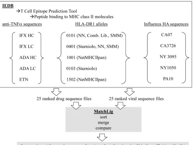
Fig 3. HA analogues in infliximab (IFX) heavy (HC) and light (LC) chains.The heavy chain was analyzed for (a) RA-associated HLA alleles (0101, 0401 and 1001) and (b) non-RA associated alleles (0103 and 1502). The light chain was also analyzed for (c) RA-associated HLA alleles (0101, 0401 and 1001) and (d) non-RA associated alleles (0103 and 1502). Matching biologic and viral ligand pairs are placed based on degrees of similarity and predicted relative binding strengths to the HLA allele. Each point represents a matching pair, identified based on their unique coordinates: the percentile ranking of a biologic sequence to the MHC allele (Pd; x-axis), percentile ranking of the HA peptide homologous to the biologic sequence, to the same allele (Pv; y-axis). Open blue
circles represent biologic sequences that share 8 (out of 15) identical and similar amino acids (as defined inTable 1) with a HA ligand, whereas closed circles in red indicate pairs with at least 9 identical or similar amino acids. Arrows labeled“NLE”or“PAV”point to analogues in biologics that mimic the influenza CD4 T cell epitope HA530–541(see alsoTable 3). Only the Fab regions were considered. Analyses for pre-2009 HA sequences (PA10 and NY1050) can be found in S4. Datasets containing the sequences can be found inS5 Fig
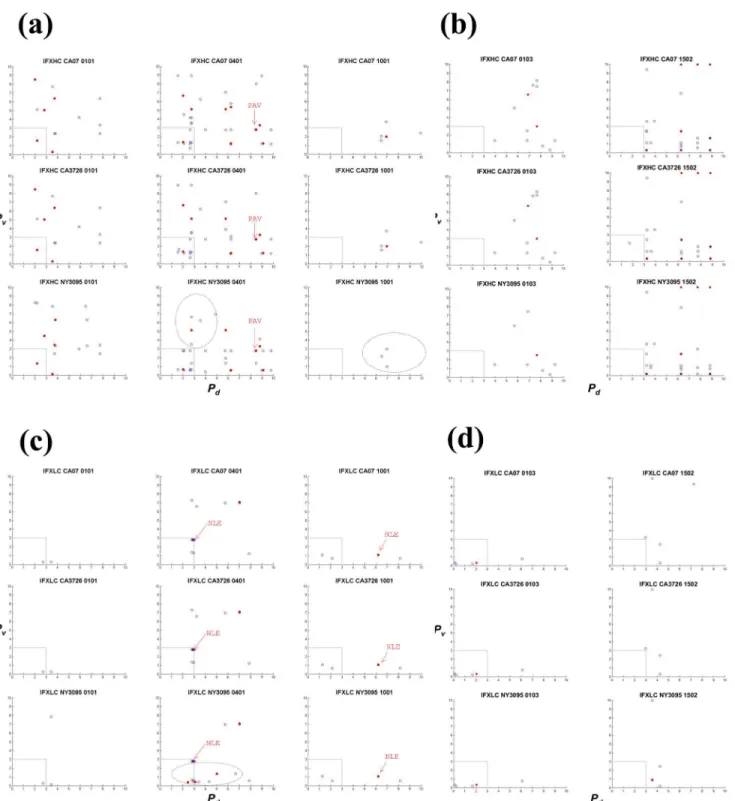
Fig 4. HA analogues in adalimumab (ADA) heavy (HC) and light (LC) chains.The heavy chain was analyzed for (a) RA-associated HLA alleles (0101, 0401 and 1001) and (b) non-RA associated alleles (0103 and 1502). The light chain was analyzed for (c) RA-associated HLA alleles (0101, 0401 and 1001) and (d) non-RA associated alleles (0103 and 1502). X axis and Y axis are defined inFig 3legend. Only the Fab regions were considered. Arrows labeled
“YCA”or“PAV”point to analogues in biologics that mimic the influenza CD4 T cell epitope HA530–541(see alsoTable 3). Analyses for pre-2009 HA sequences (PA10 and NY1050) can be found inS4 FigDatasets containing the sequences can be found inS5 Fig.
for 0401. Of the same 56 sequences, 19 (red dots) contain more than nine (out of 15) identical or similar amino acids, including three with 10-amino acids homology (Table 2;S4 Fig). Four to five analogues were found with 1001 within the 3rdpercentile (Fig 4a), while only one in 0101 (Fig 4a) and 0103 (Fig 4b). None was found within the 3rdpercentile in 1502 (Fig 4b).
The same pattern was found in pre-2009 sequences NY1050 and PA10 in that 0401 would present the most number of analogues (Table 2;S4 Fig). In ADA LC, 19 analogues are found restricted by 0401 (Fig 4c), with 15 ranked within the 3rdpercentile in binding, and one sharing nine identical or similar amino acids with an HA ligand. The allele 1001 would present the sec-ond most number of analogues in ADA LC among the alleles (Fig 4c), while 1502 would pres-ent the least (Fig 4d). While the number of potpres-ential ligands predicted for 0401 and 1502 are similar (Table 3andFig 2), only 0401 would present many more analogues within the 3rd per-centile (Figs3,4and5). Thus the analysis revealed an additional level of complexity.
Likewise, in IFX and ETN, 0401 would present more analogues than the other alleles (Figs3 and5). In IFX HC, 39 sequences bear homology to HA-derived 0401 ligands (Fig 3aand Table 2). Of the 39, 11 share more than nine identical or similar amino acids with a CA07 ligand, and 19 that are ranked within the 3rdpercentile in binding to 0401. IFX HC (Fig 3a and
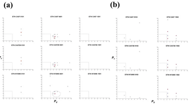
3b) and ADA HC (Fig 4a and 4b) contain more analogues compared to their respective light chains (Figs3c, 3d,4c and 4d). Overall, fewer analogues are predicted in ETN (Fig 5): 12 with 0401, three with 0101, and none with 1001. But six to seven were found with 1502 in both post-and pre-2009 HA sequences (Fig 5bandS5 Fig). Collectively, the scatter analysis provides qual-itative means for comparing cross-reactivity of the biologics across the five MHC alleles.
Several observations were made with regard to the five influenza HA sequences. Overall, substituting CA07 with NY3095 does not alter the general scattering pattern (Table 2). On the Fig 5. HA analogues in etanercept (ETN).The polypeptide was analyzed for (a) RA-associated HLA alleles (0101, 0401 and 1001) and (b) non-RA associated alleles (0103 and 1502). The entire polypeptide, inclusive of TNFR and IgG1 CH2 and CH3, were considered. X axis and Y axis are defined inFig 3legend. Analyses for pre-2009 HA sequences (PA10 and NY1050) can be found inS4 FigDatasets containing the sequences can be found inS5 Fig
other hand, NY3095 (2009) renders unique analogues that are absent in CA07 (2009) and CA3726 (2014). For example, more analogues are located below the 3rdpercentile region (dot-ted area) in NY3095 and IFX LC for 0401 compared to the same regions in CA07 and CA3726 (Fig 3c). Other NY3095 unique analogues are also detected in the IFX HC (0401 and 1001), ADA HC (0101, 0401, and 1001), ADA LC (0101, 0401, and 1001) and ETN (0401). NY3095, NY1050, and PA10 shares 80% identity with CA07, whereas CA07 and CA3726 share 98% identities (S3 Fig). Despite the relatively high degrees of identifies, dissimilarities between the California strains and the New York (and Pennsylvania) strains are manifested in the extent to which their ligands overlap (S5 Fig: m, n and o). The top three panels show scatter plots of matching CA07 against CA07 for the same MHC allele (S5 Fig: m). Symmetry is indicated by the red dots along the centerline between x and y axis, because the same sets of ligands are matched. Ligands of CA07 and CA3278 closely overlap. A high degree of symmetry exists between NY3095 and CA07 for 0401, but no symmetry can be seen in 0101 and 1001 between the two HA sequences. In comparing pre-2009 to post-2009 HA analogues, only 0401 shows Table 2. Comparison of HA ligand analogues in anti-TNFαbiologicsa.
Allele IFX ADA ETN HA sequence
HC LC HC LC
0101 13b(5c) 2(0) 19 (7) 9(0) 3(1) CA07
13 (5) 2(0) 19(7) 9(0) 3(1) CA3726
16 (5) 3(0) 24 (7) 4(0) 2(0) NY3095
17 (6) 3(0) 26 (8) 4(0) 2(0) NY1050
16 (5) 3(0) 25 (7) 4(0) 2(0) PA10
0401 39 (11) 14 (2) 56 (19) 19 (1) 12 (2) CA07
35 (10) 14 (2) 51 (15) 19 (1) 12 (2) CA3726
36 (9) 19 (5) 54 (13) 18 (1) 10 (1) NY3095
38 (10) 19 (5) 57 (14) 17 (1) 7 (1) NY1050
36 (9) 19 (5) 55 (13) 18 (1) 10 (1) PA10
1001 5(1) 4(1) 11 (3) 12 (0) 0(0) CA07
5(1) 4(1) 11 (3) 12 (0) 0(0) CA3726
3(0) 4(1) 13 (2) 9(0) 0(0) NY3095
5(1) 4(1) 15 (3) 8(0) 0(0) NY1050
3(0) 4(1) 13 (2) 9(0) 0(0) PA10
0103 12 (2) 5(1) 16 (4) 8(0) 4(0) CA07
12 (2) 5(1) 16 (4) 8(0) 4(0) CA3726
8 (1) 5(1) 12 (3) 7(0) 3(0) NY3095
9 (2) 5(1) 14 (4) 6(0) 3(0) NY1050
8 (1) 5(1) 12 (3) 7(0) 3(0) PA10
1502 42 (10) 7(0) 47 (13) 15 (0) 21 (4) CA07
44 (10) 6(0) 47 (13) 15 (0) 20 (4) CA3726
44 (9) 11 (3) 52 (12) 13 (0) 15 (3) NY3095
48 (10) 11 (3) 56 (13) 11 (0) 15 (3) NY1050
44 (9) 11 (3) 52 (12) 13 (0) 15 (3) PA10
aCorresponds in part to datasetsS4andS5Figs.
bTotal number of analogues mapped (8 of 15 identical or similar amino acids) to HA ligands; biologic and viral ligands are ranked within the 10thpercentile were tallied
cNumber of sequences containing at least 9 (out of 15) identical or similar amino acids mapped to a HA ligand.
some degrees of symmetry, for example between CA07 and NY1050, and CA07 and NY1050 (S5 Fig: n and o). These analyses served as internal check of the scripts and provide a sensitive way to compare closely related sequences with respect to antigenicity.
Analogues of a dominant HLA-DR1-restricted HA epitope
We extended the scatter analysis by identifying analogues that are more likely to cross-react with HA epitopes. This was carried out by searching in IEDB (using“Linear Epitope”search
tool at“70% blast”) for known HA-derived CD4 T cell epitopes that share homology with the analogues. The analysis identified sequences in IFX and ADA that encapsulate the HA peptide ILAIYSTVASSL (residues 530–541) conserved across all five HA sequences analyzed. This MHC-II restricted epitope was discovered in humans exposed to 2009 strains of swine-origin influenza (H1N1) [44] and induces strong CD4 T cell responses [44]. It is identified as viral ligands of 0401 (Pv= 2.79) and 1001 (Pv= 1.09) in IEDB.Table 3highlights sequences that have at least nine of the 15 amino acids that are identical or similar to HA530–451. Analogues
are found in the antigen-binding regions in heavy and light chains of IFX and ADA but not in ETN (Table 3).
IFX LC contains the analogue NLEVKRTVAAPSVFI (residues 103–117) that spans the
junction (at residues 109 and 110) between the variable (V1) and constant (CH1) domains (Table 3). This sequence (Fig 3c, labeled“NLE”), unique to IFX, is predicted as high affinity
Table 3. Highly similar sequences in biologics mapped to the known CD4 T-cell epitope HA530–541ILAIYSTVASSLa.
Corresponding anti-TNFαbiologic
Biologic (top) and viral (bottom) sequences and their alignment (shown in middle)b
Percentile rank within allele Corresponding HA strain
0101 0401 1001 0103 1502
IFX LC103–117c NLEVKRTVAAPSVFI 2.86 6.21 2.05
Qia svASII consensus
QILAIYSTVASSLVL 2.79 1.09 0.33 CA07529–543CA3726529–543
NY3095528–542NY1050528–542 PA10528–542
ADA HC95–109c YCAKVSYLSTASSLD 9.36 0.89 1.37 5.39 8.27d
YiaYtASSL consensus
YQILAIYSTVASSLV 0.28 2.79 1.72 0.79 1.13 CA07528–542CA3726528–542
NY3095527–541NY1050527–541 PA10527–541
IFX HC174–188/ ADA HC175–189
PAVLQSSGLYSLSSV 8.46 6.27
laiStaSL V consensus
ILAIYSTVASSLVLV 2.79 2.45 CA07530–544CA3726530–544
NY3095529–542eNY1050529–542e PA10529–542e
aReported by Schanen et al. inVaccine29: 3299
–3309 [44].
bOnly analogues with at least 9 of the 15 amino acids being identical or similar are shown. Biologic sequences were mapped to the viral epitope using the IEDB“Linear Epitope”search tool set at“70% blast”. Identical and similar amino acids are indicated in the consensus sequence, with upper case indicates identical, lower case indicates similar,“*”indicates neither. These sequences are indicated with arrows in Figs3and4and can be found in datasetS5 Fig
ETN analogues met the criteria were not found. cBoth peptides are unique to the biologics (S2 Fig).
The“NLE”peptide in IFX LC spans across the junction between the variable and constant region, while the“YCA”peptide in ADA HC is located in the CDR-2.
dIn ADA HC, a slightly shifted sequence, YYCAKVSYLSTASSL (residues 94
–108), is mapped to the viral epitope eThe last amino acid is a leucine in NY3095, NY1050, and PA10.
ligands of 0401 and 1001. Two ADA LC sequences in the same region, VEIKRTVAAPSVFIF (residues 104–118) and KVEIKRTVAAPSVFI (residues 103–117), are mapped to the HA pep-tide ILAIYSTVASSLVLV, with 8 identical/similar amino acids out of the 15, it is not listed in Table 3. The ADA LC peptides are both ranked at 1.79 percentile in 0401. The ADA HC pep-tide YCAKVSYLSTASSLD (residues 95–109) is also an analogue of HA530–451and is predicted
as ligands of 0101, 0401, and 1001. This peptide, labeled inFig 4cas“YCA”, spans the comple-mentarity-determining region 2 (CDR-2) region (residues 99–110) in ADA HC [33], thus
unique to the biologic. Using IEDB, YAC is partially mapped to ASQKRPSQRHGSKYLA-TAST, an HLA-DR-restricted MBP epitope implicated in multiple sclerosis in humans [45, 46]. The clinical implications of this match, however, remain to be investigated. Lastly, the sequence PAVLQSSGYSLLV in IFX HC (residues 174–188) and ADA HC (residues 175–189),
known ligands of several HLA-DR1 alleles [42,43], is identified as an HA530–451analogue
pre-sented by 0401 (Table 3). Located in the constant CH1 region, this common analogue is indi-cated inFig 3aandFig 4aas“PAV”. The analysis illustrates an approach in which candidate ligands can be triaged using IEDB based on homology to known epitopes.
Fig 6. (a) Molecular representation of an ADA analogue that resembles a known B cell epitope in HA Zhao et al. (2011) [49]. The ADA CDR3 peptide was mapped to the B cell epitope by searching for known epitopes in IEDB. Images were generated using Molecular Operating Environment (MOE). Crystal structures of HA (PDB entry: 4M4Y, residues 174–182) and ADA (PDB entry: 3WD5, heavy chain residues 102–111) were used in the modeling. (b) The distances between the Cβof the first and last amino acids are 14.55 and 15.64 Angstroms in ADA HC and HA, respectively. Locations of the side chains were labeled with single amino acid letter codes.
A potential HA-cross reactive B cell epitope in adalimumab
The positional relationship of helper T- and B-cell epitopes may influence immunogenicity [47,48]. Peptides in which a T cell epitope overlaps with a B cell epitope tend to generate anti-bodies of higher affinities than co-immunization of positional-separated epitopes. Thus we searched using IEDB to determine if analogues in the anti-TNFαbiologics overlap with B cell
epitopes. We mapped an ADA analogue to a known HA-derived B cell epitope. The ADA pep-tide, AKVSYLSTASSLDYW, located in the CDR-3 region (residues 97–111), shares homology to the B cell epitope core LSTASSWSY (HA86–92) discovered in humans exposed to the
influ-enza viruses [49]. The core is located in CA07 and CA3726 but not in the other three sequences analyzed; NY3095, PA10 and NY1050 all have the“STAS”tract replaced by“ISKE”(S3 Fig), rendering different electrostatic properties.
Patients exposed to H1N1 influenza viruses during and since the 2009 pandemic would likely have circulating anti-HA78–92antibodies [49]. While it remains to be determined if such
antibodies can cross-recognize LSTASSLDY on ADA, it should be pointed out that crystal structures show the two fragments adapt similar conformations (Fig 6). Both sequences adopt outward bend conformations with distances between the Cβatoms of the first and last residues being 14.55 Å and 15.64 Å, respectively (Fig 6b). Thus generation of ADA-reactive antibodies may be more efficient than expected, as repeated intradermal administrations of ADA would be akin to booster vaccinations with AKVSYLSTASSLDYW in which helper T cell and B cells are reactivated concomitantly. The ADA peptide is predicted as high affinity ligands of 0401 (Pd= 1.37) and 1001 (Pd= 0.89), and as having a good chance of being presented. Memory HA78–92-specific B cell clones could internalize ADA via LSTASSLDY and could present
AKV-SYLSTASSLDYW to naïve CD4 T cells restricted by 0401 or 1001. HA-primed CD4+ T cells could also recognize AKVSYLSTASSLDYW; it shares 8 of 15 amino acids with in HA residues 87–101 (CA07) and binds 0401 at 11.54 percentile (thus not included in our scatter analysis). With 1001, the segment 83–97 that encompasses LSTASSWSY is predicted to bind at 30.09 percentile, a relatively weak binding peptide. These scenarios illustrate a strategy in which potential 0401 ligands in biologics can be selected for experimental testing.
Discussion
Plasma drug concentration predicts the success of anti-TNFαbiologics in RA patients [12,50].
The advent of specific assays that exclude interferences by anti-IgG antibodies helped establish that clearance of IFX, ADA, and ETN is a function of anti-drug antibody response [4]. Given that all recombinant proteins are potentially immunogenic, we investigated a scenario in which elimination of the biologics is accelerated through exposure to influenza viruses. Patients exposed to influenza viruses could harbor memory lymphocytes that produce cross-reactive antibodies upon repeated exposure to biologics. A key variable is the vast number of HA sequences in the population; influenza viruses undergo sequence variations over time, leading to emergence of strains that are temporally unique [23]. Generation of anti-drug antibodies may be enhanced by the presence of helper T cells that recognize HA epitopes homologous with drug ligands restricted by HLA-DRB1 alleles.
an autoimmune disease and the number of analogues found. The data suggest 0401 would present more HA-mimicking analogues compared to the other four alleles analyzed (Fig 4). The number of biological ligands predicted in IEDB for 0401 and 1502 (Fig 2b) are similar, and more than each of the other three alleles. This comparison might suggest the two alleles carry more or less similar immunogenic risk for the anti-TNFαbiologics. Our cross-reactivity
analy-sis, however, reveals that only 0401, but not 1502, would present high affinity biologic ligands that mapped to high affinity viral ligands. For 0401, the 3rdpercentile regions across all HA
sequences in both IFX and ADA (including HC and LC) are well populated (Figs3and4). Conversely, for 1502, ADA LC contains three or four analogues in the 3rdpercentile region,
with one high affinity analogue found in IFX HC mapped to a peptide in CA3726. These results highlight 0401 as a potentially unique HLA allele that warrants further analyses in relating IFX and ADA treatment outcomes with history of influenza infections.
The analysis focused on an underexplored relationship between biologics and viral immu-nity. But cross-reactivity of viral immunity has been well documented in several diseases. Pro-miscuous CD4 T cells may contribute to H1N1 immunity to greater extent than previously thought [44]. Treatment outcomes in RA patients being managed with Disease-Modifying Anti-rheumatic Drugs (DMARDs) correlate with previously acquired immunity to cytomega-lovirus (CMV) [51]. The severity of joint destruction in RA correlates with the degree of preex-isting immunity to CMV and EBV [52,53]. Cross-recognition of self-antigens in autoimmune diseases can be precipitated by specific viral infections [54]. The Coxsackie B virus is implicated in eliciting autoimmune myocarditis and Type I diabetes [55,56]. Systemic lupus erythemato-sus is associated with high titers of anti-Epstein-Barr virus (EBV) antibodies in affected joints and skin [57]. Multiple sclerosis may originate in some cases by cross-recognition of EBV-spe-cific T cells with an epitope in myelin basic protein (MBP) restricted by HLA-DRB11501 [58].
The MBP epitope, ENPVVHFFKNIVTPR (residues 85–99), shares sequence homology with the immunodominant EBV peptide, TGGVYHFVKKHVHES [59]; seven of the 15 amino acids are identical or similar with respect to their physiochemical properties. The extent of homology, seven of fifteen identical or similar amino acids, between the viral and MPB epi-topes in this documented case support the criterion that only sequences that share at least eight of the fifteen amino acids are considered as analogues.
Generation of anti-drug antibodies can be influenced by a multitude of drug, patient, and environmental factors. With respect to drug product quality, differences in glycosylations may lead to recogition of a biologic by antibodies. Impurities associated with manufacturing pro-cesses (e.g. adventitious viruses) can act as adjuvant, thereby boosting an apparent non-immu-nogenic protein to become immunon-immu-nogenic by inducing local inflammation[14]. Altered protein structures and conformations resulting from physical (e.g. denaturation, aggregration) and chemical (e.g. deamination, oxidation) damages are associated with increased immunogenicity. Duration, frequency, and route of adminstration are other factors implicated. Though it is not yet feasible to unify these interactive factors for a priori determination [14,60,61], a common criterion is that the drug polypeptide should contain epitopes of T and B cells.
of a core sequence in the B cell epitope HA78–92embedded in the CDR3 region of ADA. The
15-mer ADA-derived peptide AKVSYLSTASSLDYW, containing both T and B cell epitopes, may be useful as a tool for screening patients for anti-ADA antibodies. But only CA07 and CA3726 contain the core LSTASSWSY; the other three HA sequences have the“STAS”tract broken by“ISKE”, rendering cross-reactivity of these strains with ADA-specific B and T cells unlikely. Thus it may be important to specifically identify the strain of HA in analyzing cross-reactivity between influenza and ADA.
The tenth percentile cutoff may be a conservative limit; ligands ranked above the threshold may render cross-reactivity. The datasets likely have omitted sub-dominant epitopes, given the degeneracy of T cell recognition [63]. The magnitude of the response would depend on the quality and frequency of memory T and B cells. Responses toward ligands with intermediate binding affinities may be amplified from repeated injections. The abundance of the ligands are not considered in our analysis, but anti-TNFαbiologics administered through subcutaneous
and intramuscular routes are akin to vaccinations without inflammation-inducing adjuvant. Another consideratin is that some MHC-II ligands, even high affinity ones, may be tolerogenic to T cells rather than stimulatory [42,43]. Denatured biologics tend be more immunogenic [64], partly because protein aggregates are taken up by APCs more efficiently than native, solu-ble proteins. And chemical degradation can modify the antigenicity of innocuous proteins, resulting in altered binding affinity to MHC and recognition by TCRs.
Despite these caveats, the analysis generated sequences in anti-TNFαbiologics that warrant further analyses. The strategy can be used in optimizing clinical studies aimed at understanding the variability of anti-drug antibodies in RA patients; MHC alleles and exposure to viral may be included as weighed variables. Select analogues may serve as molecular tools for predicting non-responders, and may be eliminated in engineering new biologics [65,66]. The custom script MatchLig can be modified for analysis of proteins of other pathogens that may predis-pose anti-drug antibodies toward biologics. The principle lies in authenticating the relevancy of analogues by mapping to known T cell epitopes using IEDB, a dynamically updated public resource.
As of early 2015, IEDB archives more than 130,000 peptidic epitopes, including more than 30,000 T cell epitopes validated experimentally. Using this strategy, several biologic-derived analogues were mapped to HA530–541, an immunodominant epitope validated in human
PBMC. In addition, an analogue we identified in the CDR3 region of ADA is mapped to a B cell epitope in HA (residues 86–92).
Conclusion
Immunogenicity is a critical quality attribute of pharmaceutical biologics. The present paper demonstrates an approach for analyzing T cell cross-reactivity of biologics and HA, a major antigen of a common virus. The methods enhance the analysis of data retrieved from IEDB by systematic treatments of multiple viral strains, MHC alleles, and biologics. The analogues gen-erated represent candidate cross-reactive epitopes prioritized for testing in T cell response assays. The datasets can be triaged further by mapping analogues to known T and B cell epi-topes, as shown inTable 3andFig 6. These original findings thus illustrate the overarching essence of our approach: dozens (or more) MHC ligands predicted for a given pharmaceutical biologic can be narrowed to a few candidates interlinked with experimental data.
Supporting Information
S1 Fig. Matlab scripts: MatchLig.m and Compare.m.
S2 Fig. Global alignment of infliximab and adalimumab using the Needleman-Wunsch
algorithm via the function‘nwalign’in MATLAB Bioinformatics tools.“”indicates
junc-tions between variable and constant regions in the polypeptide. Boxed regions indicate HA ligands that are mapped to biologic ligands (Table 3).
(PDF)
S3 Fig. Global alignment of HA strains using the Needleman-Wunsch algorithm via the
function‘nwalign’in MATLAB Bioinformatics Tools, with CA07 as reference for
compari-son.The purple box in CA07-CA3726 enclosed the core sequence of B and T epitopes mapped to ADA HC CDR-3 (Fig 6). The core LSTASSWSY is broken in NY3095, NY1050, and PA10. Boxed regions in blue indicate common analogues, biologic sequences mapped to viral ligands (Table 3). Despite the>80% identities, cross-matching CA07 with ligands of the four strains
(using MatchLig),S4 Fig(m-o) shows different patterns of ligands for all alleles except 0401 (with respect to their percentile rank in binding).
(PDF)
S4 Fig.Predicted ligands placed along the polypeptides of (a) IFX, (b) ADA, and (c) ETN.
Ana-logues in 2009 H1N1 influenza HA for five HLA-DR1 alleles: (d) IFX heavy chain pre-sented by alleles associated with RA, (e) and not associated with RA; (f) IFX light chain presented by alleles associated with RA, (g) and not associated with RA; (h) ADA heavy chain presented by alleles associated with RA, (i) and not associated with RA; (j) ADA light chain presented by alleles associated with RA, (k) and not associated with RA; (l) ETN presented by alleles associated with RA, (m) and not associated with RA (j). (n), (o), and (p) show cross-matching of ligands between four HA sequences against CA07.Pddenotes percentile ranking of a given ligand in a biologic, andPvdenotes percentile ranking of a given ligand in a viral HA sequence.
(PPTX)
S5 Fig. Analogue sequence datasets generated using MatchLig in Excel spreadsheets.
(ZIP)
Acknowledgments
We are grateful for Dr. Huynh-Hoa Bui in reviewing the manuscript and providing valuable insights. We would like to thank Drs. Khalid Kamal and Pamela Koerner for helpful discussions.
Author Contributions
Conceived and designed the experiments: WSM LAO BJA. Performed the experiments: BJA WSM AIS BC. Analyzed the data: WSM LAO BJA AIS. Contributed reagents/materials/analy-sis tools: AIS WSM. Wrote the paper: WSM LAO.
References
1. Elliott MJ, Maini RN. New directions for biological therapy in rheumatoid arthritis. Int Arch Allergy Immu-nol. 1994; 104(2):112–25. Epub 1994/01/01. PMID:8199454.
2. Tracey D, Klareskog L, Sasso EH, Salfeld JG, Tak PP. Tumor necrosis factor antagonist mechanisms of action: a comprehensive review. Pharmacology & therapeutics. 2008; 117(2):244–79.
3. Greenberg JD, Reed G, Decktor D, Harrold L, Furst D, Gibofsky A, et al. A comparative effectiveness study of adalimumab, etanercept and infliximab in biologically naive and switched rheumatoid arthritis patients: results from the US CORRONA registry. Annals of the rheumatic diseases. 2012; 71(7):1134–
4. Bendtzen K. Is there a need for immunopharmacologic guidance of anti–tumor necrosis factor thera-pies? Arthritis & Rheumatism. 2011; 63(4):867–70.
5. Aarden L, Ruuls SR, Wolbink G. Immunogenicity of anti-tumor necrosis factor antibodies—toward improved methods of anti-antibody measurement. Current opinion in immunology. 2008; 20(4):431–5. doi:10.1016/j.coi.2008.06.011PMID:18619538
6. Liang S, Dai J, Hou S, Su L, Zhang D, Guo H, et al. Structural basis for treating tumor necrosis factor alpha (TNFalpha)-associated diseases with the therapeutic antibody infliximab. J Biol Chem. 2013; 288 (19):13799–807. Epub 2013/03/19. doi:10.1074/jbc.M112.433961PMID:23504311; PubMed Central PMCID: PMCPmc3650416.
7. Bendtzen K, Geborek P, Svenson M, Larsson L, Kapetanovic MC, Saxne T. Individualized monitoring of drug bioavailability and immunogenicity in rheumatoid arthritis patients treated with the tumor necro-sis factor alpha inhibitor infliximab. Arthritis Rheum. 2006; 54(12):3782–9. Epub 2006/11/30. doi:10. 1002/art.22214PMID:17133559.
8. Wolbink GJ, Vis M, Lems W, Voskuyl AE, De Groot E, Nurmohamed MT, et al. Development of antiin-fliximab antibodies and relationship to clinical response in patients with rheumatoid arthritis. Arthritis & Rheumatism. 2006; 54(3):711–5.
9. Kaymakcalan Z, Xiong L. Compositions and methods comprising binding proteins for adalimumab. Google Patents; 2008.
10. Bartelds GM, Krieckaert CL, Nurmohamed MT, van Schouwenburg PA, Lems WF, Twisk JW, et al. Development of antidrug antibodies against adalimumab and association with disease activity and treatment failure during long-term follow-up. Jama. 2011; 305(14):1460–8. doi:10.1001/jama.2011.406
PMID:21486979
11. de Vries MK, van der Horst-Bruinsma IE, Nurmohamed MT, Aarden LA, Stapel SO, Peters MJ, et al. Immunogenicity does not influence treatment with etanercept in patients with ankylosing spondylitis. Ann Rheum Dis. 2009; 68(4):531–5. Epub 2008/04/01. doi:10.1136/ard.2008.089979PMID:
18375542.
12. Mok C, van der Kleij D, Wolbink G. Drug levels, drug antibodies, and clinical efficacy of the anti-TNFαbiologics in rheumatic diseases. Clinical rheumatology. 2013; 32(10):1429–35. doi:10.1007/ s10067-013-2336-xPMID:23887439
13. Dore RK, Mathews S, Schechtman J, Surbeck W, Mandel D, Patel A, et al. The immunogenicity, safety, and efficacy of etanercept liquid administered once weekly in patients with rheumatoid arthritis. Clin Exp Rheumatol. 2007; 25(1):40–6. Epub 2007/04/10. PMID:17417989.
14. De Groot AS, Scott DW. Immunogenicity of protein therapeutics. Trends Immunol. 2007; 28(11):482–
90. Epub 2007/10/30. doi:10.1016/j.it.2007.07.011PMID:17964218.
15. Novak EJ, Liu AW, Nepom GT, Kwok WW. MHC class II tetramers identify peptide-specific human CD4<sup>+ T cells proliferating in response to influenza A antigen. The Journal of clinical investiga-tion. 1999; 104(12):R63–R7. PMID:10606632
16. Li J, Uetrecht JP. The danger hypothesis applied to idiosyncratic drug reactions. Adverse Drug Reac-tions: Springer; 2010. p. 493–509.
17. Solau-Gervais E, Laxenaire N, Cortet B, Dubucquoi S, Duquesnoy B, Flipo RM. Lack of efficacy of a third tumour necrosis factor alpha antagonist after failure of a soluble receptor and a monoclonal anti-body. Rheumatology (Oxford). 2006; 45(9):1121–4. Epub 2006/03/03. doi:10.1093/rheumatology/ kel054PMID:16510526.
18. Svenson M, Geborek P, Saxne T, Bendtzen K. Monitoring patients treated with anti-TNF-alpha biophar-maceuticals: assessing serum infliximab and anti-infliximab antibodies. Rheumatology (Oxford). 2007; 46(12):1828–34. Epub 2007/11/23. doi:10.1093/rheumatology/kem261PMID:18032541.
19. Baert F, Noman M, Vermeire S, Van Assche G, DH G, Carbonez A, et al. Influence of immunogenicity on the long-term efficacy of infliximab in Crohn's disease. N Engl J Med. 2003; 348(7):601–8. Epub 2003/02/14. doi:10.1056/NEJMoa020888PMID:12584368.
20. Xue L, Rup B. Evaluation of pre-existing antibody presence as a risk factor for posttreatment anti-drug antibody induction: analysis of human clinical study data for multiple biotherapeutics. AAPS J. 2013; 15 (3):893–6. doi:10.1208/s12248-013-9497-zPMID:23761225; PubMed Central PMCID: PMC3691441.
21. Lv Q, Yin Y, Li X, Shan G, Wu X, Liang D, et al. The status of rheumatoid factor and anti-cyclic citrulli-nated peptide antibody are not associated with the effect of anti-TNFαagent treatment in patients with rheumatoid arthritis: a meta-analysis. PloS one. 2014; 9(2):e89442. doi:10.1371/journal.pone. 0089442PMID:24586782
23. Palese P, Wang TT. Why do influenza virus subtypes die out? A hypothesis. MBio. 2011; 2(5):e00150–
11. doi:10.1128/mBio.00150-11PMID:21878571
24. Zimmer SM, Burke DS. Historical perspective—emergence of influenza A (H1N1) viruses. New England Journal of Medicine. 2009; 361(3):279–85. doi:10.1056/NEJMra0904322PMID:19564632 25. Brankston G, Gitterman L, Hirji Z, Lemieux C, Gardam M. Transmission of influenza A in human beings.
The Lancet infectious diseases. 2007; 7(4):257–65. PMID:17376383
26. Raychaudhuri S, Sandor C, Stahl EA, Freudenberg J, Lee HS, Jia X, et al. Five amino acids in three HLA proteins explain most of the association between MHC and seropositive rheumatoid arthritis. Nat Genet. 2012; 44(3):291–6. Epub 2012/01/31. doi:10.1038/ng.1076PMID:22286218; PubMed Central PMCID: PMCPmc3288335.
27. Nielsen M, Lund O. NN-align. An artificial neural network-based alignment algorithm for MHC class II peptide binding prediction. BMC Bioinformatics. 2009; 10:296. Epub 2009/09/22. doi: 10.1186/1471-2105-10-296PMID:19765293; PubMed Central PMCID: PMCPmc2753847.
28. Nielsen M, Lundegaard C, Lund O. Prediction of MHC class II binding affinity using SMM-align, a novel stabilization matrix alignment method. BMC Bioinformatics. 2007; 8:238. Epub 2007/07/05. doi:10. 1186/1471-2105-8-238PMID:17608956; PubMed Central PMCID: PMCPmc1939856.
29. Sturniolo T, Bono E, Ding J, Raddrizzani L, Tuereci O, Sahin U, et al. Generation of tissue-specific and promiscuous HLA ligand databases using DNA microarrays and virtual HLA class II matrices. Nat Bio-technol. 1999; 17(6):555–61. Epub 1999/06/29. doi:10.1038/9858PMID:10385319.
30. Wang P, Sidney J, Kim Y, Sette A, Lund O, Nielsen M, et al. Peptide binding predictions for HLA DR, DP and DQ molecules. BMC bioinformatics. 2010; 11(1):568.
31. Nielsen M, Lundegaard C, Blicher T, Peters B, Sette A, Justesen S, et al. Quantitative predictions of peptide binding to any HLA-DR molecule of known sequence: NetMHCIIpan. PLoS computational biol-ogy. 2008; 4(7):e1000107. doi:10.1371/journal.pcbi.1000107PMID:18604266
32. Van Loan CF, Fan K-YD. Insight through computing: a MATLAB introduction to computational science and engineering. Philadelphia, PA: Society for Industrial and Applied Mathematics; 2010.
33. Hu S, Liang S, Guo H, Zhang D, Li H, Wang X, et al. Comparison of the inhibition mechanisms of adali-mumab and infliximab in treating tumor necrosis factor alpha-associated diseases from a molecular view. J Biol Chem. 2013; 288(38):27059–67. Epub 2013/08/15. doi:10.1074/jbc.M113.491530PMID:
23943614; PubMed Central PMCID: PMCPmc3779706.
34. Osslund TD, Clogston CL, Crampton SL, Bass RB. Crystals of etanercept and methods of making thereof. Google Patents; 2007.
35. Hennecke J, Carfi A, Wiley DC. Structure of a covalently stabilized complex of a human alphabeta T-cell receptor, influenza HA peptide and MHC class II molecule, HLA-DR1. Embo j. 2000; 19(21):5611–
24. Epub 2000/11/04. doi:10.1093/emboj/19.21.5611PMID:11060013; PubMed Central PMCID: PMCPmc305780.
36. Lang HL, Jacobsen H, Ikemizu S, Andersson C, Harlos K, Madsen L, et al. A functional and structural basis for TCR cross-reactivity in multiple sclerosis. Nature immunology. 2002; 3(10):940–3. PMID:
12244309
37. Vaughan K, Peters B, O'Connor KC, Martin R, Sette A. A molecular view of multiple sclerosis and experimental autoimmune encephalitis: What can we learn from the epitope data? Journal of neuroim-munology. 2014; 267(1):73–85.
38. Schueler-Furman O, Altuvia Y, Sette A, Margalit H. Structure-based prediction of binding peptides to MHC class I molecules: application to a broad range of MHC alleles. Protein Science. 2000; 9 (09):1838–46.
39. Stern LJ, Brown JH, Jardetzky TS, Gorga JC, Urban RG, Strominger JL, et al. Crystal structure of the human class II MHC protein HLA-DR1 complexed with an influenza virus peptide. 1994.
40. Jones EY, Fugger L, Strominger JL, Siebold C. MHC class II proteins and disease: a structural perspec-tive. Nature Reviews Immunology. 2006; 6(4):271–82. PMID:16557259
41. Fernando MM, Stevens CR, Walsh EC, De Jager PL, Goyette P, Plenge RM, et al. Defining the role of the MHC in autoimmunity: a review and pooled analysis. PLoS genetics. 2008; 4(4):e1000024. doi:10. 1371/journal.pgen.1000024PMID:18437207
42. Cousens LP, Tassone R, Mazer BD, Ramachandiran V, Scott DW, De Groot AS. Tregitope update: mechanism of action parallels IVIg. Autoimmun Rev. 2013; 12(3):436–43. Epub 2012/09/05. doi:10. 1016/j.autrev.2012.08.017PMID:22944299.
44. Schanen BC, De Groot AS, Moise L, Ardito M, McClaine E, Martin W, et al. Coupling sensitive in vitro and in silico techniques to assess cross-reactive CD4(+) T cells against the swine-origin H1N1 influ-enza virus. Vaccine. 2011; 29(17):3299–309. Epub 2011/02/26. doi:10.1016/j.vaccine.2011.02.019
PMID:21349362; PubMed Central PMCID: PMCPmc3130614.
45. Joshi N, Usuku K, Hauser SL. The T-cell response to myelin basic protein in familial multiple sclerosis: Diversity of fine specificity, restricting elements, and T-cell receptor usage. Annals of neurology. 1993; 34(3):385–93. PMID:7689820
46. Shanmugam A, Copie-Bergman C, Caillat S, Bach JF. In vivo clonal expansion of T lymphocytes spe-cific for an immunodominant N-terminal myelin basic protein epitope in healthy individuals. Journal of neuroimmunology. 1995; 59(1):165–72.
47. Shaw DM, Stanley CM, Partidos CD, Steward MW. Influence of the T-helper epitope on the titre and affinity of antibodies to B-cell epitopes after co-immunization. Molecular immunology. 1993; 30 (11):961–8. PMID:7688851
48. Senpuku H, Iizima T, Yamaguchi Y, Nagata S, Ueno Y, Saito M, et al. Immunogenicity of peptides cou-pled with multiple T-cell epitopes of a surface protein antigen of Streptococcus mutans. Immunology. 1996; 88(2):275–83. PMID:8690461
49. Zhao R, Cui S, Guo L, Wu C, Gonzalez R, Paranhos-BaccalàG, et al. Identification of a highly con-served H1 subtype-specific epitope with diagnostic potential in the hemagglutinin protein of influenza A virus. PloS one. 2011; 6(8):e23374. doi:10.1371/journal.pone.0023374PMID:21886787
50. Daïen CI, Morel J. Predictive factors of response to biological disease modifying antirheumatic drugs: towards personalized medicine. Mediators of inflammation. 2014; 2014.
51. Davis JM, Knutson KL, Strausbauch MA, Green AB, Crowson CS, Therneau TM, et al. Immune response profiling in early rheumatoid arthritis: discovery of a novel interaction of treatment response with viral immunity. Arthritis Res Ther. 2013; 15(6):R199. Epub 2013/11/26. doi:10.1186/ar4389PMID:
24267267; PubMed Central PMCID: PMCPmc3978471.
52. Davis JM 3rd, Knutson KL, Skinner JA, Strausbauch MA, Crowson CS, Therneau TM, et al. A profile of immune response to herpesvirus is associated with radiographic joint damage in rheumatoid arthritis. Arthritis Res Ther. 2012; 14(1):R24. Epub 2012/02/02. doi:10.1186/ar3706PMID:22293286; PubMed Central PMCID: PMCPmc3392817.
53. Kivity S, Agmon-Levin N, Blank M, Shoenfeld Y. Infections and autoimmunity–friends or foes? Trends Immunol. 2009; 30(8):409–14. Epub 2009/08/01. doi:10.1016/j.it.2009.05.005PMID:19643667.
54. Cusick MF, Libbey JE, Fujinami RS. Molecular mimicry as a mechanism of autoimmune disease. Clini-cal reviews in allergy & immunology. 2012; 42(1):102–11.
55. Rose NR, Hill SL. The pathogenesis of postinfectious myocarditis. Clin Immunol Immunopathol. 1996; 80(3 Pt 2):S92–9. Epub 1996/09/01. PMID:8811068.
56. Ray CG, Palmer JP, Crossley JR, Williams RH. Coxsackie B virus antibody responses in juvenile-onset diabetes mellitus. Clin Endocrinol (Oxf). 1980; 12(4):375–8. Epub 1980/04/01. PMID:6247107.
57. Zandman-Goddard G, Berkun Y, Barzilai O, Boaz M, Blank M, Ram M, et al. Exposure to Epstein-Barr virus infection is associated with mild systemic lupus erythematosus disease. Ann N Y Acad Sci. 2009; 1173:658–63. Epub 2009/09/18. doi:10.1111/j.1749-6632.2009.04754.xPMID:19758212.
58. Cheng W, Ma Y, Gong F, Hu C, Qian L, Huang Q, et al. Cross-reactivity of autoreactive T cells with MBP and viral antigens in patients with MS. Front Biosci (Landmark Ed). 2012; 17:1648–58. Epub 2011/12/29. PMID:22201827.
59. Wucherpfennig KW, Strominger JL. Molecular mimicry in T cell-mediated autoimmunity: viral peptides activate human T cell clones specific for myelin basic protein. Cell. 1995; 80(5):695–705. Epub 1995/ 03/10. PMID:7534214.
60. Woodcock J, Griffin J, Behrman R, Cherney B, Crescenzi T, Fraser B, et al. The FDA's assessment of follow-on protein products: a historical perspective. Nat Rev Drug Discov. 2007; 6(6):437–42. Epub 2007/07/20. doi:10.1038/nrd2307PMID:17633790.
61. Tangri S, Mothe BR, Eisenbraun J, Sidney J, Southwood S, Briggs K, et al. Rationally engineered ther-apeutic proteins with reduced immunogenicity. J Immunol. 2005; 174(6):3187–96. Epub 2005/03/08. PMID:15749848.
62. Jørgensen KW, Buus S, Nielsen M. Structural properties of MHC class II ligands, implications for the prediction of MHC class II epitopes. PloS one. 2010; 5(12):e15877. doi:10.1371/journal.pone.0015877
PMID:21209859
63. Mason D. A very high level of crossreactivity is an essential feature of the T-cell receptor. Immunology today. 1998; 19(9):395–404. PMID:9745202
65. Scott DW, De Groot AS. Can we prevent immunogenicity of human protein drugs? Ann Rheum Dis. 2010; 69 Suppl 1:i72–6. Epub 2010/01/09. doi:10.1136/ard.2009.117564PMID:19995750.
66. Jawa V, Cousens LP, Awwad M, Wakshull E, Kropshofer H, De Groot AS. T-cell dependent immunoge-nicity of protein therapeutics: Preclinical assessment and mitigation. Clin Immunol. 2013; 149(3):534–