Effects of Low-Intensity Pulsed Ultrasound on
New Trabecular Bone during Bone
–
Tendon
Junction Healing in a Rabbit Model: A
Synchrotron Radiation Micro-CT Study
Hongbin Lu1, Cheng Zheng1, Zhanwen Wang1, Can Chen1, Huabin Chen1, Jianzhong Hu2*
1Department of Sports Medicine, Research Center of Sports Medicine, Xiangya Hospital, Central South University, Changsha, China,2Department of Spine Surgery, Research Center of Sports Medicine, Xiangya Hospital, Central South University, Changsha, China
*jianzhonghu@hotmail.com
Abstract
This study was designed to evaluate the effects of low-intensity pulsed ultrasound on bone regeneration during the bone–tendon junction healing process and to explore the applica-tion of synchrotron radiaapplica-tion micro computed tomography in three dimensional visualizaapplica-tion of the bone–tendon junction to evaluate the microarchitecture of new trabecular bone. Twenty four mature New Zealand rabbits underwent partial patellectomy to establish a bone–tendon junction injury model at the patella–patellar tendon complex. Animals were then divided into low-intensity pulsed ultrasound treatment (20 min/day, 7 times/week) and placebo control groups, and were euthanized at week 8 and 16 postoperatively (n = 6 for each group and time point). The patella–patellar tendon specimens were harvested for ra-diographic, histological and synchrotron radiation micro computed tomography detection. The area of the newly formed bone in the ultrasound group was significantly greater than that of control group at postoperative week 8 and 16. The high resolution three dimensional visualization images of the bone–tendon junction were acquired by synchrotron radiation micro computed tomography. Low-intensity pulsed ultrasound treatment promoted dense and irregular woven bone formation at week 8 with greater bone volume fraction, number and thickness of new trabecular bone but with lower separation. At week 16, ultrasound group specimens contained mature lamellar bone with higher bone volume fraction and thicker trabeculae than that of control group; however, there was no significant difference in separation and number of the new trabecular bone. This study confirms that low-intensity pulsed ultrasound treatment is able to promot bone formation and remodeling of new tra-becular bone during the bone–tendon junction healing process in a rabbit model, and the synchrotron radiation micro computed tomography could be applied for three dimensional visualization to quantitatively evaluate the microarchitecture of new bone in bone–tendon junction.
OPEN ACCESS
Citation:Lu H, Zheng C, Wang Z, Chen C, Chen H, Hu J (2015) Effects of Low-Intensity Pulsed Ultrasound on New Trabecular Bone during Bone– Tendon Junction Healing in a Rabbit Model: A Synchrotron Radiation Micro-CT Study. PLoS ONE 10(4): e0124724. doi:10.1371/journal.pone.0124724
Academic Editor:Chunfeng Zhao, Mayo Clinic, UNITED STATES
Received:January 7, 2015
Accepted:March 4, 2015
Published:April 15, 2015
Copyright:© 2015 Lu et al. This is an open access article distributed under the terms of theCreative Commons Attribution License, which permits unrestricted use, distribution, and reproduction in any medium, provided the original author and source are credited.
Data Availability Statement:All relevant data are within the paper.
Introduction
The bone–tendon junction (BTJ) is a complex anatomical structure composed of bone,
fibro-cartilage and tendon [1,2]. This unique transitional zone is considered to transmit stress and dissipate stress concentration [3]. Unfortunately, the injury of BTJ is common in trauma and sports exercise, it often occurs in different ligaments, such as rotator cuff, patellar tendon, ante-rior cruciate ligament, and Achilles tendon [4–6]. Direct surgical reconstruction of bone to
ten-don is usually indicated for repairing the patella–patellar tendon (PPT) after transverse
fractures or comminuted fractures [7,8]. Since the healing of BTJ includes dissimilar tissues, its repair is slower and more difficult than bone-to-bone or tendon-to-tendon healing [9,10]. Thus, using a therapeutic approach to enhance the healing of BTJ would be important in basic and clinical research.
As a noninvasive treatment, low-intensity pulsed ultrasound (LIPUS) is considered to be a safe and convenient physical stimulation to administer clinically following musculoskeletal in-jury [11–15]. It is able to induce osteoblast differentiation, extracellular matrix production,
en-dochondral ossification, and mineralization by mechanical stimulation [16–20]. Our previous
studies have shown that LIPUS is able to improve the BTJ healing by enhancing osteogenesis, chondrogenesis, and angiogenesis at the healing interface in a rabbit model [21–23]. We
hy-pothesized that LIPUS would not only be able to promote bone formation but also restore new bone microarchitecture in the BTJ.
Although many detection methods enable observation of the two dimensional (2D) struc-ture of specimens to evaluate bone repair, traditional histology methods with complex section processing steps may destruct the three dimensional (3D) structure of specimens, resulting in limited information to be obtained during and after histological section preparation [24]. In fact, it is impossible to acquire a series of intact and sequential slices using traditional histology methodologies for 3D reconstruction. Therefore, application of a noninvasive and high resolu-tion detecresolu-tion system to evaluate the microarchitecture of new bone is needed. Recently, with the development of imaging technologies and experimental facilities, more internal 3D struc-tural characteristics of specimens could be visualized by noninvasive X-ray computed tomogra-phy imaging methods [25,26]. Compare with the conventional X-ray, the monochromatic X-ray beam produced by synchrotron radiation facility has many advantages, such as high co-herent photon flux, brilliant and small angular beam divergence [27,28]. The synchrotron ra-diation micro computed tomography (SR-μCT) has received increased attention as a novel imaging measurement with noninvasive and high resolution, able to provide a 3D image and allow quantitative analysis of the microarchitecture of internal structures of tested samples [27, 29–31].
We postulated that LIPUS treatment could promote new trabecular bone formation and re-modeling at the BTJ, and the new trabecular bone microarchitecture could be 3D-visualized and quantitatively evaluated using SR-μCT. In the present study, the PPT complexes in the par-tial patellectomy rabbit model were used for radiographic, histological and SR-μCT detection. The first aim of this study was to evaluate the effects of LIPUS on new trabecular bone during the BTJ healing process in an established partial patellectomy model in rabbits. The second aim was to explore the feasibility of application of SR-μCT in the 3D-visualization of the BTJ, and the quantitative evaluation of the new bone microarchitecture at its healing interface.
Materials and Methods
All experimental procedures conformed to the Chinese National Health and were approved by the Ethics Committee of the Center for Scientific Research with Animal Models of Central South University (Permit Number: 2012-06-04). All rabbits were maintained under specific Competing Interests:The authors have declared
pathogen free conditions and fed standard rodent chow ad libitum. The animals were moni-tored daily for feeding and LIPUS treament throughout the experimental period. All surgery was performed under sodium pentobarbital anesthesia. A pain relief drug (Tramadol, Gru-nenthal GmbH, Aachen, Germany) and antibiotic (penicillin sodium, North China pharma-ceutical Co, Shijiazhuang, China) were administered daily within the first 3 days after surgery, and all efforts were made to minimize suffering. There was no unintended death of animals during this study.
Animal model and surgery
Twenty four skeletal mature female New Zealand rabbits (3.5 ± 0.2 kg in weight) were provided from the Center for Scientific Research with Animal Models of Central South University. A partial patellectomy according to a previous established experimental protocol was performed on the right hind legs to establish the BTJ injury model [1]. Briefly, the rabbit was anesthetized intravenously with sodium pentobarbital (0.8 ml/kg, Sigma, St Louis, MO), the right hind leg was shaved and the PPT complex was exposed through an anterolateral skin incision. A trans-verse osteotomy was performed between the proximal 2/3 and the distal 1/3 of the patella. The distal 1/3 of the patella was removed (no remaining fibrocartilage) and two tunnels were drilled longitudinally through the proximal 2/3 patella with a diameter of 0.8mm. The patellar tendon was sutured to the proximal 2/3 patella through two drilled holes with a 3–0 non-absorbable
suture (Mersilk, ETHICON Ltd, Edinburgh, UK). A figure-of-eight tension band wire (0.8 mm diameter) was used around the superior pole of the patella and the tibial tuberosity was achieved to preserve surgical reconstruction from overstretching. The knee was immobilized for 4 weeks with a long leg cast with an open window for LIPUS treatment at the resting posi-tion. Animals were divided into the LIPUS group (treated with LIPUS, n = 12) and control group (treated with placebo, but no ultrasound, n = 12). Six PPT complexes were harvested from each group at week 8 and 16 postoperatively.
LIPUS Treatments
Before treatment, animals were anaesthetized with 3% sodium pentobarbital injected by ear vein. An ultrasound signal with a 1.5 MHz frequency, 1:4 duty cycle and 30mW/cm2spatial and temporal average incident intensity was used (Exogen; Smith and Nephew, San Francisco, CA, USA). A daily 20 min/session of LIPUS treatment was given to the surgical animals. The ultrasound signal was delivered by a 2.5 cm diameter ultrasound transducer which was placed on the skin above the surgery site with coupling gel. Treatment was initiated at day 7 and up to week 8 or week16 postoperatively. The control group received placebo treatment only (without ultrasound signal).
Sample preparation
Radiographic measurement
Anteroposterior (AP) high-resolution radiographs of the PPT complexes were captured using a Faxitron MX-20 X-ray unit (Faxitron X-ray Corp., Lincolnshire, Illinois, USA) with exposure time at 3 s and tube voltage at 32 kVp. Radiographs were analyzed using an image analysis sys-tem (Image-Pro Plus 6.0; Media Cybernetics Inc., Maryland, USA), the size of newly formed bone part from the proximal remaining patella was measured using the previous measurement protocol [1,22].
Histology
One half of each specimen was decalcified using 10%EDTA for 3 weeks, followed by a dual evaporate water rinse for 2 hours. Then, specimens were dehydrated using a series of graded ethanols (rinsing in 70, 80, 95 and twice in 100% for 4 hours each) before embedding in paraf-fin. Specimens were then sectioned (7μm thick) from the mid-sagittal plane using a microtome (RM2165; Leica Microsystems, Nussloch, Germany), deparaffinized using xylene and stained with hematoxylin-eosin (H&E) for descriptive analysis of the new bone. H&E staining was ob-served by light microscopy (Olympus CX31; Olympus Inc., Tokyo, Japan).
SR-
μ
CT imaging and processing
The other half of each specimen was dehydrated using a series of graded ethanols (rinsing in 70, 80, 95 and twice in 100% for 4 hours each) followed by SR-μCT scanning. Scanning experi-ments were performed at the X-ray imaging and biomedical application beamline (BL13W1) of Shanghai Synchrotron Radiation Facility (SSRF) in China. Each sample was placed at the centre of the rotary stage to allow 180° rotation during scanning. The beam energy was set at 18.0 keV, exposure time was 0.5 s, and the sample-to-detector distance was 5.0 cm. A total of 720 initial projection images were captured by the CCD detector with a pixel size of 3.25μm. Both dark-field and flat-field images were obtained to reduce the ring artifact side effect during reconstruction. Phase retrieval of the obtained projected images was carried out and these were transformed into 8-bit slices by PITRE software written by BL13W1 [32]. Subsequently, trans-verse slices were rendered into 3D images using the VG Studio Max software (Ver. 2.1 Volume Graphics GmbH, Germany). The gray-scale images were segmented using a fixed threshold to extract the bone phase followed by a median filter to remove noise. Morphological parameters of the newly formed trabecular bone, such as BV/TV (bone volume fraction), Tb.N (trabecular number), Tb.Th (trabecular thickness), and Tb.Sp (trabecular separation) were calculated.
Statistical analysis
All quantitative data were given as mean ± standard deviation, and the probability distribution displayed the normal distribution. The results were analyzed statistically using an indepen-dent-samplesttest. Statistical analyses were performed with SPSS 13.0 software, and the signif-icance level was set atp<0.05.
Results
Measurement on Radiographs
Fig 1. The anteroposterior radiographs of the patella.The area of the new bone increased from week 8 (b, d) to week 16 (a, c) in both groups. Compraed with control (c, d) group, the LIPUS group (a, b) showed more amount of newly formed bone at each time point. The dotted line shows the osteotomy surface. Scanned bar = 1000μm.
doi:10.1371/journal.pone.0124724.g001
Table 1. The Area of new bone formation (mm2).
Week 8 Week 16
LIPUS group 6.71±0.94a 9.27±1.27a b
Control group 4.63±0.68 6.60±0.99b
a
p<0.01 between LIPUS group and Control group at the same healing time point
b
p<0.01 between postoperative week 8 and 16 at the same group.
Descriptive histology
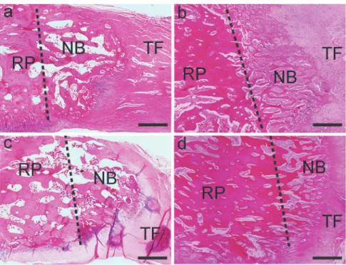
H&E staining showed that the new bone and soft tissue were structurally connected to the re-maining patella and tendon at postoperative week 8 and 16 in both groups. Newly formed tra-becular bone, with a clear boundary around the osteotomy site, was found at postoperative week 8 in both groups and was less mature compared with those at week 16. As healing time in-creased from postoperative week 8 to 16, the new trabecular bone pattern changed from woven bone to lamellar bone in the LIPUS group specimens, showing more advanced remodeling with developed lamellar bone and marrow cavity compared with the control group specimens (Fig 2).
3D visualization images by SR-
μ
CT
Because of the high resolution and signal-to-noise ratio of SR-μCT, 3D visualization images of the BTJ were acquired, which clearly distinguished the mineralization bone phase, tendon and interface between them (Fig 3), the sample resolution was about 6.5μm. The maximum size of field of view was 6.656 mm under the pixel size of 3.25μm of CCD in BL13W1 of SSRF. Imag-ing of the whole patella length was not possible because of the limitation of field of view. How-ever new bone was observed in all specimens within each field of view. As healing time increased, new bone gradually formed on the remaining patella and reconstruction was ob-served in both groups. Based on the simulation sagittal sections, the visualization images of tra-becular bone in both groups were well matched following H&E staining, and and showed that the trabecular bone at postoperative week 16 in the LIPUS group had more lamellar bone with an enlarged marrow cavity arranged in regular orientation.
Fig 2. Sagittal H&E-stained sections of the patella–patellar tendon at postoperative week 8 (b, d) and
16 (a, c).New bone formation was found at the osteotomy site in both groups (LIPUS group: a, b; control group: c, d). The dotted line shows the osteotomy surface (NB, newly formed bone; RP, remaining patella; TF, tendon fiber). The LIPUS group showed more advanced remodeling with developed lamellar bone and marrow cavity, which was more distinct and mature. Scanned bar = 1000μm.
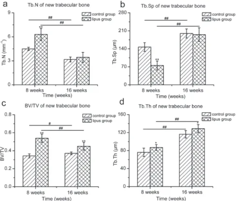
After selecting the region of interest (ROI) of newly formed trabecular bone, bone tissue was segmented and vivid 3D images were acquired (Fig 4). In order to reduce the effect of the region, the ROI was located in the central of newly formed trabecular bone. Morphological pa-rameters of each ROI were analyzed (Fig 5). At postoperative week 8, the BV/TV, Tb.Th and Tb.N of new trabecular bone in the LIPUS group were significantly higher than those of the control group (BV/TV: 0.54 ± 0.05 vs 0.34 ± 0.02,p<0.01, Tb.Th: 86.96 ± 6.38 vs
76.16 ± 9.22μm,p<0.05, Tb.N: 6.30 ± 0.75 vs 4.49 ± 0.20 1/mm,p<0.01), the Tb.Sp of new
tra-becular bone in LIPUS group was significantly lower than that of the control group
(75.27 ± 17.43 vs 147.02 ± 17.14μm,p<0.01). At postoperative week 16, the BV/TV and Tb.Th
of new trabecular bone in the LIPUS group were significantly higher than those of the control group (BV/TV: 0.45 ± 0.04 vs 0.37 ± 0.02,p<0.01, Tb.Th: 128.78 ± 39.10 vs 117.98 ± 9.02μm, p<0.05). There was no significant difference in the Tb.N and Tb.Sp between the two groups
(Tb.N: 3.44 ± 0.63 vs 3.17 ± 0.31 1/mm,p>0.05, Tb.Sp: 195.13 ± 26.75 vs 199.83 ± 17.54μm, p>0.05). With the healing time increased, the BV/TV of new trabecular bone increased from
postoperative week 8 to 16 in the control group (p<0.05), and decreased in the LIPUS group
(p<0.01). The Tb.Th and Tb.Sp increased from postoperative week 8 to 16 in both groups
(p<0.01), and the Tb.N decreased from postoperative week 8 to 16 in both groups (p<0.01).
Discussion
The present study was designed to investigate the effects of LIPUS on new trabecular bone for-mation during the BTJ healing process in an established partial patellectomy model in rabbits, and to explore the feasibility of application of SR-μCT for 3D visualization of the BTJ and quantitative evaluation of the microarchitecture of new bone at the healing interface. The histo-logic and SR-μCT results demonstrated that LIPUS accelerated new bone formation and
Fig 3. 3D visualization images (a–d) and simulation sagittal sections (e–h) of bone–tendon junction of rendered SR-μCT data with high resolution.
Primary woven bone was predominant in the LIPUS (b, f) and control (d, h) groups at postoperative week 8. At postoperative week 16, lamellar bone with marrow cavity formation was predominant in the LIPUS (a, e) and control (c, g) groups. Bone (yellow) and tendon (green) tissues could be clearly distinguished, and the marrow tissue was also seen in green. Scanned bar = 1000μm.
remodeling during the BTJ healing process, and SR-μCT could be applied for 3D visualization and quantitative evaluation of the microarchitecture of new bone.
The rabbit PPT complex in a BTJ injury model, established by partial patellectomy, was a good model to study the BTJ healing process [1]. In the present study, more new bones were formed distally from the osteotomy site of the remaining patella in the LIPUS treatment group as compared with the control group. The osteogenic effects of LIPUS have been confirmed by bothin vitroandin vivostudies [17,33]. Previous research has found that new bone formation of the remaining patella increased the patellofemoral contact area after partial patellectomy, which decreased the contact pressure [34]. New bone formation is the foundation of functional recovery after BTJ injury [35]. Our previous study found that the size of new bone at the junc-tion during BTJ healing correlated with the funcjunc-tional properties (Failure load, Ultimate strength and Energy at failure) [36], and LIPUS was able to enhance BTJ repair in a partial patellectomy model in rabbits, especially accelerated bone formation [22,23]. Thus, we be-lieved that LIPUS treatment could promote new bone formation and play a positive role during the BTJ healing process.
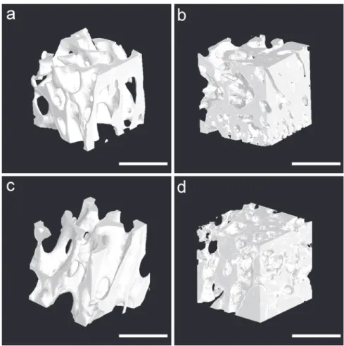
Better remodeling of the new trabecular bone occurred in the LIPUS treatment group when compared with the control group. Based on the H&E staining and SR-μCT data, the newly formed trabecular bone pattern changed from dense and irregular woven bone at the early Fig 4. Segmented 3D tomographic reconstruction images of newly formed trabecular bone in the region of interest.The new trabecular bone was dense with crisscross arrangement in the LIPUS (b) and control (d) groups at postoperative week 8. At postoperative week 16, the new trabecular bone was sparse with marrow cavity formation in the LIPUS (a) and control (c) groups. Scanned bar = 1000μm.
stage to mature lamellar bone and formation of a marrow cavity at the late stage of the healing process. In the present study, the new trabecular bone was dense with a crisscross arrangement in both groups at postoperative week 8. Furthermore, the bone volume fraction and number and thickness of new trabecular bone in the LIPUS group were greater with lower separation than those of the control group. At postoperative week 16, the new trabecular bone was thicker and had sparse marrow cavity formation, and when compared with the control group, the bone volume fraction and thickness of new trabecular bone were greater in the LIPUS group.
Bone healing is usually composed of four distinct but overlapping stages: 1) the early inflam-matory response; 2) soft callus formation; 3) hard callus formation and 4) late bone remodeling [37]. The remodeling process of new bone is crucial to restore the function of the damaged structure [38], and it is accomplished during the remodeling stage in which the irregular and disorderly woven bone callus converts into lamellar bone with orderly bone resorption, eventu-ally restoring the original trabecular bone configuration, structure and mechanical properties [37,39,40]. In the present study, quantitative analysis of the morphological parameters showed that the osteogenic effect was more active in week 8 during the healing process, and the remod-eling of new trabecular bone after week 8 was more advanced in the LIPUS group. Although the amount of new bone was augmented with healing time, the bone volume fraction of the new trabecular bone decreased with greater internal microarchitecture variation. For example, the new trabecular bone was thicker with regular arrangement, and the separation was enlarged with marrow cavity formation. These results suggested that LIPUS was also able to accelerate bone remodeling, which occurred more slowly in the control group.
Fig 5. Histogram of morphological parameters of newly formed trabecular bone.The data show that bone formation and remodeling in the LIPUS group were more advanced than in the control group (a: Tb.N; b: Tb.Sp; c: BV/TV; d: Tb.Th). (*p<0.05;**p<0.01, the difference between the LIPUS group and the control group at different time points; #p<0.05; ##p<0.01, the difference between postoperative week 8 and 16 for the LIPUS and control group; n = 6 for each group/time point).
The three-dimensionality of biological tissue is important, and new bone is augmented with 3D direction in biological tissue during the repair process. To observe the internal microarchi-tecture of new trabecular bone at the BTJ healing interface, we successfully acquired the 3D vi-sualization images, including the bone, tendon and interface between them with a pixel size 3.25μm using SR-μCT without complex sample preparation procedures. Based on the high res-olution 3D images of the BTJ, the simulation sections were highly related to the traditional his-tology slices without structure destruction, and were not restricted by angle and position.
In addition, we not only obtained high-definition 3D images of the internal microarchitec-ture of new trabecular bone in the BTJ samples by SR-μCT, but also performed quantitative analysis to evaluate the effects of LIPUS on new trabecular bone during the BTJ healing process using morphological parameters. With the development of imaging technology, the functions of imaging software have been constantly improving. Therefore the high resolution and 3D im-ages of different tissues in biological samples obtained by SR-μCT enable more detailed infor-mation to be observed from any angle and orientation using imaging software during the imaging process. In recent years, 3D visualization the images of microarchitecture of human and animal bone specimens at different sites were shown using SR-μCT, noninvasively and at high resolution, such as the femur, tibia and vertebrae [26,30,41–47]. Microarchitecture
char-acterization of bone within different bone diseases, including osteoporosis[48], osteoarthritis [43] and bone metastases[42], have also been carried using SR-μCT, which was also used to predict bone fragility or microcracks [29]. Although the microarchitecture index could be ob-tained by SR-μCT, there was a lack of research for using this method as an evaluation tool to study the effects of treatment or intervention. To our knowledge, this is the first study to per-forme 3D visualization of new trabecular bone microarchitecture in the BTJ and evaluation of the effects of LIPUS on new trabecular bone during BTJ healing in a rabbit model using SR-μCT.
In this study, some limitations exist. SR-μCT detection is not easy to perform because the fa-cility is too large and expensive for scientific research and clinical application. Furthermore, the number of facilities and the size of field of view are also limited. However, with the develop-ment of imaging technologies, we believe that these limitations will be solved prior to clinical application.
Conclusions
LIPUS treatment was able to promote BTJ healing by accelerating bone formation and remod-eling of new trabecular bone during the healing process in a rabbit model. SR-μCT was success-fully applied for 3D visualization and quantitative evaluation of the microarchitecture of new bone in the BTJ.
Supporting Information
S1 ARRIVE Checklist. ARRIVE Guidelines Checklist.
(PDF)
Acknowledgments
Author Contributions
Conceived and designed the experiments: HL JH. Performed the experiments: HL CZ ZW CC HC. Analyzed the data: HL CZ ZW. Contributed reagents/materials/analysis tools: CZ HC. Wrote the paper: HL CZ.
References
1. Qin L, Leung KS, Chan CW, Fu LK, Rosier R. Enlargement of remaining patella after partial patellect-omy in rabbits. Med Sci Sports Exerc. 1999; 31: 502–506. PMID:10211843
2. Lui P, Zhang P, Chan K, Qin L. Biology and augmentation of tendon-bone insertion repair. J Orthop Surg Res. 2010; 5: 59. doi:10.1186/1749-799X-5-59PMID:20727196
3. Benjamin MR. Fibrocartilage in tendons and ligaments—an adaptation to compressive load. J Anat. 1998; 193 (Pt 4): 481–494. PMID:10029181
4. Griffin LY, Agel J, Albohm MJ, Arendt EA, Dick RW, Garrett WE, et al. Noncontact anterior cruciate liga-ment injuries: risk factors and prevention strategies. J Am Acad Orthop Surg. 2000; 8: 141–150. PMID:
10874221
5. Cheung EV, Silverio L, Sperling JW. Strategies in biologic augmentation of rotator cuff repair: a review. Clin Orthop Relat Res. 2010; 468: 1476–1484. doi:10.1007/s11999-010-1323-7PMID:20352390
6. Hess GW. Achilles tendon rupture: a review of etiology, population, anatomy, risk factors, and injury prevention. Foot Ankle Spec. 2010; 3: 29–32. doi:10.1177/1938640009355191PMID:20400437
7. Saltzman CL, Goulet JA, McClellan RT, Schneider LA, Matthews LS. Results of treatment of displaced patellar fractures by partial patellectomy. J Bone Joint Surg Am. 1990; 72: 1279–1285. PMID:2229101
8. Hung LK, Lee SY, Leung KS, Chan KM, Nicholl LA. Partial patellectomy for patellar fracture: tension band wiring and early mobilization. J Orthop Trauma. 1993; 7: 252–260. PMID:8326431
9. Lu H, Qin L, Lee K, Wong W, Chan K, Leung K. Healing compared between bone to tendon and carti-lage to tendon in a partial inferior patellectomy model in rabbits. Clin J Sport Med. 2008; 18: 62–69. doi:
10.1097/JSM.0b013e31815c24baPMID:18185041
10. Leung KS, Qin L, Fu LK, Chan CW. A comparative study of bone to bone repair and bone to tendon healing in patella-patellar tendon complex in rabbits. Clin Biomech (Bristol, Avon). 2002; 17: 594–602. PMID:12243719
11. Waseem Z, Ford M, Syed K, Flannery J. Chronic nonunion in a patient with bilateral supracondylar dis-tal femur fractures treated successfully with twice daily low-intensity pulsed ultrasound. PM R. 2010; 2: 159–161. doi:10.1016/j.pmrj.2009.11.008PMID:20193945
12. Schofer MD, Block JE, Aigner J, Schmelz A. Improved healing response in delayed unions of the tibia with low-intensity pulsed ultrasound: results of a randomized sham-controlled trial. BMC Musculoskelet Disord. 2010; 11: 229. doi:10.1186/1471-2474-11-229PMID:20932272
13. Nolte PAvdK, Patka A, Janssen P, Ryaby IMC, Albers JP, Robert GH. Low-intensity pulsed ultrasound in the treatment of nonunions. Journal of Trauma-Injury Infection & Critical Care. 2001; 51: 693–703.
14. Bachl N, Ruoff G, Wessner B, Tschan H. Electromagnetic interventions in musculoskeletal disorders. Clin Sports Med. 2008; 27: 87–105. doi:10.1016/j.csm.2007.10.006PMID:18206570
15. Salem KH, Schmelz A. Low-intensity pulsed ultrasound shortens the treatment time in tibial distraction osteogenesis. International Orthopaedics. 2014; 38: 1477–1482. doi:10.1007/s00264-013-2254-1 PMID:24390009
16. Chen S-H, Wu C C, Wang S H, et al. Growth and Differentiation of Osteoblasts regulated by Low-Inten-sity Pulsed Ultrasound of Various Exposure Durations. J Med Biol Eng. 2014; 34: 197.
17. Angle SR, Sena K, Sumner DR, Virdi AS. Osteogenic differentiation of rat bone marrow stromal cells by various intensities of low-intensity pulsed ultrasound. Ultrasonics. 2011; 51: 281–288. doi:10.1016/j.
ultras.2010.09.004PMID:20965537
18. Katano M, Naruse K, Uchida K, Mikuni-Takagaki Y, Takaso M, Itoman M, et al. Low intensity pulsed ul-trasound accelerates delayed healing process by reducing the time required for the completion of endo-chondral ossification in the aged mouse femur fracture model. Exp Anim. 2011; 60: 385–395. PMID:
21791878
20. Wu S, Kawahara Y, Manabe T, Ogawa K, Matsumoto M, Sasaki A, et al. Low-intensity pulsed ultra-sound accelerates osteoblast differentiation and promotes bone formation in an osteoporosis rat model. Pathobiology. 2009; 76: 99–107. doi:10.1159/000209387PMID:19468249
21. Qin L, Lu H, Fok P, Cheung W, Zheng Y, Lee K, et al. Low-intensity pulsed ultrasound accelerates oste-ogenesis at bone-tendon healing junction. Ultrasound Med Biol. 2006; 32: 1905–1911. PMID:
17169702
22. Lu H, Qin L, Fok P, Cheung W, Lee K, Guo X, et al. Low-intensity pulsed ultrasound accelerates bone-tendon junction healing: a partial patellectomy model in rabbits. Am J Sports Med. 2006; 34: 1287– 1296. PMID:16567453
23. Lu H, Qin L, Cheung W, Lee K, Wong W, Leung K. Low-intensity pulsed ultrasound accelerated bone-tendon junction healing through regulation of vascular endothelial growth factor expression and carti-lage formation. Ultrasound Med Biol. 2008; 34: 1248–1260. doi:10.1016/j.ultrasmedbio.2008.01.009 PMID:18378382
24. Kuhn G, Schultz M, Müller R, Rühli F. Diagnostic value of micro-CT in comparison with histology in the qualitative assessment of historical human postcranial bone pathologies. HOMO. 2007; 58: 97–115. PMID:17451711
25. Chappard D, Retailleau-Gaborit N, Legrand E, Basle MF, Audran M. Comparison insight bone mea-surements by histomorphometry and microCT. J Bone Miner Res. 2005; 20: 1177–1184. PMID:
15940370
26. SaloméM, Peyrin Fo, Cloetens P, Odet C, Laval-Jeantet A-M, Baruchel J, et al. A synchrotron radiation microtomography system for the analysis of trabecular bone samples. Medical Physics. 1999; 26: 2194. PMID:10535638
27. Zehbe R, Riesemeier H, Kirkpatrick CJ, Brochhausen C. Imaging of articular cartilage—data matching using X-ray tomography, SEM, FIB slicing and conventional histology. Micron. 2012; 43: 1060–1067. doi:10.1016/j.micron.2012.05.001PMID:22633854
28. Bonse U, Busch F. X-ray computed microtomography (microCT) using synchrotron radiation (SR). Prog Biophys Mol Biol. 1996; 65: 133–169. PMID:9029944
29. Larrue A, Rattner A, Peter ZA, Olivier C, Laroche N, Vico L, et al. Synchrotron radiation micro-CT at the micrometer scale for the analysis of the three-dimensional morphology of microcracks in human trabec-ular bone. PLoS One. 2011; 6: e21297. doi:10.1371/journal.pone.0021297PMID:21750707
30. Nogueira LP, Braz D, Barroso RC, Oliveira LF, Pinheiro CJ, Dreossi D, et al. 3D histomorphometric quantification of trabecular bones by computed microtomography using synchrotron radiation. Micron. 2010; 41: 990–996. doi:10.1016/j.micron.2010.06.013PMID:20673730
31. Chappard C, Basillais A, Benhamou L, Bonassie A, Brunet-Imbault B, Bonnet N, et al. Comparison of synchrotron radiation and conventional x-ray microcomputed tomography for assessing trabecular bone microarchitecture of human femoral heads. Medical Physics. 2006; 33: 3568. PMID:17022253 32. Chen RC, Dreossi D, Mancini L, Menk R, Rigon L, Xiao TQ, et al. PITRE: software for phase-sensitive
X-ray image processing and tomography reconstruction. J Synchrotron Radiat. 2012; 19: 836–845. doi:
10.1107/S0909049512029731PMID:22898966
33. Xie LK, Wangrangsimakul K, Suttapreyasri S, Cheung LK, Nuntanaranont T. A preliminary study of the effect of low intensity pulsed ultrasound on new bone formation during mandibular distraction osteogen-esis in rabbits. Int J Oral Maxillofac Surg. 2011; 40: 730–736. doi:10.1016/j.ijom.2011.03.016PMID:
21550207
34. Marder RA, Swanson TV, Sharkey NA, Duwelius PJ. Effects of partial patellectomy and reattachment of the patellar tendon on patellofemoral contact areas and pressures. J Bone Joint Surg Am. 1993; 75: 35–45. PMID:8419389
35. Rodeo SA, Arnoczky SP, Torzilli PA, Hidaka C, Warren RF. Tendon-healing in a bone tunnel. A bio-mechanical and histological study in the dog. J Bone Joint Surg Am. 1993; 75: 1795–1803. PMID:
8258550
36. Lu H, Hu J, Qin L, Chan KM, Li G, Li K. Area, length and mineralization content of new bone at bone-tendon junction predict its repair quality. J Orthop Res. 2011; 29: 672–677. doi:10.1002/jor.21292 PMID:21437946
37. Schindeler A, McDonald MM, Bokko P, Little DG. Bone remodeling during fracture repair: The cellular picture. Semin Cell Dev Biol. 2008; 19: 459–466. doi:10.1016/j.semcdb.2008.07.004PMID:18692584
38. Fazzalari NL. Bone fracture and bone fracture repair. Osteoporos Int. 2011; 22: 2003–2006. doi:10.
1007/s00198-011-1611-4PMID:21523400
40. Gerstenfeld LC, Cullinane DM, Barnes GL, Graves DT, Einhorn TA. Fracture healing as a post-natal de-velopmental process: molecular, spatial, and temporal aspects of its regulation. J Cell Biochem. 2003; 88: 873–884. PMID:12616527
41. Li J, Yuan H, Wu M, Dong L, Zhang L, Shi H, et al. Quantitative assessment of murine articular cartilage and bone using x-ray phase-contrast imaging. PLoS One. 2014; 9: e111939. doi:10.1371/journal. pone.0111939PMID:25369528
42. Sone T, Tamada T, Jo Y, Miyoshi H, Fukunaga M. Analysis of three-dimensional microarchitecture and degree of mineralization in bone metastases from prostate cancer using synchrotron microcomputed tomography. Bone. 2004; 35: 432–438. PMID:15268894
43. Chappard C, Peyrin F, Bonnassie A, Lemineur G, Brunet-Imbault B, Lespessailles E, et al. Subchondral bone micro-architectural alterations in osteoarthritis: a synchrotron micro-computed tomography study. Osteoarthritis Cartilage. 2006; 14: 215–223. PMID:16504546
44. Kinney JH, Haupt DL, Balooch M, Ladd AJ, Ryaby JT, Lane NE. Three-dimensional morphometry of the L6 vertebra in the ovariectomized rat model of osteoporosis: biomechanical implications. J Bone Miner Res. 2000; 15: 1981–1991. PMID:11028451
45. Thurner PJ, Wyss P, Voide R, Stauber M, Stampanoni M, Sennhauser U, et al. Time-lapsed investiga-tion of three-dimensional failure and damage accumulainvestiga-tion in trabecular bone using synchrotron light. Bone. 2006; 39: 289–299. PMID:16540385
46. Ito M, Ejiri S, Jinnai H, Kono J, Ikeda S, Nishida A, et al. Bone structure and mineralization demonstrat-ed using synchrotron radiation computdemonstrat-ed tomography (SR-CT) in animal models: preliminary findings. J Bone Miner Metab. 2003; 21: 287–293. PMID:12928829
47. Zuluaga MA, Orkisz M, Dong P, Pacureanu A, Gouttenoire PJ, Peyrin F. Bone canalicular network seg-mentation in 3D nano-CT images through geodesic voting and image tessellation. Phys Med Biol. 2014; 59: 2155–2171. doi:10.1088/0031-9155/59/9/2155PMID:24710691