Marta Ferreira Bastos(a) Felipe Vilhena Brilhante(b) Joyce Pinho Bezerra(b) Carlos Alberto Silva(c) Poliana Mendes Duarte(a)
(a) PhD, Assistant Professor; (b)MS program – Department of Periodontics, Dental Research Division, Guarulhos University, Guarulhos, SP, Brazil.
(c) PhD, Assistant Professor, Department of Rehabilitation Sciences, Nove de Julho University, São Paulo, SP, Brazil.
Corresponding author:
Marta Ferreira Bastos
Praça Tereza, n. 229 - 1º Andar - Centro Guarulhos - SP - Brazil
CEP: 07023-070
E-mail: [email protected]
Received for publication on Sep 01, 2009 Accepted for publication on Apr 12, 2010
Trabecular bone area and bone healing
in spontaneously hypertensive rats.
A histometric study
Abstract: Clinical and experimental studies have demonstrated some negative effect of hypertension on bone mineral density. The aim of this study was to evaluate bone healing and trabecular bone area (TBA) in spontaneously hypertensive rats (SHR), a well-established model of es-sential hypertension, when compared to normotensive rats (NTR). A critical-size defect was surgically created in the right tibia of SHR (n = 12) and normotensive rats (NTR; n = 12), while the contralateral tibia was left intact. Eight days later, the animals were sacriiced and the specimens processed in order to obtain decalciied sections. The area of newly-formed bone (NB) within the defect of the right tibia and the TBA in the left tibia were histometrically evaluated. At 8 days post-operative, SHR presented a signiicantly smaller area of NB when compared to NTR (p < 0.05). In addition, SHR demonstrated a lower TBA than NTR group. In conclusion, the present study demonstrated that SHR rats pre-sented a disturbed bone healing and reduced TBA.
Descriptors: Hypertension; Bone remodeling; Bone density; Rats, Inbred SHR.
Introduction
Human essential hypertension is one of the most prevalent multi-fac-torial medical conditions, characterized by increased peripheral vascu-lar resistance to blood low, vascu-largely due to vascuvascu-lar remodeling.1 Clinical
and experimental studies have demonstrated abnormalities in calcium metabolism at the systemic level in hypertension.2,3 Essential
hyperten-sion may be linked to the increased mobilization of calcium from bone, to the increased losses of calcium from kidney, to the secondary activa-tion of the parathyroid hormone (PTH) and to the angiotensin II activat-ing bone cells.4-7
Bone healing is composed of a series of overlapping stages involving the coordinated action of several cell lineages on a cascade of biologi-cal events, and has always been one of the most important concerns in medicine and dentistry. The use of some drugs (i.e. corticosteroids, che-motherapeutic agents and anti-inlammatory drugs) and the presence of systemic and environmental factors (i.e. smoking, diabetes and osteopo-rosis) have been shown to affect bone density and healing.8,9 Although
10-14 to date, few investigations have focused on the
in-luence of hypertension on bone healing.15
Currently the hypothesis that the hypertension could lead to an increased incidence of late implant loss was evaluated by Alsaadi et al. (2008).16 The
authors found no signiicant differences in late fail-ure between normal and hypertensive individuals.16
However, the impact of hypertension on early im-plant failures rate, which is directly related to the wound healing process in the early stage of osseoin-tegration, has never been tested.
Considering the importance of bone quality and healing in several medical and dentistry situations, the aim of this study was to evaluate bone healing and trabecular bone area (TBA) in spontaneously hypertensive rats (SHR), a model of essential hy-pertension, when compared to normotensive rats (NTR).
Materials and Methods
AnimalsThe University of Guarulhos Institutional Ani-mal Care and Use Committee previously approved and supervised this study. Twelve male SHR and twelve male Wistar NTR obtained from the Fed-eral University of São Paulo were included in this study. The rats were 150 days of age and weighed approximately 259.93 ± 44 g for NTR group and 220.11 ± 15 g for SHR group, at the beginning of the study. During the acclimatization (5 days) and experimental periods (8 days), animals were housed in groups of four in plastic cages with access to food (Labina, Purina, Paulinia, SP, Brazil) and drinking water ad libitum in the Bioscience Laboratory of Guarulhos University. The rats were kept in a room with a 12-hour light/dark cycle and temperature be-tween 22°C and 24°C.
Bone healing and density
After the acclimatization period, general anesthe-sia was obtained by intraperitoneal administration of xylazine (10 mg/kg, Virbaxil; Virbac do Brasil Indústria e Comércio, Roseira, SP, Brazil) and ket-amine (10 mg/kg, Francotar; Virbac do Brasil Indús-tria e Comércio, Roseira, SP, Brazil). The skin was cleaned with iodine surgical soap and an incision of
approximately 1 cm in length was made in the up-per area of the tibia. Subsequently, a full thickness lap was relected and the bone surface of the right tibiae was exposed. Under profuse saline solution irrigation, a unicortical circumferential critical-size defect17 (3.0 x 3.0 mm) was drilled on the
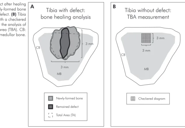
methaph-yseal area of the tibiae with a 3.0 mm wide trephine bur at a rotary speed not exceeding 1,500 rpm. The bur was introduced in the medullar area to create a drill hole with 3 mm of height. The soft tissues were then repositioned and sutured to achieve the prima-ry closure [Poly-vycril 5.0 (Ethicon, São Paulo, SP, Brazil) and Silk 4.0 (Ethicon, São Paulo, SP, Brazil)]. The left tibia remained without defect to be used in the analysis of TBA (Figure 1).
Tissue processing
Animals were euthanized by CO2 inhalation at 8 days after the creation of the bone defect. Both tibia were removed and ixed in 10% buffered for-malin for 48 hours. The specimens were decalciied in a solution containing 10% ethylene-diamine tet-raacetic acid (EDTA), dehydrated in an ascending series of ethanol solution and embedded in parafin. Semi-serial sections (5 µm thick) were obtained in a transversal direction and stained with hematoxylin and eosin solutions.
Histometric procedures
value of TA obtained was considered as 100% of the defect and the percentage of NB was calculated ac-cording to the following formula (Figure 1A):
NB and TBA, an intergroup analysis was performed by the Mann-Whitney test. The signiicance level es-tablished for all analyses was 1% (p < 0.01).
Results
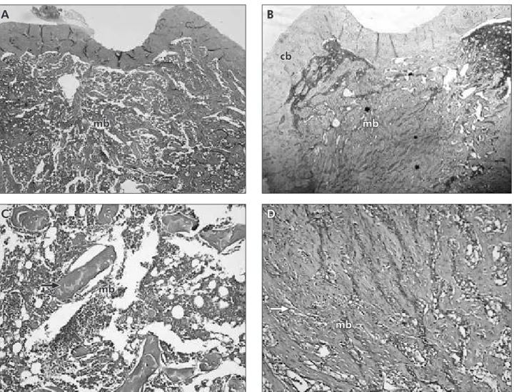
There were no statistically signiicant differ-ences between the mean body weights of SHR (304.1 ± 17 g) and NTR (307.9 ± 54 g) groups at the beginning and at the end of the experimental period (p > 0.05). The mean percentage of TBA was signiicantly lower in the SHR (15.42 ± 4.16%) when compared to the NTR group (21.46 ± 3.96%) (p = 0.0026), showing that the trabecular bone was negatively inluenced by the model of essential hy-pertension. Figures 2A-2D illustrate the histological indings of TBA for SHR and NTR. Figures 2A and 2C show a low proportion of bone trabeculae with large inter-trabecular spaces in the medullar area of the methaphyseal region in an animal from the SHR group. Figures 2B and 2D illustrate a normal can-cellous bone.
The SHR group presented a signiicantly lower percentage of NB area in the defect (25.62 ± 8.56%) when compared to the NTR group (86.76 ± 1.78%) (p < 0.0001). Figures 3A-3D illustrate the histologi-For the analysis of the TBA, a standardized
quad-rilateral area (1 mm²) was delimited in the medullar area and a checkered diagram was overlaid over this region, constituting a drawing of 314 intersections as illustrated in Figure 1B. The number of intersec-tions, under which the presence of bone tissue was observed, was then counted. The 314 intersections were considered as 100% (1 mm²) and the TBA was calculated according to the following formula (Fig-ure 1B):
NB (mm2) × 100
TA (mm2)
Statistical analysis
The mean percentage of NB and TBA of the three sections were averaged for each animal and across animals in the two experimental groups (SHR or NTR). In order to test the hypothesis that the exper-imental model of hypertension had no inluence on
(number of counted intersections) × 100 314
Figure 1 -(A) Defect after healing period with newly-formed bone
and the remained defect. (B) Tibia
without defect with a checkered diagram used for the analysis of trabecular bone area (TBA). CB: cortical bone; MB: medullar bone.
Tibia with defect:
bone healing analysis
Remained defect Newly-formed bone
Total Area (TA) 3 mm
3 mm
MB CB
Tibia without defect:
TBA measurement
Checkered diagram 3 mm
3 mm
MB CB
cal indings of bone healing for SHR and NTR, re-spectively. Figures 3A and 3C show NB restricted to areas close to the borders of the surgical defect, with immature and poorly organized bone trabecu-lae from SHR. Figures 3B and 3D present a consid-erably increased new well-organized bone growth coming from the walls of the defect in the control group.
Discussion
The outcomes of dental and orthopedic implants and the increased risk of fracture are directly re-lated to bone quality. In addition, successful bone formation after tooth extraction, osseointegration of implants, reconstruction of skeletal defects and
fracture repair are totally dependent on an adequate bone healing. As such, the inluence of systemic conditions and habits on bone tissue should be con-sidered in order to ind the groups with increased risk for deicient bone repair and poor bone density. Thus, the aim of this study was to investigate the in-luence of hypertension on bone healing and TBA in the tibia of SHR, considered to be a reliable model of essential hypertension.
In the present study, NTR with 150 days of age showed a considerably greater TBA than the age-matched SHR. In addition, at 8 days post-opera-tive, the SHR group presented a signiicantly lower bone formation than that of the NTR group within the surgically-created critical-size defects. Based
mb
Figure 2 - Photomicrographs illustrating transversal sections of the tibia without defect for SHR (A/C) and NTR (B/D) groups, showing low proportions of bone trabeculae and large inter-trabecular spaces in the medullar area of the SHR group. Lower
magnification, 25 X (A/B). Higher magnification, 100 X (C/D). cb: cortical bone; mb: medullar bone; bone trabeculae
(ar-row).
mb
A B
C D
mb
cb
on these indings, it could be suggested that hyper-tension affected not only the density of the preex-isting bone but also the newly-formed bone tissue of the medullar region. It is important to note that the histometric analyses were performed only in the medullar region of the tibia, excluding the cortical bone. This criterion was chosen based on the fact that medullar bone, which presents a great potential for repair due to the presence of large numbers of bone cells, is more responsive to the inluence of risk factors (i.e. osteoporosis, cigarette smoking), espe-cially in short-term experimental periods.8, 9
The reduced TBA observed in the SHR of the present study is consistent with some previous inves-tigations using the same animal model with
differ-ent ranges of age. Metz et al. (1990)18 demonstrated
that SHR at 6 weeks of age exhibited signiicantly lower levels of bone density than NTR and that this reduced density can be reverted by supplemental dietary calcium. Barbagallo et al. (1990)19 showed
that 24-week-old SHR presented a lower bone den-sity and calcium content than age-matched NTR. Similar indings were observed by Barbagallo et al. (1991),20 Inoue et al. (1995)21 and Wright and
DeM-oss (2000).12
Together, the results of our and these experimen-tal studies support the clinical investigations docu-menting an association between low bone mineral density (BMD) and high blood pressure. Cappuccio
et al.(1999),2 evaluating 3,676 women, showed that
Figure 3 - Photomicrographs illustrating transversal sections of the right tibia with the surgical defect for SHR (A/C) and NTR
(B/D) groups. Figure 3A shows newly-formed bone restricted to areas close to the borders of the defect, while figure 3B
illus-trates a large amount of new bone coming from the walls of the defect. Lower magnification, 25 X (A/B). Higher magnification,
100 X (C/D). cb: cortical bone; mb: medullar bone; nb: newly-formed bone; rd: remained defect.
A B
D
mb rd
mb nb
cb
C
cb
rd
elevated blood pressure in elderly white women was associated with increased bone loss at the femoral neck. Similarly, Tsuda et al. (2001)3 observed by
means of dual-energy X-ray absorptiometry a sig-niicant decrease in BMD in female hypertensive compared with normotensive women. Gotoh et al. (2005)13 demonstrated that the BMD in the
lum-bar spine in women with normotension was higher than that of women with essential hypertension. Al-though the precise pathophysiological mechanisms by which hypertension could have a harmful effect on bone density are still unclear. Some suggested hypotheses include changes in PTH and calcium metabolism and the role of angiotensin II in bone cells. Angiotensin II is the major mediator of the maintenance of extracellular luid volume and blood pressure and may decelerate osteoblastic differen-tiation and bone formation and activate osteoclasts and bone resorption.4-7, 21-23
Although some studies have already reported the inluence of hypertension on bone density, the results of the present study contribute to the original infor-mation regarding the negative effect of hypertension on the early stages of bone healing in a surgically-created critical-size defect. A mechanism that could partially explain our histometric results may also be attributed to the inluence of angiotensin II on bone cells. Although the effect of angiotensin II on osteo-blastic cells is still controversial and predominantly based on in vitro studies,23-25 there is some evidence
to suggest that this mediator is a potent suppres-sor of the differentiation of osteoblastic cells and, consequently, of the bone formation by these cells.22
Our indings are contradictory to those described
by Pereira et al. (2007),15 in which the amount of
newly-formed bone was similar in SHR and NTR animals at 7 days and higher in SHR at 21 days post-operative. In addition to the differences in the experimental periods between this study and ours, these conlicting results may also be explained by dissimilarities in rat age (data not shown) and gen-der (male and female) and by the localization (femur diaphysis) and size (2 mm) of the defects. The use of the critical-size defect is important to evaluate the real impact of local and systemic factors that could inluence bone healing. Lewandrowski et al. (1999)17
observed that a surgically created defect of 3 mm in diameter in rat tibia did not heal spontaneously in up to 7 weeks post-operative, being an appropriate model for evaluations of the newly-formed bone.
Conclusion
The results of the present study demonstrated that SHR rats presented an impairment of bone healing and reduced TBA, indicating that individu-als with essential hypertension could be a risk group for these bone disorders. Nevertheless, further stud-ies should be considered in order to clinically evalu-ate the relevance of our indings.
Acknowledgment
The authors greatly appreciated the assistance of the lab technicians from Guarulhos University: Paulo César Simões Silva, for helping with the his-tological procedures and, Rogério Tadeu Barreira, for technical support with animal care. There is no conlict of interest to declare.
References
1. Virdis A, Ghiadoni L, Versari D, Giannarelli C, Salvetti A, Taddei S. Endothelial function assessment in complicated hypertension. Curr Pharm Des. 2008;14(18):1761-70. 2. Cappuccio FP, Meilahn E, Zmuda JM, Cauley JA. High blood
pressure and bone-mineral loss in elderly white women: a prospective study. Study of Osteoporotic Fractures. Lancet. 1999 Sep 18;354(9183):971-5.
3. Tsuda K, Nishio I, Masuyama Y. Bone mineral density in wom-en with esswom-ential hypertwom-ension. Am J Hypertwom-ens. 2001 Jul;14(7 Pt 1):704-7.
4. McCarron DA, Pingree PA, Rubin RJ, Gaucher SM, Molitch M, Krutzik S. Enhanced parathyroid function in essential hypertension: a homeostatic response to a urinary calcium leak. Hypertension. 1980 Mar-Apr;2(2):162-8.
6. Young EW, McCarron DA, Morris CD. Calcium regulating hormones in essential hypertension. Importance of gender. Am J Hypertens. 1990 Aug;3(8 Pt 2):161S-166S.
7. Oshima T, Young EW. Systemic and cellular calcium me-tabolism and hypertension. Semin Nephrol. 1995 Nov;15 (6):496-503.
8. Duarte PM, César Neto JB, Gonçalves PF, Sallum EA, Nociti FH. Estrogen deficiency affects bone healing around tita-nium implants: a histometric study in rats. Implant Dent. 2003;12(4):340-6.
9. César-Neto JB, Duarte PM, Sallum EA, Barbieri D, More-no H Jr, Nociti FH Jr. A comparative study on the effect of nicotine administration and cigarette smoke inhalation on bone healing around titanium implants.J Periodontol. 2003 Oct;74(10):1454-9.
10. Izawa Y, Sagara K, Kadota T, Makita T. Bone disorders in spontaneously hypertensive rat. Calcif Tissue Int. 1985 Dec;37(6):605-7.
11. Liang H, Ma Y, Pun S, Stimpel M, Jee WS. Aging- and ovari-ectomy-related skeletal changes in spontaneously hypertensive rats. Anat Rec. 1997 Oct;249(2):173-80.
12. Wright GL, DeMoss D. Evidence for dramatically increased bone turnover in spontaneously hypertensive rats. Metabo-lism. 2000 Sep;49(9):1130-3.
13. Gotoh M, Mizuno K, Ono Y, Takahashi M. High blood pres-sure, bone-mineral loss and insulin resistance in women. Hy-pertens Res. 2005 Jul;28(7):565-70.
14. Mussolino ME, Gillum RF. Bone mineral density and hyper-tension prevalence in postmenopausal women: results from the Third National Health and Nutrition Examination Survey. Ann Epidemiol. 2006 May;16(5):395-9.
15. Pereira AC, Fernandes RG, Carvalho YR, Balducci I, Faig-Leite H. Bone healing in drill hole defects in spontaneously hypertensive male and female rats’ femurs. A histological and histometric study. Arq Bras Cardiol. 2007 Jan;88(1):104-9. 16. Alsaadi G, Quirynen M, Komárek A, van Steenberghe D.
Im-pact of local and systemic factors on the incidence of late oral
implant loss. Clin Oral Implants Res. 2008 Jul; 19(7):670-6.
17. Lewandrowski KU, Cattaneo MV, Gresser JD, Wise DL, White RL, Bonassar L, et al. Effect of a poly(propylene fu-marate) foaming cement on the healing of bone defects. Tissue Eng. 1999 Aug;5(4):305-16.
18. Metz JA, Karanja N, Young EW, Morris CD, McCarron DA. Bone mineral density in spontaneous hypertension: differ-ential effects of dietary calcium and sodium. Am J Med Sci. 1990 Oct;300(4):225-30.
19. Barbagallo M, Raddino R, Restori G, Boiardi L, Novo S, Strano A. Alterations of calcium metabolism in spontaneously hypertensive rats. Cardioscience. 1990 Jun;1(2):105-7. 20. Barbagallo M, Quaini F, Baroni MC, Barbagallo CM, Boiardi
L, Passeri G, et al. Histological evidence of increased turnover in bone from spontaneously hypertensive rats. Cardioscience. 1991 Mar;2(1):15-7.
21. Inoue T, Moriya A, Goto K, Tanaka T, Inazu M. What is the difference of bone growth in SHR and SD rats? Clin Exp Pharmacol Physiol Suppl. 1995 Dec;22(1):S242-3.
22. Hatton R, Stimpel M, Chambers TJ. Angiotensin II is gener-ated from angiotensin I by bone cells and stimulates osteoclas-tic bone resorption in vitro. J Endocrinol. 1997 Jan;152(1):5-10.
23. Hagiwara H, Hiruma Y, Inoue A, Yamaguchi A, Hirose S. Deceleration by angiotensin II of the differentiation and bone formation of rat calvarial osteoblastic cells. J Endocrinol. 1998 Mar;156(3):543-50.
24. Shimizu H, Nakagami H, Osako MK, Hanayama R, Kunugiza Y, Kizawa T, et al. Angiotensin II accelerates osteoporosis by activating osteoclasts. FASEB J. 2008 Jul;22(7):2465-75. 25. Hiruma Y, Inoue A, Hirose S, Hagiwara H. Angiotensin II