Binding Activity of Epigallocatechin
Li-Ping Bai1,2, Hing-Man Ho2, Dik-Lung Ma3, Hui Yang3, Wai-Chung Fu3, Zhi-Hong Jiang1*
1State Key Laboratory of Quality Research in Chinese Medicine, and Macau Institute for Applied Research in Medicine and Health, Macau University of Science and Technology, Taipa, Macau,2School of Chinese Medicine, Hong Kong Baptist University, Kowloon Tong, Kowloon, Hong Kong,3Department of Chemistry, Hong Kong Baptist University, Kowloon Tong, Kowloon, Hong Kong
Abstract
With the aim of enhancing G-quadruplex binding activity, two new glucosaminosides (16, 18) of penta-methylated epigallocatechin were synthesized by chemical glycosylation. Subsequent ESI-TOF-MS analysis demonstrated that these two glucosaminoside derivatives exhibit much stronger binding activity to human telomeric DNA and RNA G-quadruplexes than their parent structure (i.e., methylated EGC) (14) as well as natural epigallocatechin (EGC,6). The DNA G-quadruplex binding activity of16and18is even more potent than strong G-quadruplex binder quercetin, which has a more planar structure. These two synthetic compounds also showed a higher binding strength to human telomeric RNA G-quadruplex than its DNA counterpart. Analysis of the structure-activity relationship revealed that the more basic compound,16, has a higher binding capacity with DNA and RNA G-quadruplexes than its N-acetyl derivative,18, suggesting the importance of the basicity of the aminoglycoside for G-quadruplex binding activity. Molecular docking simulation predicted that the aromatic ring of16p-stacks with the aromatic ring of guanine nucleotides, with the glucosamine moiety residing in the groove of G-quadruplex. This research indicates that glycosylation of natural products with aminosugar can significantly enhance their G-quadruplex binding activities, thus is an effective way to generate small molecules targeting G-quadruplexes in nucleic acids. In addition, this is the first report that green tea catechin can bind to nucleic acid G-quadruplex structures.
Citation:Bai L-P, Ho H-M, Ma D-L, Yang H, Fu W-C, et al. (2013) Aminoglycosylation Can Enhance the G-Quadruplex Binding Activity of Epigallocatechin. PLoS ONE 8(1): e53962. doi:10.1371/journal.pone.0053962
Editor:Fenfei Leng, Florida International University, United States of America
ReceivedOctober 12, 2012;AcceptedDecember 4, 2012;PublishedJanuary 15, 2013
Copyright:ß2013 Bai et al. This is an open-access article distributed under the terms of the Creative Commons Attribution License, which permits unrestricted use, distribution, and reproduction in any medium, provided the original author and source are credited.
Funding:This work was financially supported by a grant from the Macao Foundation (grant numbers 0205 and 0199). The funders had no role in study design, data collection and analysis, decision to publish, or preparation of the manuscript.
Competing Interests:The authors have declared that no competing interests exist. * E-mail: zhjiang@must.edu.mo
Introduction
Nucleic acid G-quadruplexes, four-stranded helical structures held together by a core of guanine tetrads, are secondary structures formed in particular G-rich sequences. Potential nucleic acid G-quadruplex structures have been identified in telomeric DNA and RNA sequences [1–5] as well as non-telomeric chromosomal promoters [6–9] of biological significance. These higher-order structures in nucleic acids represent a new class of molecular targets for selective DNA- and RNA-interacting compounds; in view of the fact that cancer cells have high telomerase activity and abnormal overexpression of oncogenes relative to normal cells, they are promising targets for cancer drug discovery [10]. In addition, numerous compounds have been designed to inhibit telomerase or to inactivate the transcription of oncogenes, such asc-Myc,c-kit, andBcl-2[6,9,11–13], suggesting that the design of drugs targeting telomere or promoter G-quadruplexes is a rational and promising approach for generating new anticancer agents [14]. While recognition of G-quadruplex has mostly been achieved with the use of planar aromatic ligands through stacking interactions with the G-tetrad [14], grooves and negatively charged phosphate residues in G-quadruplexes are alternative binding sites to consider in the design of G-quadruplex stabilizing ligands [15–22].
Green tea catechins, the main biologically-active constituents of green tea, have gained significant recognition as cancer preventive
agents. Green tea catechins are composed of four major polyphenols: (2)-epigallocatechin gallate (EGCG), (2 )-epigalloca-techin (EGC), (2)-epicatechin gallate (ECG), and (2)-epicatechin (EC) [23]. They show a variety of pharmacological activities, including cancer-preventive, antioxidant, cancer [24], anti-angiogenesis activities [25], as well as inhibiting the fibrillogenesis of amyloid b peptide [26–27], anti-mutagenic and anti-viral activities [28–31]. It has been reported that catechins affect DNA replication, DNA repair, and transcription [32–34]. A recent study revealed that nucleic acids are binding targets of green tea catechins: nucleic acids extracted from EGCG-treated human cancer cells were catechin-colored, and direct binding of catechins with single-stranded and double-stranded DNA/RNA was observed by cold spray ionization-mass spectrometry [34]. However, a molecular docking study indicated that catechins, including EGC, are poor DNA G-quadruplex-stabilizing ligands compared with the more planar compound quercetin [35]. Therefore, structural modification of EGC is necessary for enhancing its G-quadruplex binding affinity.
aminoglycosides, such as neomycin and paromomycin, recognize the wide groove of Oxytricha nova telomeric G-quadruplex DNA [15]. These findings led to a prediction that the coupling of aminosugars with ligands that bind to G-quadruplex through stacking interactions may lead to enhanced G-quadruplex stabilizing properties. In our previous study, it was demonstrated that glycosylation of shikonin/alkannin with N-acetyl glucosamine is an effective way to generate a potent G-quadruplex DNA ligand [38]. Based on these observations, in this study we herein designed and synthesized two new glucosaminosides of EGC (16, 18) and subsequently examined their binding affinities with both telomeric DNA and RNA G-quadruplexes by ESI-TOF-MS. Furthermore, the binding of these two glucosaminosides (16,18) with oncogene G-quadruplexes was also explored. Finally, the binding mode of 16with human telomeric DNA G-quadruplex was investigated by computational docking experiments.
Results
Synthesis of EGC Glucosaminosides
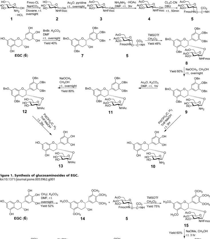
Glycosylation is an effective method for connecting saccharide units to natural products in order to obtain biologically active glycosides [39–40]. Many glycosylated natural products have been reported to show high activity against a variety of human tumors [36–37]. In this study, chemical glycosylation was employed as a key approach to acquiring EGC glucosaminosides. As illustrated in Figure 1, our initial efforts were focused on the design and synthesis of EGC-3-O-b-glucosaminoside (10) and its N-acetyl derivative (13), starting from the readily available (2)-EGC and D-(+)-glucosamine hydrochloride. The glycosyl donor 5 was prepared according to the method previously described in the literature [38,41–42]. The amino group of glucosamine was firstly blocked by 9-fluorenylmethoxycarbonyl chloride (Fmoc-Cl) and followed by acetylation of the hydroxyl groups. After selective deprotection, the anomeric hydroxyl group was transformed to trichloroacetimidate and thus activated for glycosylation [38].Tri-methylsilyl triflate (TMSOTf) was used as a catalyst for the glycosylation of the penta-benzyl ether of EGC (7). After deprotection and acetylation of the amino group, EGC glucosa-minoside [10, ESI-TOF-MSm/z(C21H26NO11)+: calcd 468.1500,
found 468.1489] and its N-acetyl product13[ESI-TOF-MSm/z
(C23H28NO12)+: calcd 510.1606, found 510.1599] were
synthe-sized. Unfortunately, we failed to purify these two products due to their instability during the course of column chromatographic purification.
To avoid this instability, we slightly modified our target products by blocking the active phenolic hydroxyl groups of EGC with methyl groups. The penta-methylated EGC (14) was subsequently glycosylated with glycosyl donor5by the action of TMSOTf, followed by deprotection to give 16. Its N-acetyl derivative 18 was synthesized by further acetylation of 16and subsequent deacetylation (Figure 2). Compounds16and18were characterized to be 5, 7, 39, 49, 59-penta-O-methyl epigallocatechin
b-D-glucosaminoside and 5, 7, 39, 49, 59-penta-O-methyl epigallo-catechin N-acetylb-D-glucosaminoside, respectively, on the basis of 1H-NMR, 13C-NMR and high resolution mass spectroscopic evidence (Figure S1, S2, S3, S4, S5, S6, S7, S8, S9) [43–44].
Analysis of Human Telomeric G-quadruplex DNA Binding by ESI-TOF-MS
Mass spectrometry coupled with the source of electrospray ionization (ESI), a soft ionization method, has played a more active role in the investigation of noncovalent complexes of nucleic acids with small organic molecules. It has the advantages of direct
assignment of the stoichiometry and gives an indication of the relative amounts of different species of complexes [45–46]. Mass spectrometry, combined with techniques of ion mobility and molecular dynamics, has demonstrated that DNA G-quadruplexes in telomeric repeats are conserved in a solvent-free environment [47].
The G-quadruplex DNA-binding activities of glucosaminosides of penta-methylated EGC (16,18), as well as their aglycone (14) and natural EGC (6), were examined with a 27 nt human telomeric sequence d[(TTAGGG)4TTA] which forms an
intra-molecular G-quadruplex, by ESI-TOF-MS. Quercetin, a flavonoid with a similar but more planar structure than EGC, was used as a reference compound for the comparison of G-quadruplex binding activity of the natural and synthetic compounds, since it was reported to be stacked with terminal tetrads of monomeric G-quadruplexes [48].
The ESI-TOF-MS spectrum of telomeric DNA showed that the addition of the NH4OAc buffer facilitated the detection of
quadruplex (Q52in Figure 3A) [49] in the25 charge state ions atm/z1697.9, 1701.3, and 1704.7. These three ions correspond to the lone oligodeoxynucleotide and the oligodeoxynucleotides with one and two NH4+ion adducts, respectively. When the drug was
added to DNA, the complex peaks with two NH4+ion adducts
became more predominant than those with one NH4+ion adduct
or none when a molar ratio of DNA/drug of 1:1 was used (Figure 3B–F). This indicated that the G-quadruplex structure stabilized by drugs holds NH4+ions inserted between G-tetrads
more tightly than free G-quadruplex in the course of being introduced into the gas phase. In other words, drug-bound G-quadruplex is more stable than when it is unbound. In order to compare the stabilization effect of different molecules on DNA G-quadruplex, the peak area ratio of all [complex]52 to [quad-ruplex]52 was used to evaluate the relative binding affinities (Figure 4) [49–51].
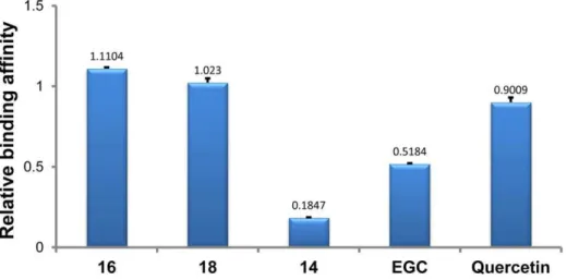
As illustrated in Figure 4, the relative binding affinities of all tested drugs with intramolecular human telomeric DNA G-quadruplex followed the descending order of 16.18. Querce-tin.EGC .14. Two synthetic glucosaminosides (16 and 18) demonstrated a more strong stabilizing effect on intramolecular human telomeric DNA G-quadruplex than their parent structure, methylated EGC (14). These two glucosaminosides also showed higher relative binding affinities than the natural catechin EGC and the even more planar flavonol quercetin. This indicated that the introduction of a glucosamine moiety into penta-methylated EGC (14), the weakest G-quadruplex binder among all tested compounds, resulted in a largely enhanced G-quadruplex stabilizing ability. This finding was further supported by the results of UV-melting study [15–22] that the melting temperature (Tm) of dAGGG(TTAGGG)3was increased 3.92, 1.86, 0.34 and
0uC?by the presence of 50mM of 16, 18, EGC and 14, respectively. On the basis of the above results, the following structure-activity relationships can be summarized. First, the more basic compound 16 demonstrated more potent G-quadruplex DNA-binding capacity than compound 18, suggesting the importance of basicity of the aminoglycoside in G-quadruplex DNA-binding activity. Secondly, the distinct binding behaviors of EGC and its penta-methylated derivative (14) to G-quadruplex DNA indicated that the hydroxyls in EGC are essential groups for its G-quadruplex DNA-binding activity.
Figure 1. Synthesis of glucosaminosides of EGC.
doi:10.1371/journal.pone.0053962.g001
Figure 2. Synthesis of glucosaminosides of penta-methylated EGC.
Figure 3. ESI-TOF-MS spectra of telomeric DNA d[(TTAGGG)4TTA] (Q) in the absence and presence of drugs.Negative ESI-TOF-MS
spectra of human telomeric DNA sequence d[(TTAGGG)4TTA] were recorded under conditions of (A) without drug, (B) with quercetin, (C) with
stoichiometry under the same condition. The multiple stoichio-metries of 16 and 18 binding with intramolecular human telomeric DNA G-quadruplex suggested that, unlike their aglycone, these two glucosaminosides of penta-methylated EGC bind to multiple sites on the human telomeric DNA G-quadruplex.
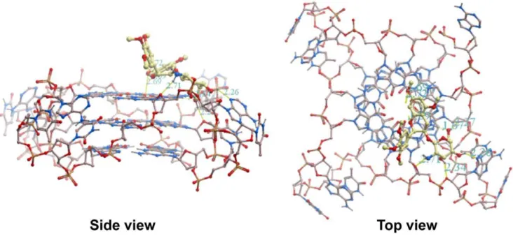
Molecular Modeling of Methylated EGC Glucosaminoside Derivatives Binding with Human Telomeric DNA G-quadruplex
Using16as a model compound, a molecular modeling study was performed on its binding with intramolecular human telomeric G-quadruplex (PDB code: 1KF1 [52]) to provide insight into the binding mode of aminoglucosides of methylated EGC. As illustrated in Figure 5, the aglycone moiety of16is predicted to bind to the 59terminal face of the G-quadruplex through stacking interactions between the aromatic rings of methylated EGC and guanine nucleotides. The part of the glucosamine moiety of16is predicted to reside in the grooves of the G-quadruplex through hydrogen bonding interactions between donors in the G-quad-ruplex and hydrogens in both the amino and hydroxyl groups of 16. The amino hydrogen of16forms a hydrogen bond with the oxygen atom of the deoxyribose in the phosphate backbone. The hydroxyl hydrogens of 16 form hydrogen bonds with oxygen atoms from the phosphate sugars and adenine residues of the G-quadruplex. This kind of binding mode, towards the top of the 59
terminus of the G-quadruplex, is the most favorable binding interaction, with a binding energy of235.99 kJ/mol.
Analysis of Binding with Oncogene G-quadruplex DNA by ESI-TOF-MS
In addition to human telomeric G-quadruplex DNA, the binding of two synthetic glucosaminosides of methylated EGC (16, 18) with oncogene G-quadruplexes derived from the sequences of c-Myc, c-kit1, c-kit2 and Bcl-2 was further in-vestigated by the same ESI-TOF-MS technique. All the oncogene sequences displayed the ability to form G-quadruplex structures in NH4OAc buffer, as ESI-TOF-MS spectra revealed that the major
ion in25 charge state of each sequence corresponds to them/z
value of the oligodeoxynucleotide with two NH4+ ions adduct
(Figure S10). As with telomeric DNA G-quadruplex,16and 18 also showed multiple binding stoichiometries with all oncogene DNA G-quadruplexes. The relative binding affinities presented in Figure 6 show that16demonstrated comparative binding strength with c-Myc, c-kit1, c-kit2, and Bcl-2 DNA G-quadruplexes. Compound18exhibited almost equivalent binding capacity with all oncogene G-quadruplexes. These results demonstrate that glucosaminosides (16, 18) of methylated EGC exhibit binding capacity to different oncogene G-quadruplexes, though without obvious G-quadruplex selectivity.
Analysis of Interaction with Human Telomeric RNA G-quadruplex by ESI-TOF-MS
A recent finding demonstrated that telomere DNA is tran-scribed into telomeric repeat-containing RNA in mammalian cells. The telomeric repeat-containing RNA sequence, r(UUAGGG)4,
folds into a parallel G-quadruplex in solution that is more stable than its DNA counterpart [53–56]. The binding of synthetic glucosaminosides (16, 18) of methylated EGC, penta-methylated EGC (14) and EGC (6) were therefore studied with a 27 nt human telomeric RNA sequence, r[(UUAGGG)4UUA],
under the same ESI-TOF-MS conditions as used for its DNA counterpart (Figure S11) [55–56]. As shown in Figure 7, the RNA G-quadruplex binding strength of these four compounds also followed the same descending trend of16.18. EGC .14 as seen with a DNA quadruplex. By Comparing with the DNA G-quadruplex binding results (Figure 4), it was found that the relative affinity of each compound for the RNA G-quadruplex was slightly higher. In order to confirm this result, competitive binding experiments were carried out for two methylated EGC glucosa-minosides (16,18) with both DNA and RNA G-quadruplexes to further elucidate the DNA and RNA G-quadruplex binding selectivity of each compound (Figure S12). In each competition experiment, human telomeric DNA and RNA G-quadruplexes were mixed with each compound to give a final molar ratio of DNA/RNA/compound of 1:1:2. The peak area ratio of drug-bound complex to free nucleic acid of each species of G-quadruplex was calculated to give the value of relative affinity
methanol.
doi:10.1371/journal.pone.0053962.g003
Figure 4. Relative binding affinities of drugs with intramolecular human telomeric DNA G-quadruplex.The numbers indicated on the top of each column showed the mean values from two determinations.
(Figure 8), which confirmed that both 16and 18show stronger binding affinity with RNA G-quadruplex than DNA G-quad-ruplex. To provide further evidence to support the ESI-TOF-MS results of the above binding studies of these aminoglucosamino-sides, we undertook UV-melting experiments [15–22]. It was demonstrated that 16and 18 increased melting temperature of rAGGG(UUAGGG)3 6.08 and 3.26uC, respectively, under the
same tested condition with that of DNA analogue. The largerDTm
value for RNA G-quadruplex further confirmed that16and18 exhibited stronger stabilizing effects on RNA G-quadruplex than DNA counterpart. However, there are no obvious increases for the
melting temperature of a double-stranded oligodeoxynucleotide (ds-DNA) 59-AGGGTTAGGGT-39/39-TCCCAATCCCA-59 in the presence of 16 (0.22uC)?and 18 (20.15uC), indicating the selectivity of two synthetic compounds to RNA and DNA G-quadruplexex over duplex oligonucleotide (Figure S13).
Discussion and Conclusion
In this study, two glucosaminosides of methylated EGC, compounds 16 and 18, have been successfully synthesized for the first time. Both the DNA and RNA G-quadruplex binding Figure 5. Molecular modeling of 16 binding with the human intramolecular telomeric G-quadruplex (PDB code: 1KF1).Oxygen atoms are highlighted in red, nitrogen atoms in blue, phosphorus atoms in yellow and carbon atoms in beige.
doi:10.1371/journal.pone.0053962.g005
Figure 6. Relative binding affinities of 16 and 18 with different intramolecular oncogene G-quadruplexes.The experiments were conducted with a molar ratio of DNA/drug of 1:1 (100mM:100mM) in 100 mM ammonium acetate (pH 7.6) containing 50% methanol. The numbers indicated on the top of each column showed the mean values from two determinations.
activity of these two glucosaminosides has been evaluated and compared with that of green tea catechin EGC by ESI-TOF-MS analysis. The DNA and RNA G-quadruplex binding capacity of both synthetic compounds and natural EGC with human telomeric DNA and RNA sequences both followed the order of 16.18.EGC..14. This finding indicated that introduction of a glucosamine moiety to penta-methylated EGC (14), the weakest G-quadruplex binder among all tested compounds, resulted in much stronger G-quadruplex stabilizing ability that exceeds natural EGC. In addition, they exhibited stronger DNA G-quadruplex binding activity than the more planar structure quercetin. Analysis of the structure-activity relationship revealed that the more basic glucosaminoside 16generally showed more potent G-quadruplex binding capacity than that of18, indicating the importance of basicity of aminoglycoside in G-quadruplex binding activity. Furthermore, it was demonstrated that both glucosaminosides of penta-methylated EGC had a greater binding affinity with the RNA G-quadruplex than its DNA counterpart. Additionally, it has been shown that glucosaminosides16and18 can also bind to different oncogene G-quadruplexes, although without obvious G-quadruplex selectivity. Taken together, these results demonstrate that aminoglycosylation of natural products is an effective way to design and synthesize small molecules targeting G-quadruplexes in nucleic acids.
ESI-TOF-MS analysis revealed that glucosaminosides16and 18demonstrated more binding stoichiometries with an intramo-lecular human telomeric G-quadruplex than EGC under a molar ratio of DNA/drug of 1:1, suggesting that there are more binding sites for 16and 18 in the intramolecular human telomeric G-quadruplex than the natural tea catechin EGC and their aglycone (14). Subsequent molecular docking simulation predicted that the aromatic ring of compound16p-stacked with the aromatic ring of guanine nucleotides, with the glucosamine moiety residing in the groove of G-quadruplex. This prediction is consistent with the binding mode of aminoglycosides neomycin and paramonomycin [15]. This kind of binding mode is also in agreement with the multiple binding stoichiometries of 16 with the 27 nt human telomeric G-quadruplex detected by ESI-TOF-MS.
Although green tea catechins were proven to bind to normal (single-stranded and double-stranded) DNA and RNA [28–31], this is the first time they were also found to bind to DNA and RNA G-quadruplex structures. The distinct binding behaviors of EGC and its penta-methylated derivative, compound14, to DNA and RNA G-quadruplexes suggested that the hydroxyl groups in EGC
are essential for stabilizing DNA and RNA G-quadruplexes. The binding of nucleic acid G-quadruplexes by green tea catechins may be in part responsible for their cancer-preventive activities.
Materials and Methods
General
The trichloroacetimidate method was employed to conduct glycosylation reaction, which was performed in the presence of TMSOTf at240uC under an atmosphere of argon, followed by deprotection of sugar moiety with sodium methoxide in methanol and further catalytic hydrogenation with palladium hydroxide on carbon powder. Unless otherwise noted, all reactions were conducted in oven-dried glassware. Column chromatography for product purification was performed on DAVISILH chromato-graphic silica gel LC60A (40–63 micron, GRACE Davison, Germany), and Chromatorex ODS (100–200 mesh, Fuji Silysia Chemical Ltd., Japan). Purity checking of product was accom-plished on an ultra performance LC system (AcquityTM, Waters) equipped with photodiode array detector (Waters) and a Bruker micrOTOF ESI-TOF mass spectrometer by using an Acquity UPLCH BEH C18 column (17mM, 2.16100 mm, part
No. 186002352, Ireland). 1H and 13C NMR were recorded at room temperature on a Bruker 400 MHz NMR Avance-III (Switzerland) or a Varian 400 MHz NMR Inova 400 (USA) spectrometers operating at 400 MHz (1H) and 100 MHz (13C). Coupling constants were given in Hz and chemical shifts were represented ind(ppm) relative to Me4Si as internal standard.
HR-ESI-MS was performed on a Q-TOF mass spectrometer (Bruker Daltonics, MA, U.S.A). Optical rotation was measured on a JASCO P-1010 polarimeter (Japan) with a 1 dm cell (C given in g/100 mL).
3,4,5-tri-O
-acetyl-2-deoxy-2-(9-Fluorenylmethoxycarbonylamino)-a-D-glucopyranosyl Trichloroacetimidate (5)
To a solution of glucosamine chloride (8.15 g, 37.8 mmol) in water (50 mL), NaHCO3 (4.76 g, 56.7 mmol) and
9-fluorenyl-methoxycarbonylchloride (11.7 g, 45.4 mmol) in dioxane (50 mL) were added and the reaction stirred at room temperature until silica gel TLC (CHCL3-EtOAc-MeOH 7:2:3) showed complete
Figure 7. Relative binding affinities of drugs with intramole-cular human telomeric RNA G-quadruplex. The numbers in-dicated on the top of each column showed the mean values from two determinations.
doi:10.1371/journal.pone.0053962.g007 Figure 8. Relative binding affinities of drugs with DNA and RNA G-quadruplex in competitive binding experiments. The numbers indicated on the top of each column showed the mean values from two determinations.
conversion of the starting material to N-protected glucosamine (2). The reaction solution was slowly poured into ice water (900 mL) and filtered under decreased pressure after stirring at room temperature for 2 h. The residue was washed by water and dried at 50uC to afford crude2(13.8 g). The N-protected glucosamine (2, 13.8 g, 34.6 mmol) in pyridine (50 mL) was acetylated with acetic anhydride (18 mL, 173 mmol) by stirring overnight at room temperature to produce tetra-O-acetyl carbohydrate (3). The reaction solution was poured into ice water and filtered under decreased pressure, followed by silica gel column chromatography (PE-acetone 7:3) to give3(18.5 g). The anomeric acetyl group of3 (8.65 g, 15.2 mmol) in DMF (50 mL) was then deprotected with hydrazine acetate (1.54 g, 16.72 mmol) by stirring at room temperature for 5 h. The reaction solution was then diluted with EtOAc (300 mL), then washed with water (100 mL64). The organic layer was dried with anhydrous Na2SO4and concentrated
to give 4 (7.3 g). To a solution of 4 (1.844 g, 3.50 mmol) in anhydrous CH2Cl2 (60 mL), CCl3-CN (2.91 mL, 13.97 mmol)
and DBU (58.2mL, 0.35 mmol) was added. After stirring at room temperature for 50 minutes, the reaction mixture was directly loaded to silica gel chromatography column eluted with n-hexane-EtOAc (6:4) containing 0.5% triethylamine to afford 5 (2.25 g, 96% yield).
(2)-5, 7, 39, 49, 59-Penta-O-methyl Epigallocatechin (14)
To a solution of epigallocatechin (612 mg, 2.0 mmol) in DMF (20 mL), methyl iodide (1.0 mL, 16.0 mmol) and K2CO3(2.21 g,
16.0 mmol) was added. The mixture was stirred at room temperature for 15 h. 180 mL of water was poured into the reaction solution, followed by extraction with ethyl acetate (100 mL64). The organic solutions was dried with anhydrous Na2SO4 and subjected to silica gel chromatography
(n-hexane-ethyl acetate 9:1 to 5:5) to afford14(388 mg, 52% yield) as a white amorphous powder. [a]D
20
276.16 (C = 0.44, EtOAc). HRMS (ESI) m/z (C20H25O7)+: calcd 377.1595, found 377.1583.
1
H-NMR (400 MHz, CDCl3)d: 4.94 (s, H-2), 4.30 (m, H-3), 2.98 (dd, J= 1.6, 17.3 Hz, H-4b), 2.90 (dd,J= 4.3, 17.3 Hz, H-4a), 6.21 (d,
J= 2.3, H-6), 6.13 (d,J= 2.3 Hz, H-8), 6.75 (2H, s, H-29, 69), 3.86 (3H, s, 49-OCH3), 3.80, 3.78 (3H each, s, 5, 7-OCH3), 3.90 (6H, s,
39, 59-OCH3), 1.78 (d,J= 5.6 Hz, 3-OH).13C-NMR (100 MHz,
DMSO)d: 78.4 (C-2), 64.4 (C-3), 28.6 (C-4), 101.0 (C-4a), 158.9, 158.7 (C-5, C-7), 93.4 (C-6), 91.4 (C-8), 155.4 (C-8a), 135.2 (C-19), 104.4 (C-29, C-69), 152.5 (C-39, C-59), 136.7 (C-49), 55.4, 55.2 (5, 7-OCH3), 55.9 (39, 59-OCH3), 60.0 (49-OCH3).
Compound 15
A heterogeneous mixture of compound 14 (113 mg, 0.30 mmol) and trichloroacetimidate 5 (336 mg, 0.50 mmol) in anhydrous CH2Cl2(6 mL) with 4 A˚ molecular sieves (500 mg) was
stirred at room temperature for 30 min under an argon atmosphere and then was cooled to240uC. TMSOTf (600mL, 0.06 M in dry CH2CL2) was added quickly to the precooled
reaction mixture and the resulting mixture was allowed to warm to room temperature over 3 h. Triethylamine (24mL) was added to quench the reaction. The organic solution was then directly loaded to silica gel chromatograph column eluted with n-hexane-ethyl acetate (from 9:1 to 5:5) to give15(199.1 mg, 75% yield). HRMS (ESI)m/z(C47H52NO16)+: calcd 886.3281, found 886.3275.
(2)-5, 7, 39, 49, 59-penta-O-methyl Epigallocatechinb -D-glucosaminoside (16)
Compound15(177 mg, 0.20 mmol) was dissolved in methanol (14 mL) and sodium methoxide (40 mg, 0.74 mmol) was added.
After 3 h of stirring at room temperature, the reaction solution was filtered and evaporated under reduced pressure. The residue was purified by silica gel chromatography eluted with chloroform-methanol-water (from 90:10:1 to 80:20:2) to afford 16 (64 mg, 60% yield) as a white amorphous powder. Optical rotation [a]D20 230.48 (C = 0.44, MeOH). HRMS (ESI) m/z (C26H36NO11)+:
calcd 538.2283, found 538.2271. 1H-NMR (400 MHz, DMSO and D2O) d: 5.14 (s, H-2), 4.50 (m, H-3), 2.72 (dd, J= 3.6,
17.4 Hz, H-4a), 2.67 (dd, J= 4.0, 17.4 Hz, H-4b), 6.12 (d,
J= 2.3 Hz, H-6), 6.09 (d, J= 2.3 Hz, H-8), 6.84 (2H, s, H-29, 69), 3.71, 3.65 (3H each, s, 5, 7-OCH3), 3.72 (6H, s, 39, 59-OCH3),
3.74 (3H, s, 49-OCH3), 4.26 (d, J= 7.8 Hz, glc-1), 2.31 (dd, J= 8.0, 9.0 Hz, glc-2), 3.04 (t, J= 9.0 Hz, glc-3), 2.96 (t,
J= 9.0 Hz, glc-4), 3.12 (m, glc-5), 3.68 (m, overlapped with H2O peak, glc-6a), 3.34 (dd,J= 6.4, 11.6 Hz, glc-6b).13C-NMR
(100 MHz, DMSO)d: 77.5 2), 70.1 3), 23.9 4), 101.3 (C-4a), 159.6, 158.9 (C-5, 7), 93.6 (C-6), 91.9 (C-8), 155.6 (C-8a), 134.2 (C-19), 105.6 (C-29, 69), 152.6 (C-39, 59), 137.1 (C-49), 100.9 1), 57.5 2), 76.4 3), 70.6 4), 77.4 5), 61.6 (glc-6), 55.9, 55.6 (5, 7-OCH3), 56.2 (39, 59-OCH3), 60.5 (49-OCH3).
Compound 17
To a solution of compound 16(16 mg, 0.03 mmol) in DMF (2 mL), acetic anhydride (40mL, 0.39 mmol) and K2CO3(12 mg,
0.09 mmol) was added at room temperature. After 2 hours of stirring, ice water (20 mL) was poured into the mixture solution to stop reaction. Then ethyl acetate (10 mL63) was used to extract
the target product. The organic solution was dried with anhydrous Na2SO4and evaporated to give17(15.6 mg, 74% yiled). HRMS
(ESI)m/z(C34H44NO15)+: calcd 706.2705, found 706.2695.
(2) 5, 7, 39, 49, 59-penta-O-methyl Epigallocatechin N-acetylb-D-glucosaminoside (18)
Sodium methoxide (6.06 mg, 0.11 mmol) was added to a solution of 17 (15.6 mg, 0.022 mmol) in methanol (3 mL). The mixture was allowed to stir at room temperature overnight. After neutralization by formic acid (100mL), the reaction solution was loaded to an ODS column eluted with 70% methanol to give 18 (8.5 mg, 66% yield) as a white amorphous powder. [a]D20 247.12 (C = 0.42, MeOH). HRMS (ESI) m/z (C28H38NO12)+:
calcd 580.2389, found 580.2377.1H-NMR (400 MHz, CD3OD) d: 5.04 (s, H-2), 4.49 (m, H-3), 2.92 (dd,J= 3.6, 17.3 Hz, H-4a), 2.70 (dd,J= 3.9, 17.3 Hz, H-4b), 6.13 (d,J= 2.3 Hz, H-6), 6.10 (d,J= 2.3 Hz, H-8), 6.91 (2H, s, H-29, 69), 3.77, 3.74 (3H each, s, 5, 7-OCH3), 3.85 (6H, s, 39, 59-OCH3), 3.80 (3H, s, 49-OCH3),
4.58 (d,J= 7.8 Hz, 1), 3.82 (m, 2), 3.45–3.58 (3H, m, glc-6a, 6b, 3), 3.21–3.24 (2H, m, glc-4, 5), 1.58 (3H, s, -COCH3).13
C-NMR (100 MHz, CD3OD) d: 79.7 (C-2), 71.9 (C-3), 24.6 (C-4),
101.8 (C-4a), 161.2, 160.5 (C-5, 7), 94.6 (C-6), 92.4 (C-8), 157.3 (C-8a), 136.1 (C-19), 106.8 (C-29, 69), 153.9 (C-39, 59), 138.5 (C-49), 99.7 (glc-1), 57.7 (glc-2), 75.6 (glc-3), 72.5 (glc-4), 78.0 (glc-5), 63.1 (glc-6), 56.1, 56.0 (5, 7-OCH3), 56.9 (39, 59-OCH3), 61.2 (49
-OCH3), 173.7 (C = O in acetyl group), 22.7 (CH3in acetyl group).
Mass Spectrometry
All ESI-MS experiments were carried out on a Bruker MicrOTOFQ mass spectrometer in negative ion mode, with the capillary voltage set to+3500 V, the dry N2gas flow set to 4.0 L/
prepared in milli Q water at the concentration of 1 mM, and further diluted by 1 M NH4OAc buffer (pH 7.6) to the desired
concentration. All stock solutions of drugs were prepared in methanol at a concentration of 400mM. For the analysis of the noncovalent complex of oncogene DNA G-quadruplex with drug, the samples were prepared at a final concentration of 100mM DNA and 100mM drug in 100 mM NH4OAc (pH 7.6) containing
50% methanol. For the analysis of human telomeric DNA and RNA G-quadruplexes, the samples were injected at a final strand concentration of 50mM oligomer and 50mM drug in 50 mM NH4OAc (pH 7.6) containing 50% methanol. Each sample of
nucleic acid-drug complex solution was prepared in duplicate. The oligonucleotide sequences are shown as the following:
(i) human telomeric DNA: 59
-TTAGGGTTAGGGT-TAGGGTTAGGGTTA-39(M= 8496.6241 Da)
(ii) c-myc DNA: 59 -TGGGGAGGGTGGGGAGGGTGGG-GAAGG-39(M= 8687.7137 Da)
(iii) c-kit 1 DNA: 59 -AGAGGGAGGGCGCTGGGAG-GAGGGGCT-39(M= 8576.6545 Da)
(iv) c-kit2 DNA: 59 -CCCGGGCGGGCGCGAGGGAGGG-GAGGT-39(M= 8513.5935 Da)
(v) truncated bcl-2 DNA: 59
-CGGGCGCGGGAG-GAAGGGGGCGGGAGC-39(M= 8562.6312 Da)
(vi) human telomeric RNA: 59
-UUAGGGUUAGG-GUUAGGGUUAGGGUUA-39(M= 8789.3336 Da)
Molecular Modeling
A computer model to study the binding of 16 with human telomeric DNA G-quadruplex was performed by using the literature method [57]. Molecular modeling was performed using the ICM-Pro 3.4-8a program (Molsoft). The X-ray crystal structure of the intramolecular G-quadruplex DNA was obtained from the Protein Data Bank (PDB code: 1KF1) and used as the model to perform molecular modeling [57].
UV-melting Study
UV-melting profiles were recorded by using a Beckman Coulter DU800H spectrophotometer equipped with a high performance temperature controller. The absorbance was monitored at 295 nm for G-quadruplex in 25 mM Tris-HCl buffer (pH 7.0) containing 5 mM KCl and 1% DMSO, and at 260 nm for duplex oligonucleotides in 25 mM Tris-HCl buffer (pH 7.0) containing 1% DMSO. The concentration of all oligonucleotides was 5mM for the DTm measurement in the absence and presence of
compounds (50mM).
Supporting Information
Figure S1 Positive ESI-TOF-MS spectrum of compound 14.
(TIF)
Figure S2 1H-NMR spectrum of compound 14. The spectrum was taken in CDCl3at room temperature.
(TIF)
Figure S3 13C-NMR spectrum of compound 14. The spectrum was taken in DMSO-d6 at room temperature.
(TIF)
Figure S4 Positive ESI-TOF-MS spectrum of compound 16.
(TIF)
Figure S5 1H-NMR spectrum of compound 16. The spectrum was taken in the mixture of DMSO and D2O at room
temperature. (TIF)
Figure S6 13C-NMR spectrum of compound 16. The spectrum was taken in the mixture of DMSO and D2O at room
temperature. (TIF)
Figure S7 Positive ESI-TOF-MS spectrum of compound 18.
(TIF)
Figure S8 1H-NMR spectrum of compound 18. The spectrum was taken in CD3OD at room temperature.
(TIF)
Figure S9 13C-NMR spectrum of compound 18. The spectrum was taken in CD3OD at room temperature.
(TIF)
Figure S10 Negative ESI-TOF-MS spectra of oncogene G-rich sequences. (A–B) c-Myc sequence d[(TG4AG3)2
T-G4A2G2] with compound 16 and compound 18. (C–D) c-kit1
sequence d[AG(AG3)2CGCTG3AG2AG4CT] with compound16
and compound 18. (E–F) c-kit2 sequence d[C3G3CG3(CG)2
A-G3AG4AG2T] with compound 16 and compound 18. (G–H)
truncated Bcl-2 sequence d[CG3CGCG3AG2A2G5CG3AGC]
with compound16and compound 18. Q represents quadruplex oligodeoxynucleotides. Spectra were recorded with 1:1 DNA-to-drug molar ratio (C = 100mM) in 50 mM ammonium acetate buffer (pH 7.6) containing 50% methanol.
(TIF)
Figure S11 Negative ESI-TOF-MS spectra of human telomeric RNA sequence r[(UUAGGG)4UUA] (Q). (A)
without drug, (B) with compound 16, (C) with compound 18, (D) with compound14,and (E) with EGC. Spectra were recorded with 1:1 DNA-to-drug molar ratio (C = 50mM) in 50 mM ammonium acetate buffer (pH 7.6) containing 50% methanol. (TIF)
Figure S12 ESI-TOF-MS spectra of human telomeric DNA and RNA G-quadruplex in competitive binding experiments.(A) an equal molar mixture of human telomeric DNA d[(TTAGGG)4TTA] (Q) and RNA r[(UUAGGG)4UUA]
(Q) without drug, (B) with compound16, and (C) with compound 18. Spectra were recorded with 1:1:2 molar ratio of DNA:RNA:-drug (25mM: 25mM: 50mM) in 50 mM ammonium acetate buffer (pH 7.6) containing 50% methanol.
(TIF)
Figure S13 Thermal denaturation profiles of oligonu-cleotides in the absence and presence of 16. (A) UV-melting profiles of rAGGG(UUAGGG)3 (5mM) in the absence
and presence (50mM) of compound16in 25 mM Tris-HCl buffer (pH 7.0) containing 5 mM KCl and 1% DMSO, (B) UV-melting profiles of dAGGG(TTAGGG)3 (5mM) in the absence and
presence (50mM) of compound 16 in 25 mM Tris-HCl buffer (pH 7.0) containing 5 mM KCl and 1% DMSO, (C) UV-melting profiles of double-stranded oligodeoxynucleotide (Ds-DNA) 59 -AGGGTTAGGGT-39/39-TCCCAATCCCA-59 (5mM) in the absence and presence (50mM) of compound16in 25 mM Tris-HCl buffer (pH 7.0) containing 1% DMSO.
Author Contributions
Conducted molecular modeling: DLM HY WCF. Conceived and designed the experiments: ZHJ LPB. Performed the experiments: LPB HMH. Analyzed the data: LPB. Wrote the paper: LPB ZHJ.
References
1. Lipps HJ, Rhodes D (2009) G-quadruplex structures: in vivo evidence and function. Trends Cell Biol 19: 414–422.
2. Neidle S, Parkinson GN (2003) The structure of telomeric DNA. Curr Opin Struct Biol 13: 275–283.
3. Azzalin CM, Reichenbach P, Khoriauli L, Giulotto E, Lingner J (2007) Telomeric repeat containing RNA and RNA surveillance factors at mammalian chromosome ends. Science 318: 798–801.
4. Schoeftner S, Blasco MA (2009) A ‘higher order’ of telomere regulation: telomere heterochromatin and telomeric RNAs. EMBO J 28: 2323–2336. 5. Luke B, Lingner J (2009) TERRA: telomeric repeat-containing RNA. EMBO J
28: 2503–2510.
6. Maizels N (2006) Dynamic roles for G4 DNA in the biology of eukaryotic cells. Nat Struct Mol Biol 13: 1055–1059.
7. Paeschke K, Simonsson T, Postberg J, Rhodes D, Lipps HJ (2005) Telomere end-binding proteins control the formation of G-quadruplex DNA structures in vivo. Nat Struct Mol Biol 12: 847–854.
8. Siddiqui-Jain A, Grand CL, Bearss DJ, Hurley LH (2002) Direct evidence for a G-quadruplex in a promoter region and its targeting with a small molecule to repress c-MYC transcription. Proc Natl Acad Sci USA 99: 11593–11598. 9. Dexheimer TS, Sun D, Hurley LH (2006) Deconvoluting the structural and
drug-recognition complexity of the G-quadruplex-forming region upstream of the bcl2 P1 promoter. J Am Chem Soc 128: 5404–5415.
10. Hurley LH (2002) DNA and associated processes as targets for cancer therapy. Nat Rev Cancer 2: 188–200.
11. Cuesta J, Read MA, Neidle S (2003) The design of G-quadruplex ligands as telomerase inhibitors. Mini-Rev Med Chem 3: 11–21.
12. Todd AK, Haider SM, Parkinson GN, Neidle S (2007) Sequence occurrence and structural uniqueness of a G-quadruplex in the human c-kit promoter. Nucleic Acids Res 35: 5799–5808.
13. Arola A, Vilar R (2008) Stabilisation of G-quadruplex DNA by small molecules. Curr Top Med Chem 8: 1405–1415.
14. Tan J-H, Gu L-Q, Wu J-Y (2008) Design of selective G-quadruplex ligands as potential anticancer agents. Mini-Rev Med Chem 8: 1163–1178.
15. Ranjan N, Andreasen KF, Kumar S, Hyde-Volpe D, Arya DP (2010) Aminoglycoside binding toOxytricha nova telomeric DNA. Biochemistry 49: 9891–9903.
16. Arya DP (2011) New approaches toward recognition of nucleic acid triple helices. Acc Chem Res 44: 134–146.
17. Xue L, Ranjan N, Arya DP (2011) Synthesis and spectroscopic studies of the aminoglycoside (neomycin)-perylene conjugate binding to human telomeric DNA. Biochemistry 50: 2838–2849.
18. Xue L, Xi H, Kumar S, Gray D, Davis E, et al. (2010) Probing the recognition surface of a DNA triplex: binding studies with intercalator-neomycin conjugates. Biochemistry 49: 5540–5552.
19. Willis B, Arya DP (2010) Triple recognition of B-DNA by a neomycin-hoechst 33258-pyrene conjugate. Biochemistry 49: 452–469.
20. Shaw NN, Xi H, Arya DP (2008) Molecular recognition of a DNA:RNA hybrid: sub-nanomolar binding by a neomycin-methidium conjugate. Bioorg Med Chem Lett 18: 4142–4145.
21. Xue L, Charles I, Arya DP (2002) Pyrene-neomycin conjugate: dual recognition of a DNA triple helix. Chem Comm 70–71.
22. Arya DP, Xue L, Tennant P (2003) Combining the best in triplex recognition: synthesis and nucleic acid binding of a BQQ-neomycin conjugate. J Am Chem Soc 125: 8070–8071.
23. Vuong QV, Stathopoulos CE, Nguyen MH, Golding JB, Roach PD (2011) Isolation of green tea catechins and their utilization in the food industry. Food Rev Int 27: 227–247.
24. Naghma K, Hasan M (2008) Multitargeted therapy of cancer by green tea polyphenols. Cancer Lett 269: 269–280.
25. Cao Y, Cao R (1999) Angiogenesis inhibited by drinking tea. Nature 398: 381. 26. Ehrnhoefer DE, Bieschke J, Boeddrich A, Herbst M, Masino L, et al. (2008) EGCG redirects amyloidogenic polypeptides into unstructured, off-pathway oligomers. Nat Struct Mol Biol 15: 558–566.
27. Zhang H, Wang J-R, Yau L, Ho H, Chan C, et al. (2012) Cellular Lipidomic study on the Ab-induced neurotoxicity and neuroprotective Effect of EGCG by using UPLC/MS based glycerolipid profiling and multivariate analysis. Mol Biosyst 8: 3208–3215.
28. Yang CS, Wang X (2010) Green Tea and Cancer Prevention. Nutr Cancer 62: 931–937.
29. Rahman MA, Amin ARMR, Shin DM (2010) Chemopreventive potential of natural compounds in head and neck cancer. Nutr Cancer 62: 973–987. 30. Lambert JD, Elias RJ (2010) The antioxidant and pro-oxidant activities of green
tea polyphenols: A role in cancer prevention. Arch Biochem Biophys 501: 65– 72.
31. Song JM, Lee KH, Seong BL (2005) Antiviral effect of catechins in green tea on influenza virus. Antivir Res 68: 66–74.
32. Fang MZ, Wang Y, Ai N, Hou Z, Sun Y, et al. (2003) Tea polyphenol (2 )-epigallocatechin-3-gallate inhibits DNA methyltransferase and reactivates methylation-silenced genes in cancer cell lines. Cancer Res 63: 7563–7570. 33. Balasubramanian S, Eckert R (2004) Green tea polyphenol and curcumin
inversely regulate human involucrin promoter activity via opposing effects on CCAAT/enhancer-binding protein function. J Biol Chem 279: 24007–24014. 34. Kuzuhara T, Sei Y, Yamaguchi K, Suganuma M, Fujiki H (2006) DNA and
RNA as new binding targets of green tea catechins. J Biol Chem 281: 17446– 17456.
35. Phosrithong N, Ungwitayatorn J (2010) Molecular docking study on anticancer activity of plant-derived natural products. Med Chem Res 19: 817–835. 36. Ferla BL, Airoldi C, Zona C, Orsato A, Cardona F, et al. (2011) Natural
glycoconjugates with antitumor activity. Nat Prod Rep 28: 630–648. 37. Ikeda T, Fujiwara S, Araki K, Kinjo J, Nohara T, et al. (1997) Cytotoxic
glycosides fromAlbizia julibrissin. J Nat Prod 60: 102–107.
38. He H, Bai L-P, Jiang Z-H (2012) Synthesis and human telomeric G-quadruplex DNA-binding activity of glucosaminosides of shikonin/alkannin. Bioorg Med Chem Lett 22: 1582–1586.
39. Deng S, Yu B, Xie J, Hui Y (1999) Highly efficient glycosylation of sapogenins. J Org Chem 64: 7265–7266.
40. Liu L, Yi L, Yang X, Yu Z, Wen X, et al. (2008) Sythesis and spectroscopic characterization of binaphthol aminosugars for stimulation of DNA strand slippage synthesis. Tetrahedron 64: 5885–5890.
41. Wrodnigg TM, Lundt I, Stu¨tz AE (2006) Synthesis of N-protected galactosamine building blocks from D-tagatose via the Heyns rearrangement. J Carbohydr Chem 25: 33–41.
42. Xi Z, Hwang G-S, Goldberg I H, Harris JL, Pennington WT, et al. (2002) Targeting DNA bulged microenvironments with synthetic agents: lessons from a natural product. Chem Biol 9: 925–931.
43. Gunaherath GMKB, Gunatilaka AAL, Sultanbawa MUS (1981) Dulcitol and (2)-49-O-methylepigallocatechin fromKokoona zeylanica. J Nat Prod 45: 140–142. 44. Schmidt CA, Murillo R, Bruhn T, Bringmann G, Goettert M, et al. (2010) Catechin derivatives from Parapiptadenia rigida with in vitro wound-healing properties. J Nat Prod 73: 2035–2041.
45. Daniel JM, Friess SD, Rajagopalan S, Wendt S, Zenobi R (2002) Quantitative determination of noncovalent binding interactions using soft ionization mass spectrometry. Int J Mass Spectrom 216: 1–27.
46. Gavelica V, De Pauw E, Rosu F (1999) Interaction between antitumor drugs and a double-stranded oligonucleotide studies by electrospray ionization mass spectrometry. J Mass Spectrom 34: 1328–1337.
47. Baker ES, Bernstein SL, Gabelica V, De Pauw E, Bowers MT (2006) G-quadruplexes in telomeric repeats are conserved in a solvent-free environment. Int J Mass Spectrom 253: 225–237.
48. Sun HX, Tang YL, Xiang JF, Xu GZ, Zhang YZ, et al. (2006) Spectroscopic studies of the interaction between quercetin and G-quadruplex DNA. Bioorg Med Chem Lett 16: 3586–3589.
49. Bai L-P, Hagihara M, Jiang Z-H, Nakatani K (2008) Ligand binding to tandem G quadruplexes from human telomeric DNA. ChemBioChem 9: 2583–2587. 50. Wan KX, Shibue T, Gross ML (2000) Non-covalent complexes between
DNA-binding drugs and double-stranded oligodeoxynucleotides: a study by ESI ion-trap mass spectrometry. J Am Chem Soc 122: 300–307.
51. Bai L-P, Cai Z, Zhao Z-Z, Nakatani K, Jiang Z-H (2008) Site-specific binding of chelerythrine and sanguinarine to single pyrimidine bulges in hairpin DNA. Anal Bioanal Chem 392: 709–716.
52. Parkinson GN, Lee MPH, Neidle S (2002) Crystal structure of parallel quadruplexes from human telomeric DNA. Nature 417: 876–880.
53. Xu Y, Suzuki Y, Ito K, Komiyama M (2010) Telomeric repeat-containing RNA structure in living cells. PNAS 107: 14579–14584.
54. Phan AT (2010) Human telomeric G-quadruplex: structure of DNA and RNA sequences. FEBS J 277: 1107–1117.
55. Collie G, Reszka AP, Haider SM, Gabelica V, Parkinson GN, et al. (2009) Selectivity in small molecule binding to human telomeric RNA and DNA quadruplexes. Chem Commun 7482–7484.
56. Collie GW, Parkinson GN, Neidle S, Rosu F, De Pauw E, et al. (2010) Electrospray mass spectrometry of telomeric RNA (TERRA) reveals the formation of stable multimeric G-quadruplex structures. J Am Chem Soc 132: 9328–9334.
![Figure 3. ESI-TOF-MS spectra of telomeric DNA d[(TTAGGG) 4 TTA] (Q) in the absence and presence of drugs](https://thumb-eu.123doks.com/thumbv2/123dok_br/16368725.190884/4.918.93.731.89.1016/figure-esi-spectra-telomeric-ttaggg-absence-presence-drugs.webp)