Variations in the Binding Pocket of an
Inhibitor of the Bacterial Division Protein FtsZ
across Genotypes and Species
Amanda Miguel1, Jen Hsin1, Tianyun Liu1, Grace Tang1, Russ B. Altman1, Kerwyn Casey Huang1,2*
1Department of Bioengineering, Stanford University, Stanford, California, United States of America, 2Department of Microbiology and Immunology, Stanford University School of Medicine, Stanford, California, United States of America
Abstract
The recent increase in antibiotic resistance in pathogenic bacteria calls for new approaches to drug-target selection and drug development. Targeting the mechanisms of action of pro-teins involved in bacterial cell division bypasses problems associated with increasingly inef-fective variants of older antibiotics; to this end, the essential bacterial cytoskeletal protein FtsZ is a promising target. Recent work on its allosteric inhibitor, PC190723, revealedin vitroactivity onStaphylococcus aureusFtsZ andin vivoantimicrobial activities. However, the mechanism of drug action and its effect on FtsZ in other bacterial species are unclear. Here, we examine the structural environment of the PC190723 binding pocket using Pock-etFEATURE, a statistical method that scores the similarity between pairs of small-molecule binding sites based on 3D structure information about the local microenvironment, and mo-lecular dynamics (MD) simulations. We observed that species and nucleotide-binding state have significant impacts on the structural properties of the binding site, with substantially disparate microenvironments for bacterial species not from theStaphylococcusgenus. Based on PocketFEATURE analysis of MD simulations ofS.aureusFtsZ bound to GTP or with mutations that are known to confer PC190723 resistance, we predict that PC190723 strongly prefers to bindStaphylococcusFtsZ in the nucleotide-bound state. Furthermore, MD simulations of an FtsZ dimer indicated that polymerization may enhance PC190723 binding. Taken together, our results demonstrate that a drug-binding pocket can vary signifi-cantly across species, genetic perturbations, and in different polymerization states, yielding important information for the further development of FtsZ inhibitors.
Author Summary
The rise of antibiotic resistance in microbes that cause dangerous diseases necessitates the development of new drugs with novel mechanisms of antimicrobial activity. The recently identified small molecule PC190723 is a promising therapeutic candidate, as it is the only
OPEN ACCESS
Citation:Miguel A, Hsin J, Liu T, Tang G, Altman RB, Huang KC (2015) Variations in the Binding Pocket of an Inhibitor of the Bacterial Division Protein FtsZ across Genotypes and Species. PLoS Comput Biol 11(3): e1004117. doi:10.1371/journal.pcbi.1004117
Editor:Rebecca C. Wade, Heidelberg Institute for Theoretical Studies (HITS gGmbH), GERMANY
Received:August 26, 2014
Accepted:January 8, 2015
Published:March 26, 2015
Copyright:© 2015 Miguel et al. This is an open access article distributed under the terms of the
Creative Commons Attribution License, which permits unrestricted use, distribution, and reproduction in any medium, provided the original author and source are credited.
Data Availability Statement:All relevant data are within the paper and its Supporting Information files.
drug known to directly inhibit FtsZ, a cytoskeletal protein that is critical for bacterial cell division. However, little is known about the effects of PC190723 in bacterial species other than the pathogenStaphylococcus aureus. Here, we evaluate the PC190723 binding site with currently available crystallographic structures of FtsZ using the structural comparison algorithm PocketFEATURE and all-atom molecular dynamics simulations. Our investiga-tions provide insight into the mechanism of PC190723-based inhibition of FtsZ and its po-tential use as an inhibitor of as-yet untested bacterial species. We also demonstrate the utility of computational analysis of the drug-binding pockets of related proteins, which we expect to accelerate the screening and development of novel antibiotics to combat
human disease.
Introduction
Rises in bacterial antibiotic resistance have motivated the development of new classes of drugs with alternative mechanisms of action [1]. One promising target is the cytoskeletal protein FtsZ, a GTPase and homolog of eukaryotic tubulin that plays a central, conserved role in cell division in both eubacteria and archaea [2]. FtsZ forms protofilamentsin vitro[3] and assem-blesin vivointo a ring-like structure termed the“Z-ring,”which acts as a scaffold to recruit other critical division proteins [4] and constricts the cell as division progresses [5,6].In vitro
studies have shown that FtsZ causes constriction when bound to liposomes, either via a mem-brane-targeting helix or with itsin vivobinding partner FtsA, suggesting that FtsZ can generate mechanical force in addition to its scaffolding function [7–9]. All-atom molecular dynamics (MD) simulations predicted that force generation may result from a dramatic bending in GDP-bound filaments induced by nucleotide hydrolysis [10]; this conformational change was later confirmed by X-ray crystallography [6].
Several compounds have been demonstrated to inhibit FtsZ, primarily through three mech-anisms [11]: modulation of FtsZ assembly/disassembly [12,13], GTPase activity [14,15], or deg-radation [16]. However, many of these compounds have as-yet unknown interactions with FtsZ, large minimal inhibitory concentrations, or are associated with toxic effects that make them unsuitable for therapeutic use [11]. In comparison, the FtsZ inhibitor PC190723 and its derivatives are promising drug candidates. PC190723 inhibited cell division at low minimal in-hibitory concentration in many Gram-positive bacteria, such as the model organismBacillus subtilisand the pathogenStaphylococcus aureus, including the methicillin-resistantS.aureus
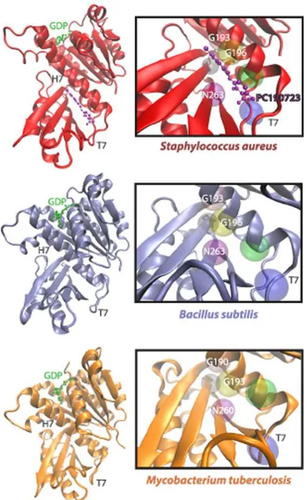
strain [12]. Encouragingly, PC190723 exhibited antimicrobial propertiesin vivoin mouse models [12], suggesting that this small molecule selectively targets bacterial cell division with-out affecting tubulin, the eukaryotic FtsZ homolog. Furthermore, single point mutations con-ferring resistance to PC190723 inS.aureuswere identified withinftsZ, suggesting that PC190723 specifically binds toS.aureusFtsZ (SaFtsZ). This observation was later confirmed via the co-crystallization of anSaFtsZ-PC190723 complex in two independent studies [17,18]; thus, currently PC190723 is the only FtsZ-targeting drug with evidence of direct binding. In both co-crystals, PC190723 binds a pocket beneath the H7 loop close to the C-terminus of FtsZ, and many of the FtsZ residues with identified resistance mutations (for example, G193, G196, and N263) lie within 6 Å of the PC190723 molecule in these structures (Fig. 1). Although the PC190723-binding pocket is quite far from FtsZ’s GTP-binding site, it is situated near the T7 loop, which is thought to contact the GTP-binding pocket of an adjacent FtsZ subunit after polymerization, forming the catalytic active site for GTP hydrolysis [19].
simulations were performed with computer time provided by the Extreme Science and Engineering Discovery Environment, which is supported by National Science Foundation Grant OCI-1053575, with allocation number TG-MCB110056 (to JH and KCH). The funders had no role in study design, data collection and analysis, decision to publish, or preparation of the manuscript.
While PC190723 inhibits cell division in a variety of bacterial species such asS.aureusand
B.subtilis, its exact mechanism remains unclear. The initial study of PC190723 showed that
ad-dition of PC190723 reducedSaFtsZ GTPase activityin vitro[12]; in contrast, a recent study re-ported that PC190723 actually increasedSaFtsZ GTPase activity, and exhibited no effect on the
GTPase activity ofB.subtilisFtsZ (BsFtsZ) [20,21].In vitro, PC190723 induced stable bundling
Fig 1. The FtsZ PC190723-binding pocket differs structurally across bacterial species.PC190723 binds a cleft beneath the H7 helix and adjacent to the T7 loop of FtsZ, as demonstrated in an
SaFtsZ-PC197023 co-crystal (PDB ID: 4DXD) [17], in aB.subtilisFtsZ (PDB ID: 2RHL) structure [42], and in aMycobacterium tuberculosisFtsZ (PDB ID: 1RQ7) structure [43]. Highlighted above are a few of the 20 amino acid residues that are within 6Åof PC190723, including G193, G196, and N263, which induce drug resistance when mutated inSaFtsZ.
ofSaFtsZ andBsFtsZ filaments; for FtsZ fromEscherichia coli, a Gram-negative bacterium whose growth is not inhibited by PC190723 treatment, no such bundling was observed [22]. The diverse behaviors observed after PC190723 treatment in FtsZ from diverse species poten-tially indicate structural complexity in the drug-binding site that has yet to be explored.
Here, we present a structural analysis of the PC190723-binding pocket of FtsZ from multiple species and across a variety of conformational states. We characterized this binding pocket using PocketFEATURE, a statistical algorithm that performs pairwise comparisons of structural information between small molecule-binding pockets in different proteins [23]. This method was previously used to predict inhibitor binding profiles between kinases [23]. PocketFEATURE divides a ligand-binding pocket into a set of microenvironments based on local structural and chemical information, and scores the similarity between this set of microenvironments with those found in other proteins. Using PocketFEATURE, we compared the PC190723-binding pocket from theSaFtsZ-PC190723 co-crystal with FtsZ crystal structures from multiple bacteri-al and archaebacteri-al species, including the Gram-positiveS.aureus,B.subtilis, andMycobacterium tuberculosis, the Gram-negativePseudomonas aeruginosa, and the archaeon Methanocaldococ-cus jannaschii(Table 1). The resulting PocketFEATURE score is a measure of the similarity of that pocket to that of theSaFtsZ-PC190723 co-crystal. PocketFEATURE scores revealed that the microenvironment of the PC190723-binding pocket is dependent on species and the nucleotide-binding state of the protein, with the best similarity scores belonging to Staphylococ-cusspecies bound to a GDP nucleotide. Similarity scores computed from the coordinates of all-atom MD simulations preserved the ranking order determined by their static crystal structures counterparts, with PC190723-resistantSaFtsZ mutants harboring pockets that were less similar to theSaFtsZ-PC190723 co-crystal than wild-typeSaFtsZ pockets. Furthermore, all FtsZ pro-teins, except those fromStaphylococcusspecies, had pockets that were no more similar to the
SaFtsZ-PC190723 co-crystal structure than PC190723-resistantSaFtsZ mutants. Finally, FtsZ polymerization increased the pocket similarity of wild-typeSaFtsZ to theSaFtsZ-PC190723 co-crystal, but not that of a PC190723-resistant mutant. Taken together, our results suggest that PC190723 binds best to polymerized, nucleotide-bound FtsZ ofStaphylococcusspecies.
Results
PC197023 pocket scores from FtsZ crystal structures are highly
species-dependent
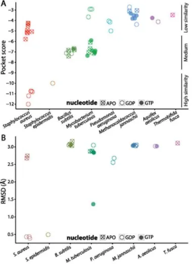
Crystal structures ofSaFtsZ bound to PC190723 (PDB ID: 4DXD and 3VOB) [17,18] suggest that PC190723 induces antimicrobial activity in this organism by specifically binding FtsZ, but no evidence of direct binding exists in other bacterial species. To probe how species-specific features of the PC190723-binding pocket’s three-dimensional microenvironment affect the drug’s affinity for FtsZ, we mapped the PC190723-binding pocket onto FtsZ molecules from various species (Fig. 1,S1 Fig,Methods) and computed scores for their similarity to the
SaFtsZ-PC190723 co-crystal (Fig. 2A,Table 1). The similarity scores roughly fell into catego-ries of high, medium, and low similarity. GDP-bound structures ofSaFtsZ andStaphylococcus epidermidisFtsZ (SeFtsZ) were highly similar to the binding pocket in theSaFtsZ-PC190723 co-crystal, consistent with a previous report that both species are sensitive to PC190723 [12]. Interestingly, crystal structures ofSaFtsZ had similar pocket scores to each other whether or not PC190723 was bound (Fig. 2A). In contrast, we obtained worse similarity scores for FtsZ from all other species;B.subtilis(BsFtsZ) andM.tuberculosis(MtFtsZ) structures had only in-termediate similarity to theSaFtsZ-PC190723 co-crystal, despite evidence of PC190723 sensi-tivity inB.subtilis[12]. FtsZ structures from all other species were associated with
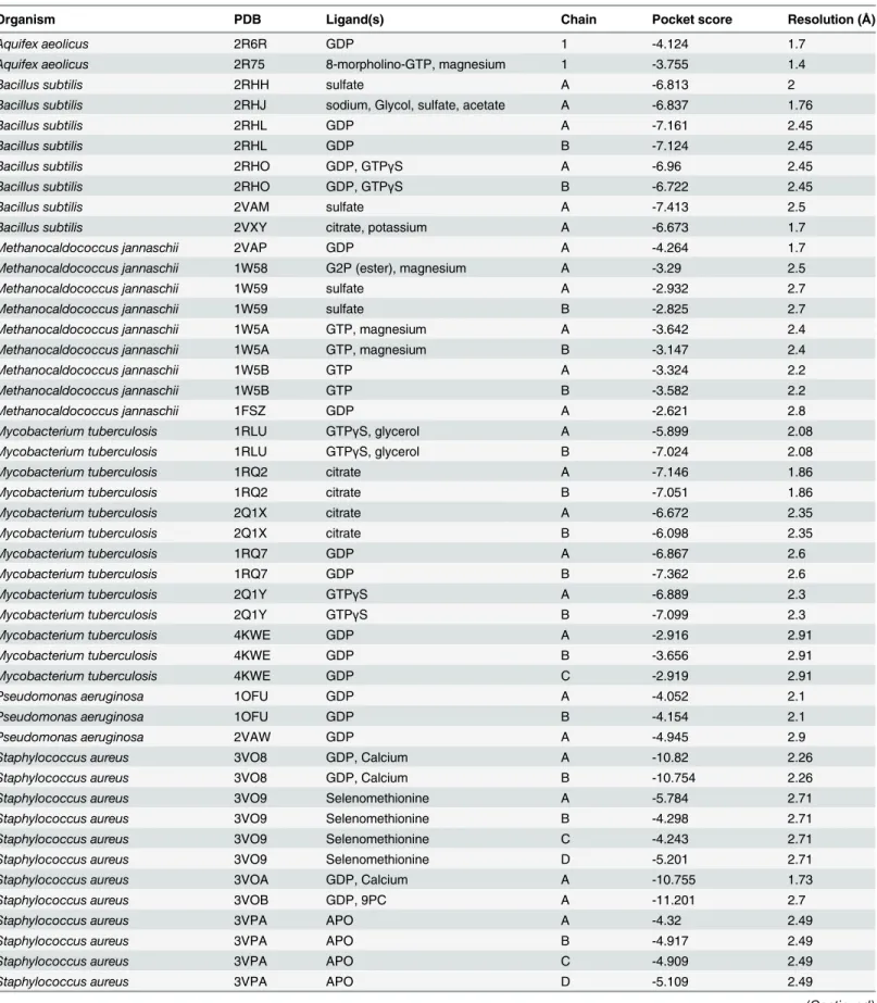
Table 1. Description of FtsZ crystal structures for which PocketFEATURE scores were computed.
Organism PDB Ligand(s) Chain Pocket score Resolution (Å)
Aquifex aeolicus 2R6R GDP 1 -4.124 1.7
Aquifex aeolicus 2R75 8-morpholino-GTP, magnesium 1 -3.755 1.4
Bacillus subtilis 2RHH sulfate A -6.813 2
Bacillus subtilis 2RHJ sodium, Glycol, sulfate, acetate A -6.837 1.76
Bacillus subtilis 2RHL GDP A -7.161 2.45
Bacillus subtilis 2RHL GDP B -7.124 2.45
Bacillus subtilis 2RHO GDP, GTPγS A -6.96 2.45
Bacillus subtilis 2RHO GDP, GTPγS B -6.722 2.45
Bacillus subtilis 2VAM sulfate A -7.413 2.5
Bacillus subtilis 2VXY citrate, potassium A -6.673 1.7
Methanocaldococcus jannaschii 2VAP GDP A -4.264 1.7
Methanocaldococcus jannaschii 1W58 G2P (ester), magnesium A -3.29 2.5
Methanocaldococcus jannaschii 1W59 sulfate A -2.932 2.7
Methanocaldococcus jannaschii 1W59 sulfate B -2.825 2.7
Methanocaldococcus jannaschii 1W5A GTP, magnesium A -3.642 2.4
Methanocaldococcus jannaschii 1W5A GTP, magnesium B -3.147 2.4
Methanocaldococcus jannaschii 1W5B GTP A -3.324 2.2
Methanocaldococcus jannaschii 1W5B GTP B -3.582 2.2
Methanocaldococcus jannaschii 1FSZ GDP A -2.621 2.8
Mycobacterium tuberculosis 1RLU GTPγS, glycerol A -5.899 2.08
Mycobacterium tuberculosis 1RLU GTPγS, glycerol B -7.024 2.08
Mycobacterium tuberculosis 1RQ2 citrate A -7.146 1.86
Mycobacterium tuberculosis 1RQ2 citrate B -7.051 1.86
Mycobacterium tuberculosis 2Q1X citrate A -6.672 2.35
Mycobacterium tuberculosis 2Q1X citrate B -6.098 2.35
Mycobacterium tuberculosis 1RQ7 GDP A -6.867 2.6
Mycobacterium tuberculosis 1RQ7 GDP B -7.362 2.6
Mycobacterium tuberculosis 2Q1Y GTPγS A -6.889 2.3
Mycobacterium tuberculosis 2Q1Y GTPγS B -7.099 2.3
Mycobacterium tuberculosis 4KWE GDP A -2.916 2.91
Mycobacterium tuberculosis 4KWE GDP B -3.656 2.91
Mycobacterium tuberculosis 4KWE GDP C -2.919 2.91
Pseudomonas aeruginosa 1OFU GDP A -4.052 2.1
Pseudomonas aeruginosa 1OFU GDP B -4.154 2.1
Pseudomonas aeruginosa 2VAW GDP A -4.945 2.9
Staphylococcus aureus 3VO8 GDP, Calcium A -10.82 2.26
Staphylococcus aureus 3VO8 GDP, Calcium B -10.754 2.26
Staphylococcus aureus 3VO9 Selenomethionine A -5.784 2.71
Staphylococcus aureus 3VO9 Selenomethionine B -4.298 2.71
Staphylococcus aureus 3VO9 Selenomethionine C -4.243 2.71
Staphylococcus aureus 3VO9 Selenomethionine D -5.201 2.71
Staphylococcus aureus 3VOA GDP, Calcium A -10.755 1.73
Staphylococcus aureus 3VOB GDP, 9PC A -11.201 2.7
Staphylococcus aureus 3VPA APO A -4.32 2.49
Staphylococcus aureus 3VPA APO B -4.917 2.49
Staphylococcus aureus 3VPA APO C -4.909 2.49
Staphylococcus aureus 3VPA APO D -5.109 2.49
to PC190723 binding. Notably, binding site microenvironment comparison performed by PocketFEATURE captures details that are not detected solely through structural analysis. Root mean squared deviation (RMSD) of the pockets of the crystal structures to the pocket of the
SaFtsZ-PC190723 co-crystal (Fig. 2B) clearly identified thatStaphylococcusspecies have pocket structures that are more similar to that of theSaFtsZ-PC190723 co-crystal, but in contrast to PocketFEATURE, RMSD is unable to distinguish among non-Staphylococcusspecies.
Table 1. (Continued)
Organism PDB Ligand(s) Chain Pocket score Resolution (Å)
Staphylococcus aureus 4DXD GDP, 9PC 1 -12 2.01
Staphylococcus epidermidis 4M8I GDP A -10.001 1.43
Thermobifida fusca 4E6E chloride, magnesium, sulfate A -3.471 2.12
doi:10.1371/journal.pcbi.1004117.t001
Fig 2. PC197023-binding pocket similarity scores depend on species and nucleotide-binding state. (A) PocketFEATURE scores for FtsZ structures from various species compared to the PC190723-binding pocket ofSaFtsZ (PDB ID: 4DXD). GDP-boundStaphylococcusspecies display more similarity (more negative scores) to theSaFtsZ-PC190723 co-crystal, with APOStaphylococcusstructures and structures from other bacterial species showing less similarity to theSaFtsZ-PC190723 co-crystal. (B) RMSD of the 3D coordinates of FtsZ crystal structures from the PC190723 co-crystal coarsely separate the proteins into close and distant relations of GDP-boundStaphylococcusFtsZ, but this measurement does not capture additional features of the drug pockets that distinguish between structures of medium and low similarity to the
SaFtsZ-PC190723 co-crystal.
PC197023 pocket score is dependent on nucleotide state
ForSaFtsZ, we also noted a marked decrease in similarity score for the APO state (no nucleo-tide) relative to GDP-bound structures, with APO scores similar to that ofBsFtsZ (Fig. 2A). This observation suggests that conformational changes associated with nucleotide binding may tune the binding affinity of PC190723 for FtsZ; no GTP-boundSaFtsZ structures exist, leaving it unclear whether GTP hydrolysis significantly affects pocket score. Interestingly, there did not appear to be any nucleotide dependence inBsFtsZ (Fig. 2A), andMtFtsZ displayed the opposite tendency toSaFtsZ (Fig. 2A), with worse similarity to theSaFtsZ-PC190723 co-crystal for GDP-bound structures relative to GTP-bound or APO structures. However, allBsFtsZ and
MtFtsZ binding pockets have medium to low similarity compared to GDP-SaFtsZ, and thus may not be optimal for PC190723 binding regardless of nucleotide state. These data suggest that the identity of the nucleotide bound to an FtsZ monomer has variable effects on PC190723 binding across species. In the case ofSaFtsZ, pocket scores predict that PC190723 is more likely to bind FtsZ when GDP is also bound to it.
Resistance mutations substantially reduce PC190723 pocket scores
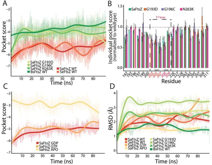
We sought to determine if FtsZ mutations distort the microenvironment of the PC190723 binding site. We therefore compared the pockets of wild-typeSaFtsZ with those of the PC190723-resistant mutants G193D, G196C, and N263K [12]. Since crystal structures of these mutants do not exist, we conducted all-atom MD simulations onSaFtsZ GDP-bound wildtype and drug-resistant (mutant) monomers in order to evaluate how the mutations affected the PC190723-binding pocket. AllSaFtsZ simulations were initialized from the crystal structure of the GDP-boundSaFtsZ without PC190723 (PDB ID: 3VO8) (Fig. 3A,Methods). We observed an initial, rapid decline in similarity to the static FtsZ-PC190723 co-crystal for all simulated monomers. Close investigation into the individual microenvironments contributing to the pocket similarity score for each frame revealed that while the majority of the 20 residues in-cluded in the pocket score computation remained within the binding pocket, some had de-creased contribution to the total similarity score. For example, over the simulation trajectory of the wild-typeSaFtsZ monomer, an average of 17 out of the 20 residues contributed to the simi-larity score at any given time point during the simulation, resulting in an overall shift in the PocketFEATURE score to less negative values (decreased similarity). Thus, small fluctuations in the amino acid functional centers within the pocket can perturb the local physiochemical properties calculated for each residue, pointing to the subtle structural balance required for the most optimal drug-binding environment that can be extremely sensitive to thermal fluctua-tions. Nevertheless, the time-scale and magnitude of this change as the simulations reached a steady state were consistent across all FtsZ monomers (Fig. 3A), indicating that comparisons between simulated protein structures can be established.
To compare pocket scores from MD trajectories across species, we also carried out MD sim-ulations ofSeFtsZ andBsFtsZ monomers initialized from their crystal structures (PDB IDs: 2RHL and 4M8I, respectively); we observed similar initial decreases in pocket similarity
(Fig. 3A). Despite the general change in pocket score to theSaFtsZ-PC190723 co-crystal in all
simulations relative to the pocket score computed from static crystal structures,SaFtsZ and
SeFtsZ monomers maintained better similarity to theSaFtsZ-PC190723 co-crystal compared to theBsFtsZ monomer. These results are consistent with the comparisons of pocket score magnitudes determined from static crystal structures (Fig. 2A), indicating that important fea-tures of the PC190723-binding pocket are conserved in MD simulations.
BsFtsZ. Further examination of the microenvironment pocket scores of theSaFtsZ mutants andBsFtsZ (Fig. 3B) revealed that a subset of residues (203–207 inS.aureus) across all four simulations had average score magnitudes below those of wild-typeSaFtsZ. Interestingly, these residues correspond to amino acids within the T7 loop, a highly conserved structural element near the site of monomer association that plays an important role in GTP hydrolysis [19]. InE.
coli, mutations affecting GTP hydrolysis occur either in the nucleotide pocket or in the T7 loop [2,19]. Truncations of this loop inSaFtsZ disrupt hydrolysis [24]. Taken together, affinity of PC190723 to FtsZ is likely closely coupled to the FtsZ structural transition that occurs during the hydrolysis cycle, and mutations in FtsZ can sufficiently disrupt the PC190723
binding environment.
Fig 3. MD simulations predict that PC190723 binding affinity is reduced in GTP-boundSaFtsZ and PC190723-resistant mutants compared with GDP-boundSaFtsZ.(A) PocketFEATURE scores of dynamic FtsZ monomer structures distinguish among the FtsZ structures that bind PC190723 (S.
aureusandS.epidermidis), PC190723-resistantS.aureusmutants (G193D, G196C, N263K), andBsFtsZ. WT, wildtype. (B) Individual residue contributions to the PocketFEATURE scores show that certain residues in the T7 loop (203, 205, 207) contribute less to the overall score in PC190723-resistant mutants compared to wildtype. (C) Pocket scores from MD trajectories depend on nucleotide, with GDP- and GTP-bound FtsZ exhibiting significantly better similarity to the co-crystal compared to the APO state. (D) RMSD of pocket residues over simulation trajectories do not capture the same hierarchy of pocket similarity scores as in (A,C). Thick lines in (A,C,D) represent a smoothed version of the raw data (Methods).
Absence of nucleotide, but not nucleotide hydrolysis, strongly affects
PC190723 pocket score
Given the large difference in pocket scores in comparisons with theSaFtsZ-PC190723 co-crystal between APO and GDP-boundSaFtsZ crystal structures, we wondered whether it was nucleotide binding or hydrolysis that had a greater effect on pocket microenvironment. Since there are no GTP-bound crystal structures ofSaFtsZ, we conducted MD simulations of the APO state and GTP-boundSaFtsZ monomers (Fig. 3C) to complement our GDP-bound monomer simulations (Fig. 3A). Due to the potential for small atomic differences between the GTP- and GDP-bound states of FtsZ, we conducted three independent simulations of each monomer (Fig. 3C). For each replicate, we observed that GTP- and GDP-boundSaFtsZ had similar drug-pocket scores, and better similarity scores than the APO state. These results illus-trate the sensitivity of the drug pocket to allosteric effects of the nucleotide bound on the oppo-site side of the protein, and suggest that occupancy of the nucleotide-binding oppo-site, rather than the identity of the nucleotide located there, alters the conformation of the pocket to favor PC190723 binding.
To test whether the chemical and physical properties evaluated by PocketFEATURE are captured by traditional structural-only metrics such as RMSD, we calculated the RMSD of the 20 pocket residues aligned to the same 20 residues of the 4DXDSaFtsZ-PC190723 co-crystal structure for each MD trajectory (Fig. 3D). Large-magnitude pocket scores did not correspond with low RMSD from theSaFtsZ-PC190723 co-crystal, withSaFtsZ andSeFtsZ having values intermediate between the low values of APOSaFtsZ andBsFtsZ and the high values of
SaFtsZG193D. Taken together, these data indicate that PocketFEATURE provides a distinctly
in-formative metric for comparing similarity between pockets, and that PC190723 binding may be unique toStaphylococcusspecies.
FtsZ polymerization improves the PC190723 pocket score
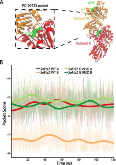
FtsZ hydrolysis and polymerization are intrinsically coupled. Key catalytic residues for GTP hydrolysis reside on the T7 loop of the adjacent subunit near the dimer interface [19], and mu-tations near the active site designed to modulate hydrolysis also affect the dynamics of polymer assembly [25]. Since the PC190723-binding pocket is located close to this interface, we hypoth-esized that it may be affected by FtsZ polymerization. We conducted MD simulations of GDP-bound dimers of wild-typeSaFtsZ (Fig. 4A) and the G193D mutant, and examined the pocket scores of each subunit of the dimers (Fig. 4B). In the subunits of the wild-type dimer, we ob-served a statistically significant difference (t-test,p<2.2e-16) in pocket scores, with the sub-unit with the pocket closest to the dimer interface having a better similarity score to the
SaFtsZ-PC190723 co-crystal (Fig. 4B). No such difference was evident in the PC190723-resis-tant G193D muPC190723-resis-tant dimer; both subunits had pocket scores throughout the trajectory that were similar to those of a wild-type subunit with a pocket away from the dimer interface (Fig. 4B).
GDP-bound dimer, we focused on the pocket similarity score during the first 120 ns of the tra-jectory (Fig. 4B), which showed that polymerization increases the pocket similarity to the
SaFtsZ-PC190723 co-crystal as compared with FtsZ monomers. While these calculations were made over a shorter timescale than that required for the system to reach convergence, our re-sults suggest that polymerization induces pocket deformations with an increased likeness to the co-crystal drug pocket.
Discussion
Our analyses of FtsZ crystal structures and MD trajectories indicate that multiple factors in-cluding species, allosteric binding, genetic perturbations, and polymerization state all contrib-ute to the conformations of the PC190723-binding pocket. In particular, polymerization and allosteric binding of a guanosine nucleotide play a pivotal role in stabilizing the PC190723 pocket; the drug pocket is also subject to species-level differences, despite the close structural similarities of FtsZ from all characterized species at the level of entire monomers [26]. Using
Fig 4. Pocket similarity score predicts that polymerization increases PC190723 binding affinity. (A) Geometry of the FtsZ dimer structure. FtsZ subunits were initialized from the GDP-boundS.aureus wild-type structure (PDB ID: 3VO8). (B) PocketFEATURE scores of the dynamic FtsZ dimer structure show that the PC190723-binding pocket close to the dimer interface has significantly more pocket similarity to the
SaFtsZ-PC190723 co-crystal than the more exposed pocket on the end of the dimer. Thick lines are smoothed versions of the raw data (Methods). WT, wildtype.
all-atom MD simulations to quantify perturbations to the PC190723-binding pocket due to re-sistance-inducing mutations, we have provided an indirect assessment of PC190723 binding af-finity that can be used as a point of comparison for at least two relevant applications. First, our results impart insight into the interpretation of experimental results suggesting different re-sponses ofSaFtsZ andBsFtsZ to PC190723 [20]. Second, our analyses illustrate how evaluation of the 3D environment of a drug-binding pocket can illuminate the molecular properties and mechanisms governing drug-protein interactions.
Our prediction that PC190723 has high affinity for its binding pocket in FtsZ structures from theStaphylococcusspecies but not in FtsZ from other species has important implications for its development as a future therapeutic. Previously, experimental evidence of PC190723’s antimicrobial properties inS.aureusandB.subtilisseemed to agree well within vitrostudies indicating that the drug acts through excess polymer stabilization [12,22]; in contrast, another
in vitrostudy reported increased enzymatic activity inSaFtsZ and no change inBsFtsZ [20]. Our analysis indicates that the PC190723-binding pocket inBsFtsZ is less similar to the
SaFtsZ-PC190723 co-crystal than the non-drug boundSaFtsZ structure, both as a static crystal structure (Fig. 2A) and in our MD trajectories (Fig. 3A). This observation, combined with evi-dence from our MD simulations thatBsFtsZ pocket scores are on par with the scores of point mutations that confer PC190723 resistance inS.aureus(Fig. 3A), argues thatBsFtsZ is unlikely to strongly bind PC190723 in this particular pocket, and therefore is likely inhibited by PC190723 either via binding to an alternate, unidentified pocket or through an unknown, indi-rect mechanism. Our analysis of the pocket’s microenvironment indicates that a major reason for the decrease in pocket similarity is the incompatibility of the residues around the catalyti-cally important T7 loop near the polymerization interface (Fig. 3D); in the case ofBsFtsZ, our data are more consistent within vitroevidence thatBsFtsZ GTPase activity is unaffected by the addition of PC190723 [20]. Taken together, the magnitude of the drop in pocket similarity for all species other thanS.aureusandS.epidermidisindicates that PC190723 may be specifically effective for binding toStaphylococcusFtsZ proteins and very close relatives. One limitation of the PocketFEATURE algorithm is that evaluation requires somea prioriknowledge or estimate of a potential binding site, either from a similar protein structure or through cavity detection software. Our PocketFEATURE analysis specifically evaluated one subset of microenviron-ments within the pocket identified in theSaFtsZ-PC190723 co-crystal; it is possible that PC190723 may bind to FtsZ from other species either through an alternative pocket or through different contacts within the same pocket. Genetic studies of mutations in FtsZ from other spe-cies that provide resistance to PC190723 would clarify whether such binding pockets exist. Evaluating whether PC190723 exerts similar antimicrobial effects on the other species we in-vestigated in this study would yield further insight into the potential efficacy of PC190723 and its derivatives as universal, cross-species bactericides. This determination is particularly impor-tant forM.tuberculosis, another virulent bacterial species that is resistant to a variety of thera-peutic drugs. Although our PocketFEATURE results indicate that the 3D microenvironments ofBsFtsZ andMtFtsZ are similar (Fig. 2A), suggesting that PC190723 does not bind to the same pocket inMtFtsZ andSaFtsZ, confirmation of the inhibition of cell division inM. tuber-culosiswould justify the investigation of PC190723 compatibility with other potential binding pockets on the surface ofMtFtsZ.
monomers (Fig. 3B) and a GDP-boundSaFtsZ dimer (Fig. 4B) capture many of the FtsZ struc-tural states at various points of this cycle, and our pocket scores indicate that the optimal PC190723-binding pocket exists when FtsZ is in a nucleotide-bound, polymerized state. Our observations that the PC190723-binding pocket is disrupted as the FtsZ dimer begins to open at the nucleotide-binding site near the dimer interface (S2 Fig) motivate future simulations of FtsZ dimers bound to PC190723 to determine whether the dimer is stabilized. Additionally, further experiments exploring PC190723 binding to anSaFtsZ dimer in various nucleotide states to validate the predictions of our MD simulations of GTP-boundSaFtsZ may shed light onto the mechanism by which PC190723 inhibits cellular division inS.aureus.
Our study also has important implications regarding the strengths and versatility of Pocket-FEATURE. By evaluating the local physical and chemical properties that dictate drug binding, we can begin to build a mechanistic understanding of the general effects of protein microenvi-ronments. Previous applications of PocketFEATURE demonstrated its ability to distinguish similar binding pockets in static, unrelated proteins [23]. However, the significance of Pocket-FEATURE scores for binding affinity has only been loosely correlated. Importantly, our study showcases PocketFEATURE’s ability to distinguish pockets in structures with close homology
(Fig. 3A,B) that had scores within a smaller range than was previously studied [22]. Our results
establish that PocketFEATURE is able to not only distinguish aspects of the drug-binding pocket in FtsZ structures from different species that are not evident from methods such as RMSD, which only considers structural information; the algorithm also can evaluate how pro-tein pockets in molecules from resistant mutants may differ from the optimal binding struc-ture. PocketFEATURE can therefore aid in the design of alternative mutants with equivalent structural and chemical features. Applications of PocketFEATURE are limited in part by the dependence of similarity score evaluation on the set of existing 3D structures of protein-drug co-crystals, a data set that determines the range of pocket variability. Excitingly, our work shows that relevant differences in pocket structure can be detected in dynamic trajectories
(Fig. 3A, Band4B), highlighting the utility of MD simulations for exploring protedrug
in-teractions, particularly in mutant proteins whose structures have yet to be solved through X-ray crystallography or nuclear magnetic resonance.
Methods
Microenvironment characterization and PocketFEATURE scores
PocketFEATURE binding site similarity score is the sum of the best-scoring, non-redundant microenvironment pairs. Further details of the method can be found in [23].
In this study, we define the“ideal”PC190723-binding pocket as the microenvironment of the 20 amino acid residues within 6 Å of the PC190723 molecule in anSaFtsZ-PC190723 co-crystal structure (PDB ID: 4DXD) [17]. For comparison with other crystal structures, we de-fined the PC197023-binding pocket as the equivalent amino acids after a structural alignment
(Fig. 1,S1 Fig) with the flexible structure alignment method jFATCAT on RCSB.org [31]. We
then used PocketFEATURE to compare the feature lists of each protein pocket to the
SaFtsZ-PC190723 binding pocket and output a similarity score. This method was applied to both static FtsZ crystal structures and to dynamic structures of FtsZ simulated via MD.
Equilibrium MD simulations
MD simulations were performed on the FtsZ crystal structures ofS.aureus(PDB ID: 3VO8),S. epidermidis(PDB ID: 4M8I), andB.subtilis(PDB ID: 2RHL) using NAMD [32] with the CHARMM27 force field [33,34] and CMAP corrections [35]. Simulations were initialized from crystal structures with missing residues at the N- and C-termini, but no missing internal resi-dues. ForS.aureusFtsZ, residues 12–316 of chain A were present; forS.epidermidisFtsZ,
resi-dues 11–321; forB.subtilisFtsZ, residues 12–316. All water molecules retained in the crystal structure were removed prior to simulation and the structures were then resolvated in a box of water molecules described by the TIP3P model [36] with 14 Å padding and neutralized with NaCl using the solvate and auto-ionize VMD extensions [37]. Single point mutations were in-troduced into theS.aureuscrystal structure with the mutate residue VMD modeling extension [37].S.aureusFtsZ dimer structures were initialized from the 3VO8 structure. All structures
underwent energy minimization prior to the equilibrium simulations. Long-range electrostatic forces were evaluated via the particle-mesh Ewald summation approach with a grid spacing of <1 Å. An integration time step of 2 fs was used [38]. Bonded terms and short-range, non-bonded terms were evaluated every time step, and long-range electrostatics were evaluated every other time step. Constant temperature (T= 310 K) was maintained using Langevin dy-namics [39], with a damping coefficient of 1 ps-1. A constant pressure of 1 atm was enforced using the Langevin piston algorithm [40] with a decay period of 200 fs and a time constant of 50 fs.
For all analyses of MD simulations, amino acid pocket scores were evaluated for the last 30 ns of the trajectories to ensure structural equilibration. Smoothing was carried out with the“geom_smooth”function using a generalized additive model method in the R package ggplot2 [41].
Supporting Information
S1 Fig. Sequence alignment of FtsZ proteins from eight prokaryotic species.Blue, strictly conserved amino acids; red, highly conserved amino acids (75% conservation). The secondary structure elements ofSaFtsZ appear above the sequence; orange and green bars denote helices and sheets, respectively.
(EPS)
co-crystal. (B) This drop is concurrent with a large change in the protein structure over the next 50 ns. By 180 ns, the GDP molecule (blue) is exposed to the solvent due to a conformational change in the T7 loop.
(EPS)
Acknowledgments
The authors thank members of the Huang and Altman labs and Jared Shaw for insightful discussions.
Author Contributions
Conceived and designed the experiments: AM JH TL GT RBA KCH. Performed the experi-ments: AM JH TL GT. Analyzed the data: AM JH. Contributed reagents/materials/analysis tools: AM JH TL GT. Wrote the paper: AM JH TL GT RBA KCH.
References
1. Fischbach MA, Walsh CT (2009) Antibiotics for Emerging Pathogens. Science 325: 1089–1093. doi:
10.1126/science.1176667PMID:19713519
2. Löwe J, Amos LA (1998) Crystal structure of the bacterial cell-division protein FtsZ. Nature 391: 203–206. PMID:9428770
3. Lu C, Reedy M, Erickson HP (2000) Straight and curved conformations of FtsZ are regulated by GTP hydrolysis. J Bacteriol. 182: 164–170. doi:10.1128/JB.182.1.164-170.2000
4. Weiss DS (2004) Bacterial cell division and the septal ring. Mol Microbiol 54: 588–597. PMID:
15491352
5. Li Z, Trimble MJ, Brun YV, Jensen GJ (2007) The structure of FtsZ filaments in vivo suggests a force‐
generating role in cell division. EMBO J 26: 4694–4708. PMID:17948052
6. Li Y, Hsin J, Zhao L, Cheng Y, Shang W, et al. (2013) FtsZ Protofilaments Use a Hinge-Opening Mech-anism for Constrictive Force Generation. Science 341: 392–395. doi:10.1126/science.1239248PMID:
23888039
7. Osawa M, Anderson DE, Erickson HP (2008) Reconstitution of Contractile FtsZ Rings in Liposomes. Science 320: 792–794. doi:10.1126/science.1154520PMID:18420899
8. Jiménez M, Martos A, Vicente M, Rivas G (2011) Reconstitution and organization ofEscherichia coli
proto-ring elements (FtsZ and FtsA) inside giant unilamellar vesicles obtained from bacterial inner membranes. Journal of Biological Chemistry 286: 11236–11241. doi:10.1074/jbc.M110.194365
PMID:21257762
9. Osawa M, Erickson HP (2013) Liposome division by a simple bacterial division machinery. Proc Natl Acad Sci USA 110: 11000–11004. doi:10.1073/pnas.1222254110PMID:23776220
10. Hsin J, Gopinathan A, Huang KC (2012) Nucleotide-dependent conformations of FtsZ dimers and force generation observed through molecular dynamics simulations. Proc Natl Acad Sci USA 109: 9432–9437. doi:10.1073/pnas.1120761109PMID:22647609
11. Sass P, Brötz-Oesterhelt H (2013) Bacterial cell division as a target for new antibiotics. Current Opinion in Microbiology 16: 522–530. doi:10.1016/j.mib.2013.07.006PMID:23932516
12. Haydon DJ, Stokes NR, Ure R, Galbraith G, Bennett JM, et al. (2008) An inhibitor of FtsZ with potent and selective anti-staphylococcal activity. Science 321: 1673–1675. doi:10.1126/science.1159961
PMID:18801997
13. Haydon DJ, Bennett JM, Brown D, Collins I, Galbraith G, et al. (2010) Creating an Antibacterial with in Vivo Efficacy: Synthesis and Characterization of Potent Inhibitors of the Bacterial Cell Division Protein FtsZ with Improved Pharmaceutical Properties. J Med Chem 53: 3927–3936. doi:10.1021/jm9016366
PMID:20426423
14. Margalit DN, Mets RB, Hebert AM, Mitchison TJ, Kirschner MW, et al. (2004) Targeting cell division: small-molecule inhibitors of FtsZ GTPase perturb cytokinetic ring assembly and induce bacterial lethali-ty. Proc Natl Acad Sci USA 101: 11821–11826. PMID:15289600
15. Domadia PN, Bhunia A, Sivaraman J, Swarup S, Dasgupta D (2008) Berberine targets assembly of
Escherichia colicell division protein FtsZ. Biochemistry 47: 3225–3234. doi:10.1021/bi7018546PMID:
16. Brötz-Oesterhelt HBO, Beyer D, Kroll H-P, Endermann R, Ladel C, et al. (2005) Dysregulation of bacte-rial proteolytic machinery by a new class of antibiotics. Nature Medicine 11: 1082–1087. PMID:
16200071
17. Tan CM, Therien AG, Lu J, Lee SH, Caron A, et al. (2012) Restoring methicillin-resistant Staphylococ-cus aureussusceptibility toβ-lactam antibiotics. Sci Transl Med 4: 126ra35. doi:10.1126/scitranslmed. 3003592PMID:22440737
18. Matsui T, Yamane J, Mogi N, Yamaguchi H, Takemoto H, et al. (2012) Structural reorganization of the bacterial cell-division protein FtsZ fromStaphylococcus aureus. Acta Cryst (2012) D 68: 1175–1188. doi:10.1107/S1744309112030023PMID:23027742
19. Scheffers D-J, de Wit JG, Blaauwen den T, Driessen AJM (2002) GTP hydrolysis of cell division protein FtsZ: evidence that the active site is formed by the association of monomers. Biochemistry 41: 521–529. PMID:11781090
20. Anderson DE, Kim MB, Moore JT, O’Brien TE, Sorto NA, et al. (2012) Comparison of Small Molecule In-hibitors of the Bacterial Cell Division Protein FtsZ and Identification of a Reliable Cross-Species Inhibi-tor. J Am Chem Soc 7: 1918–1928.
21. Elsen NL, Lu J, Parthasarathy G, Reid JC, Sharma S, et al. (2012) Mechanism of Action of the Cell-Division Inhibitor PC190723: Modulation of FtsZ Assembly Cooperativity. J Am Chem Soc 134: 12342–12345. doi:10.1021/ja303564aPMID:22793495
22. Andreu JM, Schaffner-Barbero C, Huecas S, Alonso D, Lopez-Rodriguez ML, et al. (2010) The Antibac-terial Cell Division Inhibitor PC190723 Is an FtsZ Polymer-stabilizing Agent That Induces Filament As-sembly and Condensation. Journal of Biological Chemistry 285: 14239–14246. doi:10.1074/jbc.M109. 094722PMID:20212044
23. Liu T, Altman RB (2011) Using Multiple Microenvironments to Find Similar Ligand-Binding Sites: Appli-cation to Kinase Inhibitor Binding. PLoS Comput Biol 7: e1002326. doi:10.1371/journal.pcbi.1002326
PMID:22219723
24. Matsui T, Han X, Yu J, Yao M, Tanaka I (2013) Structural change in FtsZ induced by intermolecular in-teractions between bound GTP and the T7 loop. J Biol Chem 289: 3501–3509. doi:10.1074/jbc.M113. 514901
25. Stricker J, Maddox P, Salmon ED, Erickson HP (2002) Rapid assembly dynamics of theEscherichia coliFtsZ-ring demonstrated by fluorescence recovery after photobleaching. Proc Natl Acad Sci USA 99: 3171–3175. PMID:11854462
26. Oliva MA, Cordell SC, Löwe J (2004) Structural insights into FtsZ protofilament formation. Nat Struct Mol Biol 11: 1243–1250. PMID:15558053
27. Bramhill D, Thompson CM (1994) GTP-dependent polymerization ofEscherichia coliFtsZ protein to form tubules. Proc Natl Acad Sci USA 91: 5813–5817. PMID:8016071
28. Romberg L, Mitchison TJ (2004) Rate-limiting guanosine 5‘-triphosphate hydrolysis during nucleotide turnover by FtsZ, a prokaryotic tubulin homologue involved in bacterial cell division. Biochemistry 43: 282–288. doi:10.1021/bi035465rPMID:14705956
29. Levin PA (2003) Assembly Dynamics of the Bacterial Cell Division Protein FtsZ: Poised at the Edge of Stability. Annu Rev Microbiol 57: 125–154. PMID:14527275
30. Wei L, Altman RB, Chang JT (1997) Using the radial distributions of physical features to compare amino acid environments and align amino acid sequences. Pac Symp Biocomput: 465–476. PMID:
9390315
31. Ye Y, Godzik A (2003) Flexible structure alignment by chaining aligned fragment pairs allowing twists. Bioinformatics 19 Suppl 2: ii246–ii255. doi:10.1093/bioinformatics/btg1086PMID:14534198
32. Phillips JC, Braun R, Wang W, Gumbart J, Tajkhorshid E, et al. (2005) Scalable molecular dynamics with NAMD. J Comput Chem 26: 1781–1802. PMID:16222654
33. MacKerell A D Jr, Bashford D, Bellott M, Dunbrack R L Jr, Evanseck J D, et al. (1998) All-Atom Empirical Potential for Molecular Modeling and Dynamics Studies of Proteins. J Phys Chem B 102: 3586–3616. doi:10.1021/jp973084fPMID:24889800
34. Foloppe N, MacKerell AD Jr (2000) All‐atom empirical force field for nucleic acids: I. Parameter optimi-zation based on small molecule and condensed phase macromolecular target data. J Comput Chem 21: 86–104.
35. Mackerell AD, Feig M, Brooks CL (2004) Extending the treatment of backbone energetics in protein force fields: Limitations of gas‐phase quantum mechanics in reproducing protein conformational distri-butions in molecular dynamics simulations. J Comput Chem 25: 1400–1415. PMID:15185334
37. Humphrey W, Dalke A, Schulten K (1996) VMD: Visual molecular dynamics. Journal of Molecular Graphics 14: 33–38. PMID:8744570
38. Tuckerman M, Berne BJ, Martyna GJ (1992) Reversible multiple time scale molecular dynamics. The Journal of Chemical Physics 97: 1990–2001.
39. Brünger A, Brooks CL III, Karplus M (1984) Stochastic boundary conditions for molecular dynamics simulations of ST2 water. Chemical Physics Letters 105: 495–500.
40. Feller SE, Zhang Y, Pastor RW, Brooks BR (1995) Constant pressure molecular dynamics simulation: The Langevin piston method. The Journal of Chemical Physics 103: 4613–4621.
41. Wickham H (2009) ggplot2: elegant graphics for data analysis. Springer. 1–7 p.p. doi: 10.1007/978-0-387-98141-3_1
42. Raymond A, Lovell S, Lorimer D, Walchli J, Mixon M, et al. (2009) Combined protein construct and syn-thetic gene engineering for heterologous protein expression and crystallization using Gene Composer. BMC Biotechnol 9: 37. doi:10.1186/1472-6750-9-37PMID:19383143