UDC 577.32
The mechanisms of substrates interaction with the
active site of
Mycobacterium tuberculosis
tyrosyl-tRNA
synthetase studied by molecular dynamics simulations
V. V. Mykuliak
1, 2, A. I. Kornelyuk
1, 21
Institute of High Technologies, Taras Shevchenko National University of Kyiv 64, Volodymyrska Str., Kyiv, Ukraine, 01601
2
Institute of Molecular Biology and Genetics, NAS of Ukraine 150, Akademika Zabolotnoho Str., Kyiv, Ukraine 03680
Aim. To study the mechanisms of substrates interaction with the active site of Mycobacterium tuberculosis tyro-syl-tRNA synthetase (MtTyrRS).Methods. Complexes of MtTyrRS with tyrosine, ATP and tyrosyl adenylate were constructed by superposition of the MtTyrRS structure and crystallographic structures of bacterial TyrRS. All complexes of MtTyrRS with substrates were investigated by molecular dynamics (MD) simulations in solution.
Results. It was shown the formation of network of hydrogen bonds between substrates and the MtTyrRS active center, which were stable in the course of MD simulations. ATP binds in the active site both by hydrogen bonds and via electrostatic interactions with Lys231 and Lys234 of catalytic KFGKS motif.Conclusions. The L-tyro-sine binding site in the enzyme active site is negatively charged, whereas the ATP binding site contains positive Lys231 and Lys234 residues of catalytic KFGKS motif. The occupancy of H-bonds between substrates and the en-zyme evidences a significant conformational mobility of the active site.
Keywords: tyrosyl-tRNA synthetase, Mycobacterium tuberculosis, substrate, hydrogen bond, molecular dyna-mics, grid.
Introduction. Tyrosyl-tRNA synthetase fromM. tuber-culosis (MtTyrRS) belongs to a class I of aminoacyl-tRNA synthetases (aaRSes) that catalyze the attach-ment of tyrosine to its cognate tRNATyrat the preribo-somal protein synthesis step.
The catalytic domain of MtTyrRS has the Ross-mann fold and the active center has the HIGH and KMSKS (KFGKS inMtTyrRS) motifs that catalyze the amino acid activation with ATP [1–3]. MtTyrRS is a promising antibiotic target for discovering and deve-loping new selective inhibitors [4–7]. In general, the aminoacylation reaction has two steps: L-tyrosine is ac-tivated by ATP, forming the enzyme-bound tyrosyl-adenylate intermediate, and at the second step of the
re-action, the activated tyrosine transfers to tRNATyr
to form the tyrosyl-tRNATyrcomplex [1–3].
The inhibitor SB-219383 and its analogues are a class of specific inhibitors of bacterial TyrRS, but their polarity prevents the transport across the bacterial cell wall. SB-219383 shows the competitive inhibitory acti-vity againstStaphylococcus aureusTyrRS (Ki= IC50=
= 0.6 nM forS. aureusTyrRS; IC50= 22mM for
mam-malian TyrRS) and a weak anti-bacterial activity against someStreptococcal strains in vitro(MIC = 32mg/ml) [8, 9]. Other pyranosyl and carbocyclic analogues of SB-219383 have been synthesized to reduce its overall polarity and thus improve its penetration through the bacterial cell wall, although only one compound exhi-bits a weak antimicrobial activity against Streptococ-cus pyogenes(MIC 8 mg/ml) [10].
The active sites of bacterial aaRSes have being stu-died for years. The 3D structures of TyrRS fromE. coli
(1VBM, 1VBN, 1WQ3, 1WQ4, 1X8X) [11, 12], Ther-mus thermophilus(1H3E, 1H3F) [13],S. aureus(1JII, 1JIJ, 1JIK, 1JIL) [14], Bacillus stearothermophilus
(1TYD, 2TS1, 3TS1, 4TS1) [15], andM. tuberculosis
(2JAN) [16] were solved by X-ray crystallography. Tyr RS [17–20, 26], MetRS [21], AspRS [22, 23], LysRS [24] and TrpRS [25] were studied by the MD simu-lations. The structure of full-length Bos taurusTyrRS was modeled and analyzed [26]. Mammalian TyrRS was studied by fluorescence spectroscopy [27].
Since the different bacterial TyrRSes have homolo-gous catalytic domains, their active sites are similar [4]. According to the data of the Protein database of NCBI, theMtTyrRS active center (H37Rv strain) is formed by following 20 residues: Tyr36, Gly38, Phe39, Asp40, His47, Gly49, His50, Tyr171, Gln175, Asp178, Gln191, Gly193, Gly194, Gln197, Leu223, Val224, Lys231, Phe232, Gly233, Lys234.
In this paper, we have investigated the mechanisms of the substrates interaction with the MtTyrRS active site. Specifically, we have studiedMtTyrRS in the comp-lexes with L-tyrosine, ATP and tyrosyl-adenylate by 100 ns MD simulations. The data on dynamic binding of the substrates in the active center are important to de-sign new inhibitors. The search for and development of inhibitors based on dynamic pharmacophores may help to find a new specific inhibitor of MtTyrRS, non-toxic to humans.
Materials and methods.Initial structures. Structu-re of theMtTyrRS dimer in free state was prepared ac-cording to the scheme described in our previous work [28]. The crystallographic structures of complexes of bacterial TyrRSes were used to build theMtTyRS struc-ture in the complexes with substrates. To construct the complexes we superimposed the atomic coordinates of the protein – 2JAN [16] and ligand (Tyr) – 1X8X (E. co-liTyrRS) [11], keeping the protein structure and Tyr in-variable. The same strategy was applied to generate the complex of MtTyrRS with ATP (1H3E – T. thermo-philus TyrRS) [13], and with the tyrosyl-adenylate intermediate (1VBM –E. coliTyrRS) [11]. In the latter case we replaced the atom S by P, to obtain the tyrosyl-adenylate but not its analogue.
Molecular dynamics. MD simulations were per-formed using the GROMACS 4.5 package [29]. Each system was simulated for 100 ns with the Amber ff99SB-ILDN force field [30] and three times with the CHARMM27 force field [31]. The ligand topologies for the Amber ff99SB-ILDN force field were prepared by using the acpype (AnteChamber PYthon Parser in-terfacE) scripts [32], based on the antechamber suite. The ligand topologies for the CHARMM27 force field were prepared by using the SwissParam web-service [33]. The protein was placed in a triclinic water box with the minimum distance betweenMtTyrRS and the box wall of 1 nm. The explicit TIP3P water molecules were used. All simulations were performed under perio-dic boundary conditions. Na+and Cl–counterions were added to neutralize completely the system at 150 mM NaCl salt concentration. Each system was energy-mi-nimized and then equilibrated with positioning restraints on heavy atoms of the protein before the simulations were initiated. The leap-frog integration algorithm was used, with a 2 fs timestep. All bond lengths were con-strained using the LINCS algorithm. Unless otherwise stated, the long-range electrostatic interactions were computed using the fourth-order particle mesh Ewald (PME) method with a Fourier spacing of 0.16 nm. The real space coulombic interactions and the pair-list calcu-lations were set to 1.0 nm. A twin-range cutoff of 1 nm was used for the Van der Waals interactions. The tempe-rature and pressure were maintained by coupling the temperature and pressure baths using theV-rescale and Parrinello-Rahman methods with relaxation times of 0.1 and 0.5 ps, respectively. A temperature of 310 K and pressure of 1 atm were used. All MD simulations were performed using the services of the MolDynGrid virtual laboratory (http://moldyngrid.org), at the ICYB and ISMA clusters of the Ukrainian National Grid envi-ronment [34–36].
hydro-gen bonds between the substrate and the residues of the active center [38].
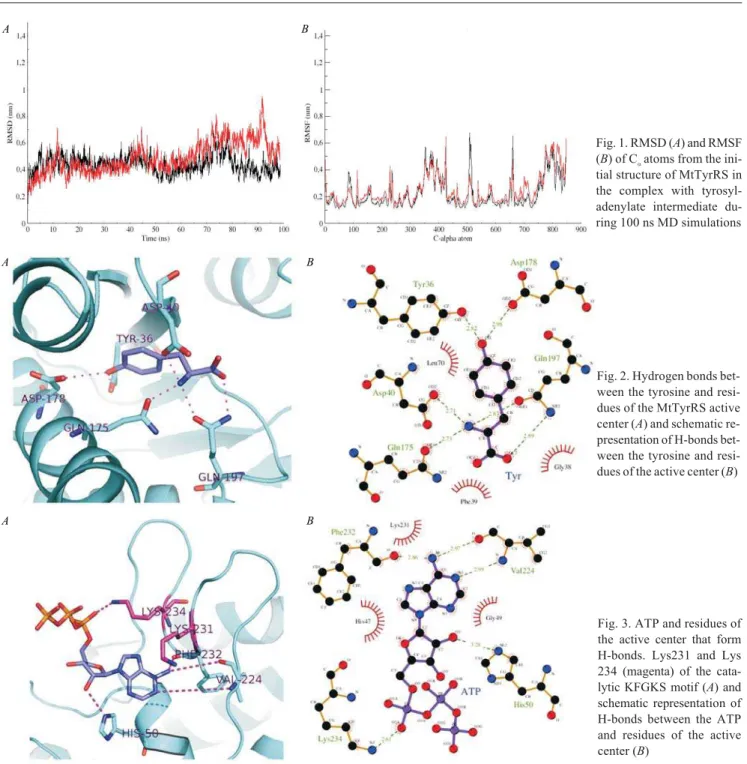
Results and discussion. To check the global struc-tural stability in the course of MD simulations the RMSD and RMSF of C-alpha atoms of MtTyrRS in complex with tyrosyl-adenylate were monitored (Fig. 1). RMSD increase up to ~ 10 ns, and then become more stable (~ 3– 7 C). After ~70 ns of simulations RMSD increase again up to ~ 9.3 C. This is due to high mobility of the
C-termi-nal domains [20]. RMSF show that besides the C-mo-dules, the catalytic KMSKS loops are also highly mobile elements of the protein [28].
In order to evaluate the substrate binding in the MtTyrRS active center the H-bonds were calculated with their occupancy over the entire 100 ns of MD simu-lations (Table). L-tyrosine in the active site forms bonds to Tyr36, Asp40, Gln175, Asp178 and two bonds with Gln197 (Fig. 2). Occupancy of these
H-A B
Fig. 1. RMSD (A) and RMSF (B) of Ñaatoms from the
ini-tial structure of MtTyrRS in the complex with tyrosyl-adenylate intermediate du-ring 100 ns MD simulations
A B
Fig. 2. Hydrogen bonds bet-ween the tyrosine and resi-dues of the MtTyrRS active center (À) and schematic re-presentation of H-bonds bet-ween the tyrosine and resi-dues of the active center (B)
A B
bonds is about ~ 30–40 % of 100 ns of MD simulations for residues of loops, and up to 99 % of 100 ns of MD simulations for residues ofa-helixes andb-strand of the enzyme active center. It is worth to note, that the L-ty-rosine binding pocket is negatively charged because of Asp40 and Asp178.
For the binding of ATP in the active center, Lys 231, Phe232 and Lys234 of the catalytic KFGKS se-quence are important. The positively charged Lys231 and Lys234 interact with the negative phosphate groups of ATP. Phe232 and Lys234 have H-bonds with ATP. Besides, one H-bond with ATP is formed by His50, and two bonds – by Val224 (Fig. 3). Due to the high mobi-lity of the catalytic loop, the occupancy of each H-bond to ATP is not more than ~ 50 % of 100 ns of MD simu-lations (Table). The catalytic loop catalyzes the forma-tion of the tyrosyl-adenylate intermediate by interac-ting with the phyrophosphate moiety of ATP [39].
The tyrosyl-adenylate intermediate occupies entire pocket of the active site interacting with the catalytic
A B
Fig. 4. Hydrogen bonds bet-ween the tyrosyl-adenylate and residues of the MtTyrRS active center (A) and schema-tic representation of H-bonds between the tyrosyl-adenyla-te and residues of the active center (B)
L-tyrosine binding site ATP binding site -Asp40
-Asp178
-Asp196
+Lys234
+Lys231
KFGKS motif Pocket of the active center
Fig. 5. Schematic representation of theMtTyrRS active center. The L-tyrosine binding site has negatively charged Asp40 and Asp178. The ATP binding site has negatively charged Asp196 and positively char-ged Lys231 and Lys234 of the catalytic sequence
Hydrogen bonds Distance, C Occupancy, %
MtTyrRS–Tyr
Tyr36-OH–OH 2.82 75.41
Asp40-OD2–H1N 2.71 30.42
Gln175-OE1–H2N 2.73 47.56
Asp178-OD2–HO 2.98 99.21
Gln197-NE2H–OC2 2.89 37.74
Gln197-OE1–H3N 2.87 39.56
MtTyrRS–ATP
His50-NE2H–O2' 3.28 20.00
Val224-O–H20N6 2.81 47.47
Val224-NH–N1 3.03 49.71
Phe232-O–H1N6 2.86 18.70
Lys234-NZHZ2–O2A 2.61 43.68
MtTyrRS–Tyr–AMP
Tyr36-OH–OH 3.11 33.44
Gly38-O–H24OAE 2.89 20.01
Asp40-NH–OAD 3.04 88.37
Gln175-OE1–H1N 2.69 89.49
Asp178-OD2–HO 2.71 99.07
Gly194-NH–O2' 2.67 26.89
Asp196-OD1–HO3' 2.62 52.80
Gln197-NE2H–O5' 3.23 21.87
Gln197-OE1–H2N 2.86 37.08
Val224-O–H1N6 3.01 60.70
Val224-NH–N1 3.28 66.12
Phe232-O–H2N6 3.12 52.38
N o t e. For each hydrogen bond the percentage occupancy was cal-culated.
loop (Fig. 4). The substrate forms H-bonds with resi-dues that interact with other substrates (tyrosine and ATP) and with Tyr38, Gly194 and Asp196 (Table). H-bonds occupancy reveals stability of the tyrosyl-ade-nylate in the enzyme active center. In general, theMt
TyrRS active center can be divided into two parts: the L-tyrosine binding site and the ATP binding site (Fig. 5). The L-tyrosine binding site involves the negatively charged Asp40 and Asp178. The ATP binding site con-tains the negative Asp196 as well as the positive Lys 231 and Lys234 of the universal catalytic KMSKS mo-tif of the aaRS of class I. In bacterial TyrRS, the Lys 231 and Lys234 of the catalytic KMSKS sequence stabilize the intermediate state for the tyrosine activa-tion by interacactiva-tion with the phyrophosphate moiety of ATP substrate [39].
Conclusions. In this study, we have investigated the mechanisms of the substrates interaction with the active center of MtTyrRS in solution. We have performed 100 ns MD simulations of the MtTyrRS dimer in complexes with L-tyrosine, ATP and tyrosyl-adenylate intermedi-ate. The L-tyrosine binding site is negatively charged, whereas the ATP binding site has the positively char-ged Lys231 and Lys234 of the catalytic sequence. The H-bonds occupancy reveals significant conformational mobility of the active center ofMtTyrRS in solution.
Ìåõàí³çì âçàºìî䳿 ñóáñòðàò³â ç àêòèâíèì öåíòðîì òèðîçèë-òÐÍÊ ñèíòåòàçèMycobacterium tuberculosisçà äàíèìè ìîëåêóëÿðíî¿ äèíàì³êè
Â. Â. Ìèêóëÿê, Î. ². Êîðíåëþê
Ðåçþìå
Ìåòà. Äîñë³äèòè ìåõàí³çìè âçàºìî䳿 ñóáñòðàò³â ðåàêö³¿ àì³íî-àöèëþâàííÿ ç àêòèâíèì öåíòðîì òèðîçèë-òÐÍÊ ñèíòåòàçè åó-áàêòå𳿠Mycobacterium tuberculosis (MtTyrRS).Ìåòîäè. Ñóïåðïî-çèö³ºþ MtTyrRS ç êðèñòàëîãðàô³÷íèìè ñòðóêòóðàìè áàêòåð³é-íèõ TyrRS ïîáóäîâàíî êîìïëåêñè ç òèðîçèíîì, òèðîçèíîì, ÀÒÔ ³ òèðîçèëàäåí³ëàòîì. Êîìïëåêñè MtTyrRS ç ñóáñòðàòàìè âèâ÷àëè ìåòîäîì ìîäåëþâàííÿ ìîëåêóëÿðíî¿ äèíàì³êè (ÌÄ) ó ðîç÷èí³. Ðå-çóëüòàòè. Ïîêàçàíî âîäíåâ³ çâ’ÿçêè ì³æ ñóáñòðàòàìè ³ àêòèâíèì öåíòðîì MtTyrRS òà ¿õíþ ñòàá³ëüí³ñòü ó ïðîöåñ³ ÌÄ. Ñòàá³ëü-í³ñòü ÀÒÔ â àêòèâíîìó öåíòð³ çàáåçïå÷óºòüñÿ âîäíåâèìè çâ’ÿçêà-ìè, à òàêîæ åëåêòðîñòàòè÷íèìè âçàºìîä³ÿìè ç Lys231 òà Lys 234 êàòàë³òè÷íîãî ìîòèâó KFGKS.Âèñíîâêè. ijëÿíêà çâ’ÿçóâàí-íÿ L-òèðîçèíó â àêòèâíîìó öåíòð³ ôåðìåíòó º íåãàòèâíî çàðÿä-æåíîþ, òîä³ ÿê ä³ëÿíêà çâ’ÿçóâàííÿ ÀÒÔ ìຠïîçèòèâíî çàðÿäæå-í³ Lys231 ³ Lys234 êàòàë³òè÷íî¿ ïîñë³äîâíîñò³ KFGKS. Ïðîöåíò-íå ñï³ââ³äíîøåííÿ òðèâàëîñò³ ³ñíóâàííÿ âîäÏðîöåíò-íåâèõ çâ’ÿçê³â, ÿê³ ôîðìóþòüñÿ ì³æ ñóáñòðàòàìè òà ôåðìåíòîì, äî çàãàëüíîãî
÷àñó ìîäåëþâàííÿ ÌÄ ñâ³ä÷èòü ïðî êîíôîðìàö³éíó ðóõëèâ³ñòü àêòèâíîãî öåíòðà.
Êëþ÷îâ³ ñëîâà: òèðîçèë-òÐÍÊ ñèíòåòàçà, Mycobacterium tu-berculosis, ñóáñòðàò, âîäíåâèé çâ’ÿçîê, ìîëåêóëÿðíà äèíàì³êà, ãðèä.
Ìåõàíèçì âçàèìîäåéñòâèÿ ñóáñòðàòîâ ñ àêòèâíûì öåíòðîì òèðîçèë-òÐÍÊ ñèíòåòàçûMycobacterium tuberculosisïî äàííûì ìîëåêóëÿðíîé äèíàìèêè
Â. Â. Ìèêóëÿê, À. È. Êîðíåëþê
Ðåçþìå
Öåëü. Èññëåäîâàòü ìåõàíèçìû âçàèìîäåéñòâèÿ ñóáñòðàòîâ ðå-àêöèè àìèíîàöèëèðîâàíèÿ ñ àêòèâíûì öåíòðîì òèðîçèë-òÐÍÊ ñèíòåòàçû ýóáàêòåðèè Mycobacterium tuberculosis (MtTyrRS). Ìå-òîäû. Ñóïåðïîçèöèåé MtTyrRS ñ êðèñòàëëîãðàôè÷åñêèìè ñòðóê-òóðàìè áàêòåðèàëüíûõ TyrRS ïîñòðîåíû êîìïëåêñû ñ òèðîçè-íîì, òèðîçèíîì è ÀÒÔ è òèðîçèëàäåíèëàòîì. Êîìïëåêñû MtTyrRS ñ ñóáñòðàòàìè èçó÷àëè ìåòîäîì ñèìóëÿöèè ìîëåêóëÿðíîé äèíà-ìèêè (ÌÄ) â ðàñòâîðå.Ðåçóëüòàòû. Ïîêàçàíû âîäîðîäíûå ñâÿçè ìåæäó ñóáñòðàòàìè è àêòèâíûì öåíòðîì MtTyrRS è èõ ñòàáèëü-íîñòü â ïðîöåññå ÌÄ. Ñòàáèëüñòàáèëü-íîñòü ÀÒÔ â àêòèâíîì öåíòðå îáå-ñïå÷èâàåòñÿ âîäîðîäíûìè ñâÿçÿìè, à òàêæå ýëåêòðîñòàòè÷åñ-êèìè âçàèìîäåéñòâèÿìè ñ Lys231 è Lys234 êàòàëèòè÷åñêîãî ìî-òèâà KFGKS.Âûâîäû. Ñàéò ñâÿçûâàíèÿ L-òèðîçèíà â àêòèâíîì öåíòðå ôåðìåíòà çàðÿæåí îòðèöàòåëüíî, â òî âðåìÿ êàê ó÷à-ñòîê ñâÿçûâàíèÿ ÀÒÔ èìååò ïîëîæèòåëüíûå Lys231 è Lys234 êà-òàëèòè÷åñêîé ïîñëåäîâàòåëüíîñòè KFGKS. Ïðîöåíòíîå ñîîò-íîøåíèå äëèòåëüíîñòè ñóùåñòâîâàíèÿ âîäîðîäíûõ ñâÿçåé, ôîð-ìèðóþùèõñÿ ìåæäó ñóáñòðàòàìè è ôåðìåíòîì, ê îáùåìó âðåìå-íè ìîäåëèðîâàâðåìå-íèÿ ÌÄ ñâèäåòåëüñòâóåò î êîíôîðìàöèîííîé ïîä-âèæíîñòè àêòèâíîãî öåíòðà.
Êëþ÷åâûå ñëîâà: òèðîçèë-òÐÍÊ ñèíòåòàçà, Mycobacterium tuberculosis, ñóáñòðàò, âîäîðîäíàÿ ñâÿçü, ìîëåêóëÿðíàÿ äèíàìè-êà, ãðèä.
REFERENCES
1.Kornelyuk A.Structural and functional investigation of mamma-lian tyrosyl-tRNA synthetase.Biopolym Cell. 1998;14(4):349–59. 2.Bonnefond L, Giege R, Rudinger-Thirion J.Evolution of the
tRNA(Tyr)/TyrRS aminoacylation systems. Biochimie. 2005;
87(9–10):873–83.
3.Bedouelle H.Recognition of tRNA(Tyr) by tyrosyl-tRNA syn-thetase.Biochimie. 1990;72(8):589–98.
4.Odynets’ KO, Korneliuk OI.A model of three-dimensional struc-ture ofMycobacterium tuberculosistyrosyl-tRNA synthetase.
Ukr Biokhim Zh. 2008;80(5):62–75.
5.Hoffmann M, Torchala M.Search for inhibitors of aminoacyl-tRNA synthases by virtual click chemistry.J Mol Model.2009;
15(6):665–72.
6.Eitner K, Gaweda T, Hoffmann M, Jura M, Rychlewski L, Bar-ciszewski J.eHiTS-to-VMD interface application. The search for tyrosine-tRNA ligase inhibitors.J Chem Inf Model.2007;
47(2):695–702.
7.Manning J, Vincent J. Tyrosyl tRNA synthetase: A new site for antibiotics. Literature Seminar. Univ. of Alabama, 2006; 10 p. 8.Stefanska AL, Coates NJ, Mensah LM, Pope AJ, Ready SJ, Warr
aMicromonosporasp. I. Fermentation, isolation and properties.
J Antibiot (Tokyo). 2000;53(4):345–50.
9.Houge-Frydrych CS, Readshaw SA, Bell DJ. SB-219383, a no-vel tyrosyl tRNA synthetase inhibitor from aMicromonospora
sp. II. Structure determination.J Antibiot (Tokyo). 2000;53(4): 351–6.
10.Jarvest RL, Berge JM, Brown P, Hamprecht DW, McNair DJ, Mensah L, O’Hanlon PJ, Pope AJ. Potent synthetic inhibitors of tyrosyl tRNA synthetase derived from C-pyranosyl analogues of SB-219383.Bioorg Med Chem Lett.2001;11(5):715–8. 11.Kobayashi T, Takimura T, Sekine R, Kelly VP, Kamata K,
Saka-moto K, Nishimura S, Yokoyama S. Structural snapshots of the KMSKS loop rearrangement for amino acid activation by bacte-rial tyrosyl-tRNA synthetase.J Mol Biol.2005;346(1):105–17. 12.Kobayashi T, Sakamoto K, Takimura T, Sekine R, Kelly VP,
Ka-mata K, Nishimura S, Yokoyama S. Structural basis of nonnatu-ral amino acid recognition by an engineered aminoacyl-tRNA synthetase for genetic code expansion.Proc Natl Acad Sci USA. 2005;102(5):1366–71.
13.Yaremchuk A, Kriklivyi I, Tukalo M, Cusack S.Class I tyrosyl-tRNA synthetase has a class II mode of cognate tyrosyl-tRNA recogni-tion.EMBO J. 2002;21(14):3829–40.
14.Qiu X, Janson CA, Smith WW, Green SM, McDevitt P, Johanson K, Carter P, Hibbs M, Lewis C, Chalker A, Fosberry A, Lalonde J, Berge J, Brown P, Houge-Frydrych CS, Jarvest RL. Crystal structure ofStaphylococcus aureustyrosyl-tRNA synthetase in complex with a class of potent and specific inhibitors.Protein Sci.2001;10(10):2008–16.
15.Brick P, Bhat TN, Blow DM.Structure of tyrosyl-tRNA synthe-tase refined at 2.3 A resolution. Interaction of the enzyme with the tyrosyl adenylate intermediate.J Mol Biol.1989;208(1):83–98. 16. Hartmann MD, Shkolnaya LA, Bourenkov GP, Strizhov NI,
Bartunik HD. The structure of tyrosyl-tRNA synthetase from
Mycobacterium tuberculosis. DOI:10.2210/pdb2jan/pdb. 17.Li T, Froeyen M, Herdewijn P.Comparative structural
dyna-mics of Tyrosyl-tRNA synthetase complexed with different substrates explored by molecular dynamics.Eur Biophys J.2008;
38(1):25–35.
18.Yesylevskyy SO, Savytskyi OV, Odynets KA, Kornelyuk AI. Inter-domain compactization in human tyrosyl-tRNA synthetase stu-died by the hierarchical rotations technique. Biophys Chem. 2011;154(2–3):90–8.
19. Savytskyi OV, Yesylevskyy SO, Kornelyuk AI. Asymmetric structure and domain binding interfaces of human tyrosyl-tRNA synthetase studied by molecular dynamics simulations.J Mol Re-cognit. 2013;26(2):113–20.
20.Mykuliak VV, Kornelyuk AI. Conformational mobility of tyrosyl-tRNA synthetase fromM. tuberculosiseubacteria according to computer modeling of molecular dynamics data. Physics of the Alive. 2011;19(2):4–8.
21.Budiman ME, Knaggs MH, Fetrow JS, Alexander RW.Using molecular dynamics to map interaction networks in an amino-acyl-tRNA synthetase.Proteins. 2007;68(3):670–89. 22.Thompson D, Plateau P, Simonson T.Free-energy simulations
and experiments reveal long-range electrostatic interactions and substrate-assisted specificity in an aminoacyl-tRNA synthetase.
Chembiochem. 2006;7(2):337–44.
23.Thompson D, Simonson T.Molecular dynamics simulations show that bound Mg2+
contributes to amino acid and aminoacyl adeny-late binding specificity in aspartyl-tRNA synthetase through long range electrostatic interactions.J Biol Chem. 2006;281(33): 23792–803.
24.Hughes SJ, Tanner JA, Miller AD, Gould IR. Molecular dynamics simulations of LysRS: an asymmetric state.Proteins. 2006;62
(3):649–62.
25.Kapustina M, Carter CW Jr. Computational studies of tryptopha-nyl-tRNA synthetase: activation of ATP by induced-fit.J Mol Biol.2006;362(5):1159–80.
26.Pydiura NA, Kornelyuk AI.Flexible 3D structure ofBos taurus
tyrosyl-tRNA synthetase suggests the existence of hinge mecha-nism provided by conservative Gly353 at interdomain linker.
Biopolym Cell. 2012;28(5):397–403.
27.Kornelyuk AI, Klimenko IV, Odynets KA.Conformational chan-ge of mammalian tyrosyl-tRNA synthetase induced by tyrosyl adenylate formation.Biochem Mol Biol Int.1995;35(2):317–22. 28.Mykuliak VV, Kornelyuk AI.Dynamic formation of theb-strand
structure in the active site of tyrosyl-tRNA synthetase from Mu-cobacterium tuberculosiseubakteria according to the molecular dynamics. Reports of the National Academy of Sciences of Uk-raine. 2012; (5):158–162.
29.Hess B, Kutzner C, van der Spoel D, Lindahl E.GROMACS 4: Algorithms for highly efficient, load-balanced, and scalable mo-lecular simulation.J Chem Theory Comput.2008;4(3):435–47. 30.Hornak V, Abel R, Okur A, Strockbine B, Roitberg A,
Simmer-ling C.Comparison of multiple Amber force fields and develop-ment of improved protein backbone parameters.Proteins. 2006;
65(3):712–25.
31.Bjelkmar P, Larsson P, Cuendet M, Hess B, Lindahl E. Imple-mentation of the CHARMM force field in GROMACS: analysis of protein stability effects from correction maps, virtual interac-tion sites, and water models.J Chem Theory Comput.2010;6
(2):459–66.
32.Wang J, Wang W, Kollman PA, Case DA.Automatic atom type and bond type perception in molecular mechanical calculations.
J Mol Graph Model. 2006;25(2):247–60.
33.Zoete V, Cuendet MA, Grosdidier A, Michielin O.SwissParam: a fast force field generation tool for small organic molecules.J Comput Chem. 2011;32(11):2359–68.
34.Salnikov A, Sliusar I, Sudakov O, Savytskyi O, Kornelyuk A. Vir-tual laboratory MolDynGrid as a part of scientific infrastructure for biomolecular simulations. Int J Computing. 2010; 9(4): 295–301.
35.Savytskyi OV, Sliusar IA, Yesylevskyy SO, Stirenko SG, Korne-lyuk AI.Integrated tools for molecular dynamics simulation data analysis in the MolDynGrid virtual laboratory.Intelligent Data Acquisition and Advanced Computing Systems (IDAACS), 2011 IEEE 6thInternational Conference on.2011;1:208–211. 36.Salnikov AO, Sliusar IA, Sudakov OO, Savytskyi OV, Kornelyuk
AI.MolDynGrid virtual laboratory as a part of Ukrainian Acade-mic Grid infrastructure.Intelligent Data Acquisition and Advan-ced Computing Systems: Technology and Applications, IDAACS 2009. IEEE International Workshop on.2009:237–240. 37. The PyMOL Molecular Graphics System, Version 1.5.
Schro-dinger, LLC.
38.Laskowski RA, Swindells MB. LigPlot+: multiple ligand-protein interaction diagrams for drug discovery. J Chem Inf Model.
2011;51(10):2778–86.
39.Austin J, First EA.Comparison of the catalytic roles played by the KMSKS motif in the human andBacillus stearothermophi-lus trosyl-tRNA synthetases. J Biol Chem. 2002; 277(32): 28394–9.