R E S E A R C H
Open Access
In silico analysis of binding interaction of
conantokins with NMDA receptors for
potential therapeutic use in Alzheimer
’
s
disease
Maleeha Waqar and Sidra Batool
*Abstract
Background:The N-methyl-D-aspartate (NMDA) receptors are glutamate receptors that play vital roles in central nervous system development and are involved in synaptic plasticity, which is an essential process for learning and memory. The subunit N-methyl D-aspartate receptor subtype 2B (NR2B) is the chief excitatory neurotransmitter receptor in the mammalian brain. Disturbances in the neurotransmission mediated by the NMDA receptor are caused by its overexposure to glutamate neurotransmitter and can be treated by its binding to an antagonist. Among several antagonists, conantokins from cone snails are reported to bind to NMDA receptors.
Methods:This study was designed to analyze the binding mode of conantokins with NMDA receptors in both humans and rats. To study interactions, dockings were performed using AutoDock 4.2 and their results were further analyzed using various computational tools.
Results:Detailed analyses revealed that these ligands can bind to active site residues of both receptors as reported in previous studies.
Conclusions:In light of the present results, we suggest that these conantokins can act as antagonists of those receptors and play an important role in understanding the importance of inhibition of NMDA receptors for treatment of Alzheimer’s disease.
Keywords: N-methyl-D-aspartate, Glutamate, Synaptic plasticity, NR2B, Neurotransmitter, Antagonists, Conantokins, Docking, In silico, Alzheimer’s dieases
Background
The N-methyl-D-aspartate (NMDA) receptors are ino-tropic glutamate receptors that are gated cation channels [1, 2]. The NMDA receptors (NMDAR) play vital roles in central nervous system (CNS) development [2]. These receptors are highly permeable to Ca2+ions and the cal-cium flux is critically important for synaptic plasticity, which is an essential neurochemical process for learning and memory [2–4]. The receptor itself has many sub-units and their variants have numerous functions in the brain. The subunit N-methyl-D-aspartate receptor sub-type 2B (NR2B) is the chief excitatory neurotransmitter
receptor in the mammalian brain [5]. The glutamate neurotransmitter allows for a transmembrane ion flow through the receptor to increase the action potential of the neuron. This characteristic makes the synapsis among these neurons to be the main memory storage unit and hence associates them with learning and memory [6]. Due to their functioning in the CNS, the potential of these re-ceptors as drug targets for various neurodegenerative dis-eases has been highlighted in the literature.
NMDAR antagonists have emerged as potential lead com-pounds for Alzheimer’s patients [7]. The cognitive symp-toms associated with deficits in learning and menory have been attributed to disturbances in glutaminergic neurotrans-mission [8]. The excessive stimulation by the glutamate neurotransmitter of neurons causes excitotoxicity and
* Correspondence:[email protected]
Department of Biosciences, COMSATS Institute of Information Technology, Park Road, Chak Shahzad, Islamabad 45550, Pakistan
results in damage and death of neurons [8]. Blocking the glutaminergic neurotransmission mediated by NMDA re-ceptors can alleviate the excitotoxicity and prevent further neuron damage and death.
Several venom toxins have made their way in scientific studies and clinical trials for their therapeutic potential against various diseases. Having mostly inhibitory ef-fects, these toxin peptides target different receptors across the body, the hyperactivity of which is associated with the pathophysiology of many diseases. Numerous toxins have been reported to target and block receptors used for the treatment of Alzheimer’s disease, such as neurotoxins that target acetylcholinesterase enzymes and certain toxins from Mamba snakes that were reported to inhibit the muscarinic acetylcholine receptors [9, 10]. A few toxins with antagonistic properties against NMDA receptor have been reported as well. The conantokins are powerful and potent blockers of the NMDA receptor, with particularly high selectivity for the NR2B subunit [11]. These toxins have been reported to block these
glutamine receptors and therefore have therapeutic potential for treating Alzheimer’s disease.
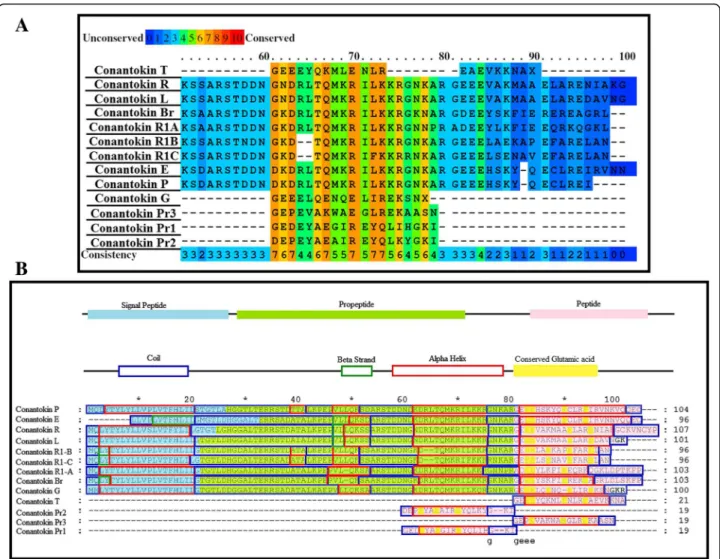
The objective of this study was to perform and look into the in silico analysis of the binding interaction of conanto-kins with the NMDA receptor NR2B subunit. The mode of interaction and the binding residues for both the ligand dataset and the receptor dataset were collected. Due to unavailability of the crystal structures of the NMDA receptor in humans and most of conantokins, their three-Fig. 1aConserved residues highlighted in conantokins.bPredicted secondary structures of all conantokins aligned
Table 1Docking parameters used for docking studies on AutoDock 4.2
Grid Parameters Docking Parameters
Spacing 0.375 Å Energy evaluations 2.5 × 10⁶
Grid center 80X Å Iterations 27,000 80Y Å Mutation rate 0.02 80Z Å Crossover rate 0.80
dimensional structures were predicted via computational homology modeling methods and the predicted models were validated to continue their further use.
Docking studies provided insights into binding pattern of receptors and ligands. A number of in silico studies investigated many computational approaches ranging from construction of structural models to investigation and discovery of potential drug candidates [12–14]. The analysis of the binding interactions of the receptor and the ligand peptides produced results that helped us dem-onstrate the pharmacological importance of conantokins and their potential use as NMDA receptor antagonists for treatment of Alzheimer’s disease.
Methods
Receptor dataset collection
The first step of methodology included collection of receptor proteins. As the structure of NMDA receptor in humans is not available, it had to be
predicted via computational homology modeling, which allowed the construction of a three-dimensional structure of a protein based on the known structures of similar protein templates. SWISS-MODEL was used for this purpose, as it is a fully automated protein structure homology modeling server [15]. The protein template used for structure prediction of NMDAR in humans was the structure of NMDAR in Rattus norvegicus (brown rat) [16].
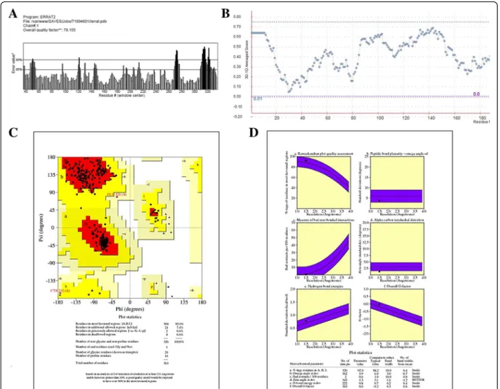
The structure of the template (pdb id: 3JPW) was re-trieved from the Research Collaboratory for Struc-tural Bioinformatics (RSCB) [17]. After structure prediction, the Structure Analysis and Verification Server (SAVES) was used for validation by generating Ramachandran plot [18] and ERRAT [19], which gives a factor of overall quality of the predicted structure. Whereas Verify_3D [20] was employed to analyze the compatibility of the atomic model of the protein with its own amino acid sequence.
Ligand dataset collection
The ligand dataset comprised reported conantokins that are a class of conopeptides (17–27 amino acids) without cysteine residues that selectively influence NMDA recep-tors [21]. Among them, only three-dimensional struc-tures of conantokin G and conantokin T are available on Protein Data Bank (PDB ID: 1ONU and 1ONT, respect-ively). The crystal structures of other conantokins Br, L, P, R, E, Pr1, Pr2, Pr3, R1A, R1B and R1C are not avail-able on the Protein Data Bank and therefore were sub-jected to three-dimensional structure prediction. Homology modeling was used initially, but due to ab-sence of any homologues for the conantokins, the struc-ture prediction was carried out via fold recognition (threading) on the Iterative Threading ASSEmbly Refine-ment (iTASSER) server [22], which detects structure templates from the Protein Data Bank and constructs full-length structure models by reassembling structural fragments from threading templates. The predicted structures were then validated on the SAVES metaserver using Procheck, ERRAT and Verify_3D. Multiple se-quence alignment (MSA) using PRALINE [23] multiple sequence alignment toolbox was performed on all the
conantokins to find out residues and secondary structure conservation. The residues responsible for the signal peptides, peptide precursors and active protein peptides were also highlighted for each conantokin showing con-siderable conservation in these peptide regions as well, as shown in Fig. 1.
The conserved glutamic acid residues in each conanto-kin are the positions for residue modification, where the glutamic acid is converted to 4-carboxyglutamate. Suffi-cient conservation of secondary structures was observed among all the conantokins.
Experimental background reported for conantokins and NMDAR
and NMR spectroscopy have allowed the understanding of how similar the inhibition of NMDAR by conantokins in humans is to previously reported inhibition of NMDAR in rats by conantokins [25]. Using chemically synthesized conantokin variants (created by techniques of point muta-tion and chimeric proteins), the subtype selectivity of NMDAR has been extensively studied for most conanto-kins [26]. In addition, conantoconanto-kins represent a class of NMDA antagonists with an improved safety profile com-pared to other antagonists that show psychotomimetic,
amnesic and motor-impairing actions and neurotoxicity, which limit their usefulness in humans [21]. These wet lab studies revealed the pharmacological importance of con-antokins as antagonists of NMDAR for their therapeutic applications in many neuropathologies and have led us to investigate this property computationally.
Docking studies
Molecular dockings are performed to predict the binding orientation between a receptor and its ligand to form a Table 2Docking results of conantokin ligands in complex with NMDA receptor in humans
Conantokin Hydrogen bonds Hydrophobic bonds
Receptor residues Ligand residues Distance (Å) Atoms Receptor residues Ligand residues Conantokin G – – – – Glu236
Gln110 Phe114 Ile111 Pro177 Ile12 Leu5 Ser16 Gln9 Conantokin L Gln110 Asp96
Ala93 Leu65 2.36 2.25 2.81 N-OD1 OE1-N NE2-O Pro177 Ile111 Glu236 Phe114 Met88 Thr24 Val85 Gly81 Conantokin E Pro177
Glu236 Arg67Asp18 Gly13 Gly12 3.09 2.32 2.23 3.19 O-NH1 OE2-N N-O OE2-N Ile111 Phe114 Gln110 Leu41 Gln59 Lys66 Conantokin Pr1 Glu236 Gln13 3.13 OE2-NE2 Pro177
Gln110
Asp3 Ala6 Conantokin Pr2 Glu236 Lys15 3.11 OE2-NZ Gn110
Ile111 Phe114
Glu4 Ala8 Tyr5 Conantokin Pr3 – – – – Gln110
Ile111 Phe114 Pro177 Glu2 Gly1 Glu4 Trp8 Conantokin T Glu236
Gln110 Arg13Gln6 2.85, 2.922.88 OE2-NH2OE2-NE OE1-NE2
Ile111 Glu16
Conantokin R1A Glu236 Gln110 Arg54 Asp58 2.70 2.82 O-NH1 N-O Ile111 Phe114 Pr0177 Leu13 Leu18 Glu33 Conantokin R1B Gln110 Tyr8 3.22 N-OH Ile111
Pro177 Glu236
Lys62 Lys71 Ala95 Conantokin R1C Gln110 Val11 2.06 NE2-O Glu236
Ile111 Pro177 Phe114 Asp77 Ala39 Pro12 Leu13 Conantokin R Glu236
Phe114
Gln50 Ala98
3.33 2.01
OE2-NE2 N-O Ile111 Pro177 Gln110
Ser52 Leu65 Met68 Conantokin Br Gln110 Ala94 1.93 NE2-O Pro177
Ile111 Glu236
Thr22 Ile82 Asp77 Conantokin P Glu236 Leu65 2.33 N-OE2 Gln110
stable molecular complex [27]. This allows a detailed insight of the three-dimensional structure formed be-tween two biomolecules and to correlate this informa-tion to find a potential drug candidate for these receptors [28]. Molecular docking was performed for NMDA receptors for both humans and rats. AutoDock 4.2 [29] was used to perform automated docking runs, in order to find the binding mode of each receptor with each conantokin ligand. The number of runs for each docking was set to 50 to allow each ligand 50 different conformations with the receptors in order to let them bind freely anywhere on the receptor. The grid size was set to cover the entire receptor in order to find a poten-tial binding site for each ligand and to analyze if each ligand indeed occupies the site on the receptor that is crucial for its functioning in Alzheimer’s and could po-tentially inhibit it. The docking parameters used are shown in Table 1.
Post-docking analysis of the results was carried out on AutoDock 4.2 and later visualized on Chimera [30].
Ligplot+ [31] was used for further validation of the protein-protein interactions, which generates two-dimensional schematics on the basis of hydrogen bonds and hydrophobic interactions. The three-dimensional structural analysis was performed on PyMol [32] that allowed the residues in the protein-protein interaction of the receptor-ligand complex to be highlighted. The en-ergy of each complex that was achieved via docking allowed the receptor-ligand bindings to be confirmed and refined and used for further analysis and results discussion.
Results and discussion
indicated by the Ramachandran plot, 92% of the residues were in favored regions. Moreover, the main chain pa-rameters such as peptide bond planarity, measure of non-bonded interactions, α carbon tetrahedral distor-tion, H-bond energy and overall G factor for the struc-ture were found within favorable regions. ERRAT and Verify_3D further validated the structure by scoring it with an overall quality factor of 79.155 and by passing it with 80% of the amino acids having scored≥0.2
respect-ively (Fig. 2). The validation results suggested that the predicted model of the NMDA receptor in humans was of good quality and can be used for further analysis.
Structure prediction and validation of conantokins The predicted structure of each conantokin was also val-idated in order to check the quality of their three-dimensional structures. All predicted structures were passed by Ramachandran plot, ERRAT and Verify_3D, suggesting that these structures were of good quality and could be used further study.
Binding site residue information
After structure prediction and evaluation, binding site residue information for NMDA receptor in humans was
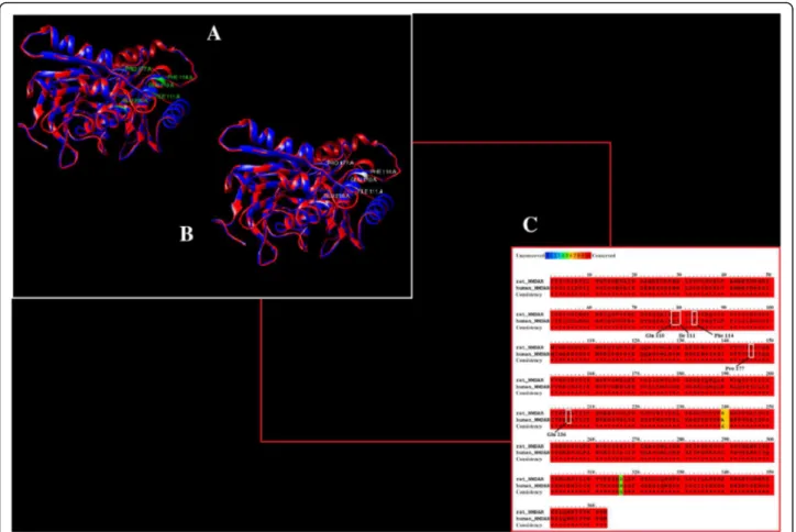
gathered. Since this is a computational based analysis, it is very important to identify binding site residues correctly and verify the results. As no previous information for binding residues of NMDAR in humans was reported, the binding site was retrieved by using the binding site resi-dues of NMDAR reported in the literature that showed residues specific for the NR2B subunit in rats. The two structures were aligned and it was observed that the binding site residues reported for NMDAR in rats are conserved in humans. The binding residues for NMDAR in both humans and rats are shown in Fig. 3. These identified residues have been taken as refer-ences to further evaluate the docking results.
Binding residue information for conantokin G only suggests Glu2, Gln6, Asn8, Arg13, Asn17 and Lys15 as predominant residues that are involved in interac-tions with the NMDAR subtype selectivity for NR2B subunit. Moreover, the first five amino acids of con-antokins, especially Glu2 and Gla4 and a hydrophobic residue at position 12 are critical for functional activ-ity in vitro [21]. It has been observed that the peptide region has shown importance in interactions. These residues are also found to be conserved in other con-antokins as well.
Docking results
After structure prediction and binding site identification, molecular dockings were performed on NMDAR receptors with conantokin ligands. Docking results were run based on their energy values and were ranked as such with each run showing results for receptor-ligand complex with the lowest energy. Detailed analysis of each run for each com-plex showed that all the conantokins showed bindings with the conserved active sites of NMDA receptor in both humans and rats. All the conantokins seemed to bind with the glutamine and glutamic acid residues in the NMDA re-ceptors in both humans and rats. Table 2 shows the
detailed analysis retrieved from plotting the docking results on LigPlot+, showing the binding of NMDA receptor in humans with conantokins. These data include information about respective hydrogen bond residues of both receptors and ligands, their bond distances, bond atoms and the interacting hydrophobic residues.
Glutamic acid at position 236 and glutamine at pos-ition 110 were predominantly involved in the binding of the NR2B subunit of NMDAR in human with all the conantokins. Isoleucine at position 111, phenylalanine at position 114 and proline at position 177 were mostly found in hydrophobic interactions. The binding patterns Table 3Docking results of conantokin ligands in complex with NMDA receptor in rats
Conantokin Hydrogen bonds Hydrophobic bonds
Receptor residues Ligand residues Distance (Å) Atoms Receptor residues Ligand residues Conantokin G Gln110 Arg13 2.64 OE1-NE Ile111
Phe114
Ile12 Gln9 Conantokin L Gln110 Pro46 1.52 NE2-O Ile111
Phe114 Pro177 Glu236 Pro12 Ala30 Asp77 Val42 Conantokin E Gln110
Ile111 Ile11 Cys83 2.94 1.90 OE1-N N-O Phe114 Pro177 Glu236 Lys61 Ser28 Leu64 Conantokin Pr1 – – – – Glu236
Phe114 Pro177 Gln110 Ile15 His16 Asp3 Lys18 Conantokin Pr2 Gln110 Lys15 2.88 OE1-NZ Glu236
Pro177 Phe114
Glu14 Gly1 Ala6 Conantokin Pr3 Gln110 Lys7 2.96 OE1-NZ Ile111
Phe114 Pro177
Trp8 Glu2 Glu4 Conantokin T Glu236 Arg13 2.69, 2.71 OE2-NE,
OE2-NH2 Ile111 Glu16 Conantokin R Gln110
Ile111 Lys73 Lys87 2.13 3.35 OE1-N O-NZ Phe114 Pro177 Glu236 Glu95 Lys99 Asn62 Conantokin R1A Gln110
Glu236 Leu7, Leu9, Thr5 Thr65 2.22 2.15, 2.80 1.79 3.14 OE1-N N-O, NE2-O NE2-O OE1-N Ile111 Phe114 Pro177 Ile19 Arg63 Thr56 Conantokin R1B – – – – Gln110
Ile111 Pro177 Glu236 Pro12 Ala77 Asp63 Thr76 Conantokin R1C Gln110
Glu236 Val14 Ser52 2.47 2.54 OE1-N OE2-OG Ile111 Phe114 Pro177 Gln65 His17 Leu49 Conantokin Br Gln110 Ala94 1.93 NE2-O Ile111
Pro177 Glu236
Asn59 Ile82 Asp77 Conantokin P Glu236
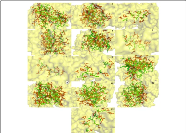
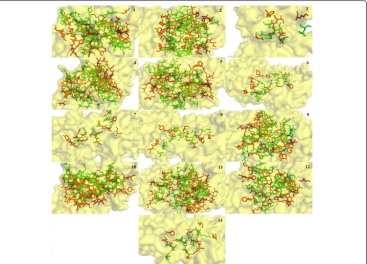
of all the conantokin ligands individually in complex with the NR2B subunit of NMDAR in human are shown in Fig. 4.
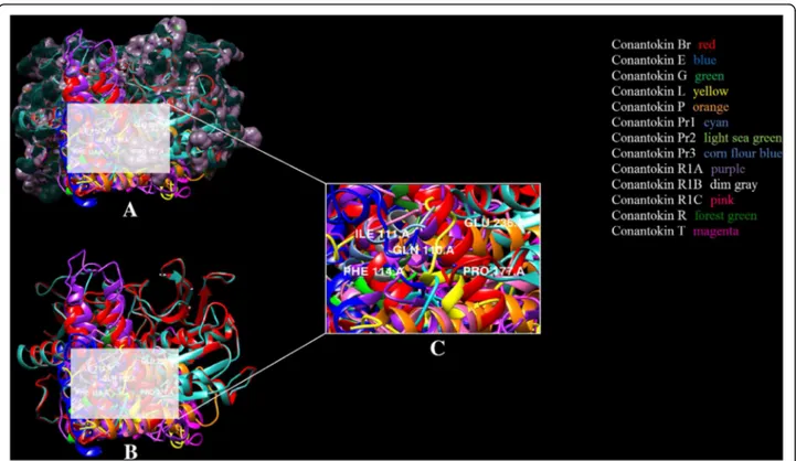
It is clear in Fig. 4 that due to difference in sizes and structure, each conantokin occupies the binding site on NMDAR in humans in its own respective orientation to form the most stable complex. However, each ligand is shown to bind to the same binding residues as re-ported. The position of binding pocket of the NR2B subunit of NMDAR in humans resided by conanto-kins is shown in Fig. 5. It is shown clearly that all the ligands occupied the same binding pocket in structure of NMDAR as they showed binding with the same residues that have been reported.
Similar binding pattern was observed for the NR2B subunit of NMDAR in rat. Table 3 shows the binding formation for NMDAR in rats, highlighting residues in-volved in hydrogen bonding, their atoms and their bond distances, along with the hydrophobic residues for both the receptor and the ligands.
It has been observed that likewise for human NR2B, Glu236 and Gln110 residues of NR2B subunit in rats were found interacting with the residues of every conantokin lig-and. Ile111, Phe114 and Pro177 were mostly found in hydrophobic interactions. This binding pattern was con-served among all the conantokins showing their similarity in potency for the NR2B subunit. The binding pattern of all conantokins for NMDAR in rats is shown in Fig. 6.
The binding pocket of conantokins with the receptor is highlighted in Fig. 7. As in the case of NMDAR in rats, Fig. 7 clearly demonstrates that each conantokin occupies the same binding pocket as NMDAR in humans.
Energy values
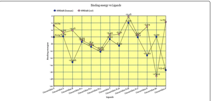
successive energy values for the ligand-receptor complex formed by each toxin with the NMDA receptor in both humans and rats are shown in Fig. 8.
The energy values for all the complexes fall below the value of 0, as clearly observed from the graphical represen-tation. This suggests the formation of relatively stable com-plexes between NMDA receptor in both rats and humans with all conantokins. NMDAR in humans showed most af-finity for conantokin P, conantokin E, conantokin R and conantokin Pr3, whereas NMDAR in rats showed more af-finity for Conantokin Br and Conantokin Pr3.
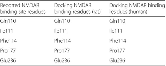
Comparison of reported residues for NMDAR to docking results
A comparative analysis was performed of the docking re-sults of NMDA receptors in both humans and rats with all the conantokins. The reported residues were inspected to verify if the docking results of this study were indeed in line with the reported information regarding the binding site residues of NR2B subunit of NMDAR. It is clearly dis-played in Table 4 that the binding site residues of NMDAR reported in the literature were achieved in the docking experiments for both humans and rats.
Fig. 8Graphical plot of binding energies versus toxin ligands
Conclusions
Conantokins had been reported as selective antagonists of NR2B subtype of the NMDA receptor. The NR2B subunit is directly involved in the excitotoxicity caused by the over-exposure to glutamate neurotransmitter. Therefore, inhibiting its activity has become a possible target for treatment of Alzheimer’s. We designed an in silico study and analysis of the binding interaction of conantokins with the NMDA receptor in both humans and rats. After the binding site residues for the receptors were understood, docking studies were performed and the residues achieved via docking were compared to the binding residues reported in the literature. The similarity of the results suggests the potential successful binding of the toxin ligands with both the receptors, and their sub-sequent function as their antagonists. Our results helped us to demonstrate the potential of these receptors in complex with the conantokins for the symptomatic treatment of Alzheimer’s patients.
Abbreviations
CNS:Central nervous system; MSA: Multiple sequence alignment; NMDA: N-methyl-D-aspartate; NMDAR: NMDA receptor; NR2B: N-methyl D-aspartate re-ceptor subtype 2B; RSCB: Research collaboratory for structural bioinformatics; RSMD: Root mean square deviation; SAVES: Structure analysis and verification server
Acknowledgements
The authors would like to thank the facilities provided by the Department of Biosciences, COMSATS Institute of Information Technology, Pakistan.
Funding Not applicable
Authors’contributions
MW conducted the research and prepared the draft. SB developed the study concept and refined the draft. Both authors read and approved the final manuscript.
Ethics approval and consent to participate Not applicable
Consent for publication Not applicable
Competing interests
Both authors declare that they have no competing interests.
Publisher’s Note
Springer Nature remains neutral with regard to jurisdictional claims in published maps and institutional affiliations.
Received: 6 April 2017 Accepted: 13 September 2017
References
1. Blanke ML, VanDongen AMJ. Activation mechanisms of the NMDA receptor. In: Van Dongen AM, editor. Biology of the NMDA receptor. Boca Raton (FL): CRC Press; 2009.
2. Li ST, Ju JG. Functional roles of synaptic and extrasynaptic NMDA receptors in physiological and pathological neuronal activities. Curr Drug Targets. 2012;13(2):207–21.
3. Hunt DL, Castillo PE. Synaptic plasticity of NMDA receptors: mechanisms and functional implications. Curr Opin Neurobiol. 2012;22(3):496–508. 4. Martin SJ, Grimwood PD, Morris RG. Synaptic plasticity and memory: an
evaluation of the hypothesis. Annu Rev Neurosci. 2000;23:649–711. 5. Loftis JM, Janowsky A. The N-methyl-D-aspartate receptor subunit NR2B:
localization, functional properties, regulation, and clinical implications. Pharmacol Ther. 2003;97(1):55–85.
6. Yang JL, Sykora P, Wilson DM, Mattson MP, Bohr VA. The excitatory neurotransmitter glutamate stimulates DNA repair to increase neuronal resiliency. Mech Ageing Dev. 2011;132(8–9):405–11.
7. Farlow MR. NMDA receptor antagonists. A new therapeutic approach for Alzheimer's disease. Geriatrics. 2004;59(6):22–7.
8. Danysz W, Pasons CG. Alzheimer’s disease,β-amyloid, glutamate, NMDA receptors and memantine–searching for the connections. Br J Pharmacol. 2012;167(2):324–52.
9. Servent D, Blanchet G, Mourier G, Marquer C, Marcon E, Fruchart-Gaillard C. Muscarinic toxins. Toxicon. 2011;58(6–7):455–63.
10. Waqar M, Batool S.In silicoanalysis of binding of neurotoxic venom ligands with acetylcholinesterase for therapeutic use in treatment of Alzheimer’s disease. J Theor Biol. 2015;372:107–17.
11. Waqar M, Kamal MA, Batool S.In silicoanalysis of binding interaction of mamba toxins with M4 and M2 muscarinic acetylcholine receptors for therapeutic use in Alzheimer’s disease. CNS Neurol Disord Drug Targets. 2015;14(8):1031–40.
12. Amin SA, Bhattacharya P, Basak S, Gayen S, Nandy A, Saha A.
Pharmacoinformatics study of Piperolactam a from Piper Betle root as new lead for non steroidal anti fertility drug development. Comput Biol Chem. 2017;67:213–24.
13. Amin SA, Adhikari N, Agrawal RK, Jha T, Gayen S. Possible binding mode analysis of Pyrazolo-triazole hybrids as potential anticancer agents through validated molecular docking and 3D-QSAR modeling approaches. Letters in Drug Design & Discovery. 2017;14(5):515–27. www.eurekaselect.com/145590 14. Amin SA, Bhargava S, Adhikari N, Gayen S, Jha T. Exploring pyrazolo [3, 4-d]
pyrimidine phosphodiesterase 1 (PDE1) inhibitors: a predictive approach combining comparative validated multiple molecular modelling techniques. J Biomol Struct Dyn. 2017;13:1–19.
15. Schwede T, Kopp J, Guex N, Peitsch MC. SWISS-MODEL: an automated protein homology-modeling server. Nucleic Acids Res. 2003;31(3):3381–5. 16. Karakas E, Simorowski N, Furukawa H. Structure of the zinc-bound amino-terminal
domain of the NMDA receptor NR2B subunit. EMBO J. 2009;28(24):3910–20. 17. Berman HM, Westbrook J, Feng Z, Gilliland G, Bhat TN, Weissig H, et al. The
Protein Data Bank. Nucleic Acids Res. 2000;28(1):235–42.
18. Laskowski RA, Rullmannn JA, MacArthur MW, Kaptein R, Thornton JM. AQUA and PROCHECK-NMR: programs for checking the quality of protein structures solved by NMR. J Biomol NMR. 1996;8(4):477–86. 19. Colovos C, Yeates TO. Verification of protein structures: patterns of
nonbonded atomic interactions. Protein Sci. 1993;2(9):1511–9. 20. Bowie JU, Lüthy R, Eisenberg D. A method to identify protein
sequences that fold into a known three-dimensional structure. Science. 1991;253(5016):164–70.
21. Lebbe EKM, Tytgat J. In the picture: disulfide-poor conopeptides, a class of pharmacologically interesting compounds. J Venom Anim Toxins incl Trop Dis. 2016;22:30. doi: 10.1186/s40409-016-0083-6.
22. Roy A, Kucukural A, Zhang Y. I-TASSER: a unified platform for automated protein structure and function prediction. Nat Protoc. 2010;5(4):725–38.
Table 4Comparative tabulation of binding residues of reported NMDAR versus post-docking results
Reported NMDAR binding site residues
Docking NMDAR binding residues (rat)
23. Simossis VA, Heringa J. PRALINE: a multiple sequence alignment toolbox that integrates homology-extended and secondary structure information. Nucleic Acids Res. 2005;33:W289–94.
24. Teichert RW, Jimenez EC, Twede V, Watkins M, Hollmann M, Bulaj G, et al. Novel conantokins fromConus pariusvenom are specific antagonists of N-methyl-D-aspartate receptors. J Biol Chem. 2007;282(51):36905–13. 25. Nielsen KJ, Skjaerbaek N, Dooley M, Adams DA, Mortensen M, Dodd PR, et
al. Structure-activity studies of conantokins as human N-methyl-D-aspartate receptor modulators. J Med Chem. 1999;42(3):415–26.
26. Sheng Z, Prorok M, Castellino FJ. Specific determinants of conantokins that dictate their selectivity for the NR2B subunit of N-methyl-D-aspartate receptors. Neuroscience. 2010;170(3):703–10.
27. Lengauer T, Rarey M. Computational methods for biomolecular docking. Curr Opin Struct Biol. 1996;6(3):402–6.
28. Mondal C, Halder AK, Adhikari N, Saha A, Saha KD, Gayen S, et al. Comparative validated molecular modeling of p53-HDM2 inhibitors as antiproliferative agents. Eur J Med Chem. 2015;90:860–75.
29. Morris GM, Huey R, Lindstrom W, Sanner MF, Belew RK, Goodsell DS, et al. AutoDock4 and AutoDockTools4: automated docking with selective receptor flexiblity. J Comput Chem. 2009;30(16):2785–91.
30. Pettersen EF, Goddard TD, Huang CC, Couch GS, Greenblatt DM, Meng EC, et al. UCSF chimera–a visualization system for exploratory research and analysis. J Comput Chem. 2004;25(13):1605–12.
31. Wallace AC, Laskowski RA, Thornton JM. LIGPLOT: a program to generate schematic diagrams of protein-ligand interactions. Protein Eng. 1995;8(2):127–34. 32. DeLano WL. Pymol: An open-source molecular graphics tool. CCP4
Newsletter On Protein Crystallography. 2002;40:82–92.
• We accept pre-submission inquiries
• Our selector tool helps you to find the most relevant journal
• We provide round the clock customer support
• Convenient online submission
• Thorough peer review
• Inclusion in PubMed and all major indexing services
• Maximum visibility for your research Submit your manuscript at
www.biomedcentral.com/submit