J of Evolution of Med and Dent Sci/ eISSN- 2278-4802, pISSN- 2278-4748/ Vol. 4/ Issue 60/ July 27, 2015 Page 10460
SERUM MAGNESIUM IN ACUTE MYOCARDIAL INFARCTION
Nambakam Tanuja Subramanyam1, Girish P. Vakrani2
HOW TO CITE THIS ARTICLE:
Nambakam Tanuja Subramanyam, Girish P. Vakrani Serum Magnesium in Acute Myocardial Infarction. Journal
of Evolution of Medical and Dental Sciences 2015; Vol. 4, Issue 60, July 27; Page: 10460-10467, DOI: 10.14260/jemds/2015/1508
ABSTRACT: BACKGROUND: In myocardial infarction, there occurs functional deficit of available magnesium due to trapping of free magnesium in adipocytes. Magnesium has been implicated in the pathogenesis of acute myocardial infarction and its complications. Magnesium ions are considered essential for the maintenance of functional integrity of myocardium. The serum magnesium concentration was found to have great significance in acute myocardial infarction. The present study was undertaken to evaluate the prognostic value of serum magnesium in acute myocardial infarction.
AIMS AND OBJECTIVES: 1. To evaluate serum magnesium concentration in patients with acute myocardial infarction within 24 hours of presentation and on 7th day post myocardial infarction. 2. To evaluate validity of serum magnesium as prognostic indicator in acute myocardial infarction.
METHODS AND MATERIALS: All cases admitted over a period of 2 years were taken as time bound study (Approximately 50-70 cases by hospital statistics). Serum magnesium was estimated by spectrophotometric method. Difference in serum magnesium levels within 24 hours and on the 7th day were calculated. These values were compared with the control values. RESULTS: We observed statistically significant lower serum magnesium concentration at onset in comparison to control. Serum magnesium gradually raised to normal value by 7th day. Further, mean serum magnesium on the 1st and 7th days in complicated cases was lesser than uncomplicated cases. Difference in serum magnesium levels within 24 hours and on the 7th day were calculated using Z test and P value was then calculated. These values were compared with the control values. CONCLUSIONS: Serum magnesium levels were found to be lower in acute myocardial infarction cases at presentation within 24 hours as compared to matched controls. Serum magnesium levels were lower in acute myocardial infarction with complication, when compared with acute myocardial infarction without complication. Serum magnesium levels were very low in patients of acute myocardial infarction who expired. Serum magnesium levels raised towards normal values by 7th day. So it can be concluded that measurement of serum magnesium levels is of prognostic significance in acute myocardial infarction.
KEYWORDS: Magnesium, Myocardial infarction, Complication.
INTRODUCTION: Coronary heart disease is the most important form of heart disease and single most cause of death. By 2020, it is estimated that it will become the major cause of death in all regions of the world.(1)
Acute myocardial infarction (MI) continues to be a major public health problem in the industrialised world and is becoming an increasingly important problem in developing world. It is a fatal event in approximately one third of third of patients with acute MI. Atherosclerotic vascular disease may manifest as coronary heart disease.(1)
J of Evolution of Med and Dent Sci/ eISSN- 2278-4802, pISSN- 2278-4748/ Vol. 4/ Issue 60/ July 27, 2015 Page 10461 Magnesium ions are considered essential for the maintenance of functional integrity of myocardium. The serum magnesium concentration was found to have great significance in acute MI. The present study was undertaken to evaluate the prognostic value of serum magnesium in acute MI.
AIMS AND OBJECTIVES:
1. To evaluate serum magnesium concentration in patients with acute MI within 24 hours of presentation and on 7th day post MI.
2. To evaluate validity of serum magnesium as prognostic indicator in acute MI.
Materials and Methods: Source Of Data: Cases admitted to CCU with clinical features of MI along with age and sex matched healthy controls. Cases taken in this study were not included in any other study group.
Method of Collection of Data: Study Design: Acute MI cases were enrolled in the study after taking into consideration of inclusion and exclusion criteria. Detailed history was taken, detailed clinical examination and appropriate investigations were done.
Inclusion Criteria:
1. Patients presenting within 24 hours of onset of MI confirmed by clinical features, electrocardiography showing ST segment elevation of 1 mm in at least two contiguous leads and echocardiography.
2. Age and sex matched healthy controls.
Exclusion Criteria:
1. Alcoholism (Based on history).
2. Cirrhosis (Based on history, clinical examination, liver function test).
3. Renal failure (Based on history, clinical examination, biochemical investigation like haemoglobin level, urine examination, blood urea, serum creatinine, serum electrolytes). 4. Chronic diarrhoea (Based on history).
5. Long term vomiting (Based on history).
6. On magnesium compound antacids (Based on history). 7. Anti-cancer drugs (Based on history).
SAMPLE SIZE: At prevalence of 10.9% and with 20% of permissible error, size of sample ought to be 300. Since all cases may not come to our hospital and considering inclusion/exclusion criteria, all cases admitted over a period of 2 years were taken as time bound study (approximately 50-70 cases by hospital statistics).
METHOD OF SERUM MAGNESIUM ESTIMATION: Serum Mg was estimated by spectrophotometric method (Gindler and Heth method) using semi autoanalyser (ERBA CHEM 5 plus) in clinical biochemistry laboratory. Brand name of the kit used was Raichem (Mg Liquid Reagent order no. 85207 and 85244).
J of Evolution of Med and Dent Sci/ eISSN- 2278-4802, pISSN- 2278-4748/ Vol. 4/ Issue 60/ July 27, 2015 Page 10462
STATISTICS: Difference in serum Mg levels within 24 hours and on the 7th day were calculated using Z test and P value was then calculated. These values were compared with the control values.
RESULTS:
Fifty cases of acute MI admitted over a period of 2 years were studied:
1. AGE: Maximum number of patients were seen in the age group of 51-60 years forming 38% of all cases of acute MI. Further, it was seen that majority of patients of acute MI (90%) belonged to age group of 41 and above. The incidence of acute MI in adults (<40 years age) was 14%.
2. SEX: Out of the total cases taken in the study, 38(76%) were males and 12 (24%) were females. So, incidence of MI in males was higher.
3. RELIGION: 39 (80%) were Hindus, 10(19.99%) were Muslims and 1(0.01%) were Christians. The latest census showed population of Hindus to be 85% and that of Muslims 15%. Hence the apparent higher incidence amongst Hindus is not a fact, but reflects the difference in population.
4. OCCUPATION: There were 28 businessmen, 11 house wives, 9 agriculturist and 2 clerical job holders. It was noted that 54.9% did sedentary work, 27.5% did moderate work and 17.6% did heavy work. Therefore, it seems that 82.4% of the individual’s belonged to mild to moderate activity suffered MI, in contrast to 17.6% involved in heavy work.
5. DIET: 38(76%) patients consumed mixed diet, and 12 (24%) were vegetarians. Non-vegetarians have a higher risk of acute MI owing to higher content of cholesterol in their diet.
6. RISK FACTORS: The cases with smoking habit increased risk of 1.28 times of suffering from MI compared to Controls. The cases with diabetes mellitus had increased risk of 1.06 times of MI compared to controls. The cases with hypertension had increased risk of 1.09 times of MI compared to controls. The cases with BMI> 25 had increased risk 2.49 times of MI compared to controls. The cases with hypercholesterolaemia had increased risk of 3.17 times of MI compared to controls.
7. CLINICAL FEATURES: Chest pain was the most commonest symptom followed by breathlessness and sweating.
8. CLINICAL SIGNS: Tachycardia was the most common clinical sign followed by hypertension.
9. COMPLICATIONS: 1(2%) patient of anterior wall MI had ventricular premature complexes. Left bundle branch block (LBBB) was present in 2(4%) of anterior wall MI, right bundle branch block (RBBB) in 2(4%) of anterior wall MI and 2(4%) of inferior wall MI and left anterior hemi block (LAHB) was present in 1(2%) of anteroseptal wall MI.
10.STATIFICATION OF CASES IN KILLIP CLASS: 39(78%) were in Killip class 1 while 7(14%) in class 2, 1(2%) in class 3 and 3(6%) in class 4 at presentation.
LABORATORY INVESTIGATION; BLOOD:
a. Leucocytosis was present in 16 (32%) of the cases.
b. Erythrocyte sedimentation rate ranged from 10 – 60 mm1st hour, and average rate at the time of presentation being 22mm1st hour.
c. Random blood sugar was > 200mg/dl in 5 patients.
d. Serum cholesterol < 200 in 21 (42%), between 200- 239 in 15(30%) and >240 in 14(28%) of patients.
J of Evolution of Med and Dent Sci/ eISSN- 2278-4802, pISSN- 2278-4748/ Vol. 4/ Issue 60/ July 27, 2015 Page 10463 f. Serum electrolytes; Serum sodium was within normal limits, whereas 5 patients had
hypokalemia of 3.4mmols/dl at presentation.
Radiological Investigation: Chest X-ray was done for all except for one patient who expired due to acute inferior wall MI with RV infarction and CHB. 16 patients showed cardiomegaly, out of which 2 patients had signs suggestive of pulmonary edema. X-ray of 1 patient showed signs of chronic obstructive airway disease.
Electrocardiogram: It was found that incidence of anterior wall MI constituted 76% out of 50 cases of MI, whereas inferior wall MI contributed to 24% of the total.
Echocardiography: Out of 50 patients, in 47(94%) patients, echocardiography was done, as the other three died during the first day of admission. Out of 47 cases, echocardiography was suggestive of wall motion abnormality in all, and with ejection fraction ranging from 40% to 64%.
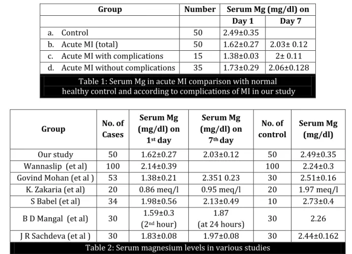
Serum Magnesium: Serum Mg of the 50 age and sex matched controls was done. It ranged from 1.88 mg/dl to 3.11mg/dl with a mean value of 2.49±0.35mg/dl.
Serum Mg of 50 acute MI cases within 24 hours ranged from 1.2 –3.3 mg/dl with a mean value of 1.62±0.27mg/dl and that on the 7th day ranged from 1.75 –2.52 mg/dl with a mean value of 2.03±0.12 mg/dl.
Mortality: In this study, 3(6%) patient died within 48 hours of admission. One patient who was known diabetic and hypertensive had acute inferior wall MI with RV infarction with serum Mg on admission being 1.2 mg/dl. Patient had developed CHB. Second patient had extensive anterior wall MI with congestive cardiac failure with admission serum Mg level being 1.33 mg/dl. Third patient had acute anteroseptal wall MI with LVF and LAHB, with admission serum Mg level being 1.41 mg/dl.
SERUM MAGNESIUM AS A PROGNOSTIC INDICATOR;
Out of 50 patients, in 41 patient’s serum Mg value was low during first 24 hours. Serum Mg levels (mean 1.628 mg/dl ) gradually raised to normal (mean 2.03 mg%) by 7th day in patients who survived. 3 patients died within 48 hrs of admission had serum Mg values of 1.2 mg%, 1.33 mg% and 1.41mg%.
Hence, serum Mg value acts as prognostic indicator and low levels act as a bad prognostic index.
DISCUSSION:
Age and Sex Distribution: In this study, maximum number of patients were seen in age group 51 -60 years. Coronary artery disease in India is said to peak between 51- 60 years of age.(3) The sex distribution in the study group was 76% male patients and 24% female. This was similar to that described in stamler J et al in 1991(4) and kedarnath et al in 1969.(5)
J of Evolution of Med and Dent Sci/ eISSN- 2278-4802, pISSN- 2278-4748/ Vol. 4/ Issue 60/ July 27, 2015 Page 10464
Risk Factors: In this study, smoking was in 22(44%) cases, where as it has been calculated that in countries where smoking has been a widespread habit, it is responsible for 25% of CAD deaths.
In the study group, diabetes mellitus was present in 17(34%) cases. CAD is responsible for 30- 50% of deaths in diabetes over the age of 40 years in industrialised countries.(3)
Total Leucocyte Count: This was found to be raised (>11000/mm3) in about 16(32%) of cases. Thomson SP et al in 1995 found and described the increase in total leucocyte and differential leucocyte count.(8)
ESR: In our study, mean ESR in acute MI was to be 22mm 1st hour. In S.Babel et al study in 1983, mean serum ESR was found to be 9.8 mm 1st hour.(9)
Serum Cholesterol: In our study, mean serum cholesterol in acute MI was found to be 220.1±0.16 mg/dl. In S. Babel et al study in 1983, mean serum cholesterol was found to be 286.3±0.81 mg/ dl.(9)
SGOT: In our study, mean serum SGOT was found to be 38.2±2.11 IU/L. in S. Babel etal study in 1983 and B.D. Mangal et al 1981 mean serum SGOT was found to be 12.8±9.54 IU/L, and 26.03 IU/L respectively.(9,10)
Serum Magnesium in Acute Myocardial Infarction: In this study, serum Mg levels were estimated in 50 cases of acute MI within 24 hours of onset and on the 7th day of MI. We observed statistically significant lower serum Mg concentration of 1.62±0.27mg/dl at onset in comparison to control mean of 2.49±0.35mg/dl. Serum Mg gradually raised to normal value by 7th day with mean value of 2.0±0.12mg/dl. Further, mean serum Mg in this study on the 1st day in complicated cases was 1.38±0.03 mg/dl and in uncomplicated cases was 1.73±0.29mg/dl and 7th day, in complicated cases 2±0.1mg/dl and uncomplicated cases was 2.06±0.12mg/dl.
Wannasilp N et al 2001 demonstrated that CAD patients may be associated with Mg deficiency and contribute to the pathogenesis of CAD.(11) Mean value of serum Mg level in 100 CAD patients was 2.14±0.39 mg/dl (p= 0.052) with 100 healthy controls (mean value 2.24± 0.3 mg/dl) in the study. The prevalence of Mg deficiency did not differ significantly between the study groups, however it tended to be higher in CAD patients.
GQ Khan et al 2002 reported statistically significant (p < 0.001) fall of serum Mg in 50 cases of acute MI with mean serum Mg in controls being 2.2±0.24 mg/dl. Further, the serum Mg level was found comparatively lower in the patients getting cardiac arrhythmias. They concluded that, the fall in serum Mg in acute MI can be taken as sensitive diagnostic index, especially in early hours of postinfarction period when cardiac enzymes and ECG may not be significant.(12)
Simmikharb et al 1999 demonstrated mean serum Mg levels in22 acute MI to be 1.27±0.57 mg/dl (p<0.001) compared to mean value in 15 controls to be 2.41±0.54 mg/dl. They concluded that Mg deficiency in MI patients can potentiate oxidative injury to post ischemic myocardium.(13)
J of Evolution of Med and Dent Sci/ eISSN- 2278-4802, pISSN- 2278-4748/ Vol. 4/ Issue 60/ July 27, 2015 Page 10465 complications raised to 2.36 ±0.12 mg/dl compared to mean serum Mg values in acute MI without complications of 2.29±0.16 mg%. It was observed that serum Mg were lowest in patients who died due to major arrhythmias and cardiogenic shock followed by pump failure.(14)
CONCLUSION: Serum Mg levels were found to be lower in acute MI cases at presentation within 24 hours as compared to matched controls. Serum magnesium levels were lower in acute MI with complication, when compared with acute MI without complication. Serum magnesium levels were very low in patients of acute MI who expired. Serum Mg levels raised towards normal values by 7th day. So it can be concluded that measurement of serum magnesium levels is of prognostic significance in acute MI.
REFERENCES:
1. Boon N.A, K.A.A. Fox, P. Bloomfield, A. Brad Bury: Cardiovascular disease in Davidson’s Principles and practice of Medicine, 19th Edition, Churchill Living Stone, 2002, Pg. 420, 422, 424, 441, 443. 2. Elliot M Antman, Eugene Braunwald: Acute Myocardial infarction in Heart Disease, 6th edition,
Philadelphia Saunders, 2001, p1114-1137, 1171-1172.
3. Parks Text book of Preventive and Social Medicine, 15th edition, 1997, p. 269-273.
4. Stamler J: Epidemiology, established major risk factors and the primary presentation of coronary heart disease . )n Chatterjee K, Cheitlin MP, Kaslines J et al Eds cardiology: an illustrated text, reference vol 2; 0-1 Philadelphia, JB Lippincott 1991.
5. Kedarnath, K.K. Sikki, B.K. Sur: Serum magnesium in clinical experimental myocardial infarction, Ind Jour Med Res Feb 1969, 57, 2, p317-323.
6. Huggins GS, OGara PT: Clinical presentation and diagnostic evaluation. In Fister V, Ross R and Topol ET (Eds): Atherosclerosisand coronary artery disease , Philadelphia, Lippincott, Raven 1996.
7. Muller JE, Tofler GH, Mittleman H: Triggering of onset of MI and sudden cardiac death. In Fister V, Ross R and Topol ET (Eds): Atherosclerosisand coronary artery , Philadelphia, Lippin cott, Raven 1996, 819- 834.
8. Thompson SG; (emostatic factors and the risk of myocardial infarction or sudden death in patients with angina pectoris , N Eng J Med ; 332; 635-641.
9. Babel S, H. N. S. Bhatnagar, L. K. Bhatnagar: Serum magnesium levels in cases of acute myocardial infarction and its prognostic significance, JAPI 1983, Vol. 31, 12, 755-757.
10. B.D. Mangal, R. Prasad, K.S. Sikka: Evaluation of serial magnesium and SGOT in acute infarction, Jr Asso Phys Ind. April 1981, vol. 29 p. 263-266.
11.Wannaslip N, Poungvarin N, Pokums: Seum magnesium in Thai coronary artery disease
patients , J Med Assoc Thai , Dec: , Suppl : -9.
12. G Q Khan, G Mohammad, G (assan; Study of serum magnesium in acute myocardial infarction , J Asso Phys Ind Jan 2002, vol. 50, p101.
13.Simmikharb, Veena Singh: Magnesium deficiency potentiates free radical production associated
with myocardial infarction , J Asso Phys )nd ; ; -485.
J of Evolution of Med and Dent Sci/ eISSN- 2278-4802, pISSN- 2278-4748/ Vol. 4/ Issue 60/ July 27, 2015 Page 10466
Group Number Serum Mg (mg/dl) on Day 1 Day 7
a. Control 50 2.49±0.35
b. Acute MI (total) 50 1.62±0.27 2.03± 0.12
c. Acute MI with complications 15 1.38±0.03 2± 0.11 d. Acute MI without complications 35 1.73±0.29 2.06±0.128
Table 1: Serum Mg in acute MI comparison with normal healthy control and according to complications of MI in our study
Group No. of Cases
Serum Mg (mg/dl) on
1st day
Serum Mg (mg/dl) on
7th day
No. of control
Serum Mg (mg/dl)
Our study 50 1.62±0.27 2.03±0.12 50 2.49±0.35
Wannaslip (et al) 100 2.14±0.39 100 2.24±0.3
Govind Mohan (et al ) 53 1.38±0.21 2.351 0.23 30 2.51±0.16 K. Zakaria (et al) 20 0.86 meq/l 0.95 meq/l 20 1.97 meq/l
S Babel (et al) 34 1.98±0.56 2.13±0.49 10 2.73±0.4
B D Mangal (et al) 30 1.59±0.3 (2nd hour)
1.87
(at 24 hours) 30 2.26
J R Sachdeva (et al ) 30 1.83±0.08 1.97±0.08 30 2.44±0.162 Table 2: Serum magnesium levels in various studies
TABLES SHOWING THE STATISTICAL DIFFERENCE BETWEEN TWO GROUPS:
Group Mean value Z- value P value Comment
Control 2.49
13.66 < 0.001 Highly significant Cases within 24 hours 1.62
Table 3
Group Mean value Z- value P value Comment
Control 2.49
8.63 < 0.001 Highly significant Cases on 7th day 2.03
Table 4
Group Mean value Z - value P value Comment
Cases within 24 hours 1.64
15.70 < 0.001 Highly significant Cases within 7th day 2.03
J of Evolution of Med and Dent Sci/ eISSN- 2278-4802, pISSN- 2278-4748/ Vol. 4/ Issue 60/ July 27, 2015 Page 10467 AUTHORS:
1. Nambakam Tanuja Subramanyam
2. Girish P. Vakrani
PARTICULARS OF CONTRIBUTORS:
1. Assistant Professor, Department of General Medicine, Vydehi Institute of Medical Sciences and Research Centre. 2. Assistant Professor, Department of
Nephrology, Vydehi Institute of Medical Sciences and Research Centre.
FINANCIAL OR OTHER
COMPETING INTERESTS: None
NAME ADDRESS EMAIL ID OF THE CORRESPONDING AUTHOR:
Dr. Nambakam Tanuja Subramanyam, A-29, Vydehi Hospital Staff Quarters, # 82, EPIP Area, Whitefield,
Bangalore-66, India.
Email: [email protected]