Universidade Federal de Minas Gerais
Programa de Pós-Graduação em Ciências da Saúde
Avaliação dos níveis urinários de citocinas
pró-inflamatórias e do fator de crescimento e
transformação do tipo beta em pacientes com
hidronefrose diagnosticada intraútero
MARIANA AFFONSO VASCONCELOS
MARIANA AFFONSO VASCONCELOS
Avaliação dos níveis urinários de citocinas pró-inflamatórias e do fator de
crescimento e transformação do tipo beta em pacientes com hidronefrose
diagnosticada intraútero
Dissertação apresentada ao Programa de Pós-Graduação da Faculdade de Medicina da Universidade Federal de Minas Gerais, área de concentração em saúde da Criança e do Adolescente
Orientador: Eduardo Araújo de Oliveira
Co-Orientadora: Ana Cristina Simões e Silva
UNIVERSIDADE FEDERAL DE MINAS GERAIS FACULDADE DE MEDICINA
PÓS-GRADUAÇÃO EM CIÊNCIAS DA SAÚDE
ÁREA DE CONCENTRAÇÃO: SAÚDE DA CRIANÇA E DO ADOLESCENTE
UNIVERSIDADE FEDERAL DE MINAS GERAIS
Reitor: Professor Ronaldo Tadêu Pena
Vice-Reitora: Professora Heloisa Maria Murgel Starling
Pró-Reitora de Pós-Graduação: Professora Elizabeth Ribeiro da Silva Pró-Reitor de Pesquisa: Professor Carlos Alberto Pereira Tavares
FACULDADE DE MEDICINA:
Diretor: Professor Francisco José Penna Vice-Diretor: Professor Tarcizo Afonso Nunes
PROGRAMA DE PÓS-GRADUAÇÃO EM CIÊNCIAS DA SAÚDE – ÁREA DE
CONCENTRAÇÃO EM SAÚDE DA CRIANÇA E DO ADOLESCENTE:
Coordenador: Professor Joel Alves Lamounier
Subcoordenadora: Professora Ana Cristina Simões e Silva
COLEGIADO DO PROGRAMA DE PÓS-GRADUAÇÃO EM CIÊNCIAS DA SAÚDE –
ÁREA DE CONCENTRAÇÃO EM SAÚDE DA CRIANÇA E DO ADOLESCENTE:
Professor Ivani Novato Silva Professor Jorge Andrade Pinto
Professora Lúcia Maria Horta Figueiredo Goulart Professora Maria Cândida Ferrarez Bouzada Viana Professor Marco Antônio Duarte
Professora Regina Lunardi Rocha
AGRADECIMENTOS
__________________________________________________________________
Ao professor Eduardo Araújo Oliveira, pelos incentivos, desde a graduação, a seguir a carreira acadêmica, pelos ensinamentos, paciência e confiança.
À professora Ana Cristina Simões e Silva por estar sempre presente, pelo apoio, compreensão e pelo detalhismo na revisão da dissertação.
Ao professor José Maria Penido Silva, meu mestre, meu muito obrigada por acreditar no meu potencial e por ter me apoiado em momentos difíceis da carreira.
À professora Maria Candida Ferrarez Bouzada Viana, pela dedicado cuidado clínico dos pacientes e pela coordenação do projeto junto às fontes financiadoras.
À equipe de nefrologia pediátrica, em especial, à professora Eleonora Moreira Lima pelos ensinamentos.
À doutora Roberta Maia de Castro Romanelli pela amizade e por sempre me estimular a seguir meus ideais.
Às acadêmicas Flávia, Juliana e Letícia, pelo bom desempenho nas tarefas atribuídas. À pesquisadora Kátia Daniela Silveira, sempre disposta, pela a ajuda incondicional nas dosagens.
Aos funcionários do laboratório central que cederam espaço e auxiliaram nas coletas. À minha irmã Marisa por me despertar o interesse pela pesquisa.
Ao meu irmão Daniel pelo carinho.
À minha avó Hercília, uma fortaleza, exemplo de pessoa.
Aos pacientes que participaram do estudo, sem eles essa dissertação não seria possível. A todas as pessoas que, direta ou indiretamente, contribuíram para a execução dessa dissertação de mestrado.
APOIO:
Este projeto foi aprovado pelo edital Universal 2007 da FAPEMIG, sob a coordenação da Professora Maria Candida Ferrarez Bouzada Viana
LISTA DE ABREVIATURAS E SIGLAS
_________________________________________________
- AUC: area under the curve.
- CCL2/MCP-1: Monocyte chemotactic protein-1
- CCL3/MIP-1α: Macrophage inflammatory protein 1 alfa
- CCL5/RANTES: regulated on activation, normal T Expressed and Secreted.
- CEMEFE: Centro de Medicina Fetal.
- COEP: Comitê de Ética em Pesquisa.
- DAP: diâmetro anteroposterior.
- DMSA: dimercaptosuccicinic acid/ cintilografia estática.
- DTPA: dietilenoaminopentacetic acid/ cintilografia dinâmica.
- ELISA: enzyme-linked immunoassay/ ensaio imunoenzimático.
- IGF-1: insulin-growth factor-1
- IL-1: interleucina-1/ interleukin-1.
- IL-6: interleucina-6/ interleukin-6.
- IL-8/CXCL-8: interleucina-8/ interleukin-8.
- IP-10: ɣ-interferon-inducible protein-10. - JUP: junção ureteropélvica.
- MIP-2: macrophage inflammatory protein-2.
- mRNA: RNA mensageiro
- NGF: nerve growth factor
- PNH: prenatal hydronephrosis.
- ROC: Receiver-operating curve.
- RPD: renal pelvic dilatation.
- RVU: refluxo vesicoureteral.
- SFU: Society of Fetal Urology.
- TGF-β1: fator de crescimento e transformação-β1/ transforming and growth factor-β1
- UFMG: Universidade Federal de Minas Gerais
- UCM: uretrocistografia miccional.
- UE: urografia excretora
- UPJO: ureteropelvic junction obstruction.
- US: ultra-sonografia/ ultrasound
- UTI: urinary tract infection.
- VCUG: voiding cystourethrogram.
- VEGF: vascular endothelial growth factor.
- VUP: válvula de uretra posterior.
SUMÁRIO
_________________________________________________
1 – Introdução --- 11
2 – Artigo de revisão --- 22
Artigo: Cytokines in congenital anomalies of kidney and urinary tract 3 – Objetivos --- 54
4- Pacientes e Métodos --- 55
4.1 – Delineamento, população e local de estudo--- 55
4.1.1 – Período do estudo--- 55
4.1.2 – Critérios de inclusão--- 55
4.1.3 – Critérios de exclusão--- 55
4.1.4 – Aspectos éticos--- 55
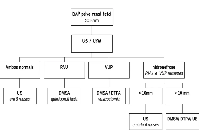
4.2 – Definições e classificação da dilatação da pelve renal--- 56
4.3 – Investigação clinica e por imagens de fetos e recém-nascidos portadores de dilatação da pelve renal 58 4.4 – Determinação das concentrações urinárias de citocinas e creatinina 66 4.5 – Conduta clínica 69 4.6 – Análise Estatística --- 70
1.
INTRODUÇÃO
_________________________________________________
O diagnóstico intrauterino é definido, segundo a OMS, como "todas aquelas ações no pré-natal que tenham como objetivo o diagnóstico de um defeito congênito, entendido como toda anomalia do desenvolvimento morfológico, estrutural, funcional ou molecular presentes ao nascimento (ainda que possa manifestar tardiamente), externa ou interna, familiar ou esporádica, hereditária ou não, única ou múltipla" 1. As anormalidades envolvendo o trato geniturinário podem ser suspeitadas em 1 a cada 100 gestações, dependendo do critério adotado 2-3, 5. A dilatação da pelve renal é a uropatia mais comum detectada intraútero 4.
Ressalte-se que dentre as etiologias da insuficiência renal crônica, são as uropatias, possivelmente, as únicas em que o tratamento precoce, evitando uma sobrecarga de pressão e episódios infecciosos sobre o parênquima renal, pode prevenir ou adiar uma perda da função renal 7. Modelos experimentais foram utilizados para demonstrar essa possível prevenção do dano renal com o tratamento precoce das uropatias obstrutivas. Em uma série de estudos, em fetos de carneiro, foi demonstrada que a obstrução no primeiro trimestre de gestação resulta em marcante fibrose intersticial e uma desorganização do parênquima características dos quadros de displasia. A recuperação da função renal foi diretamente proporcional ao tempo de alívio da obstrução e inversamente proporcional ao tempo que o rim permaneceu obstruído 14-17.
Poucos estudos abordam os fatores associados à evolução da função renal que, em última análise, serão os determinantes de uma boa qualidade de vida para essas crianças. Neste estudo, vários fatores relacionados ao ultrassom pré-natal, às condições maternas e do recém-nascido foram sistematicamente obtidos durante o seguimento desses pacientes. Esses fatores também têm sido relacionados a eventos adversos, tais como insuficiência renal e óbito 12, 13.
interleucina-6 (IL-6) e IL-8/CXCL-8 tem sido observada em pacientes com infecção urinaria 41-44. Haraoka et al 45 demonstraram que os níveis urinários de IL-8/CXCL8 eram maiores em crianças com refluxo vesicoureteral e dano renal. Outros marcadores também têm sido utilizados nas anomalias do trato urinário. O fator de transformação e crescimento do tipo beta (TGF-β1) 1 é um mediador de fibrose renal em uropatia obstrutiva. O TGF-β1 é uma citocina que estimula a síntese de matriz extracelular e inibe sua degradação 48. A fibrose intersticial desenvolve como resultado de uma ausência de balanço entre a síntese de matriz extracelular, seu depósito e degradação 48. O sistema renina-angiotensina (RAS) encontra-se ativado após o início de obstrução ureteral, e a angiotensina II pode contribuir para a perda precoce do rim obstruído 47. Angiotensina II induz diretamente e indiretamente a produção de TGF-β1 47. A expressão do RNA mensageiro (mRNA) para TGF-beta1 aumenta em rins obstruídos 48. Alguns autores têm demonstrado que há um consistente aumento de TGF-β1 em crianças com uropatia obstrutiva 49-51. No entanto, não há estudos de lactentes portadores de uropatia identificada por meio da detecção de hidronefrose fetal.
A proposta deste estudo transversal, desenvolvido pela Unidade de Nefrologia Pediátrica do Departamento de Pediatria da UFMG foi de avaliar marcadores capazes de auxiliar no diagnóstico e na definição do prognóstico de pacientes com hidronefrose diagnosticada intraútero e assim facilitar o seu manejo clínico e cirúrgico.
científicos a serem submetidos a revistas médicas. Sendo assim, a apresentação do trabalho seguiu a seguinte estrutura:
1. Seção de Introdução
2. Seção de Revisão da Literatura, apresentada sob a forma do artigo de revisão: Cytokines in congenital anomalies of kidney and urinary tract.
3. Seção de Objetivos 4. Seção de Metodologia
5. Seção de Resultados e Discussão, apresentada sob a forma do artigo original: Urinary levels of transforming growth factor beta-1 and interleukins in patients with prenatally detected uropathies.
Observações:
As tabelas e figuras estão dispostas no final de cada artigo.
REFERÊNCIAS BIBLIOGRÁFICAS
_________________________________________________
1. Carrera JM, Alegre M, Torrents M. Anomalías nefrourológicas. In: Carrera JM, ed. Diagnóstico prenatal. Barcelona: Salvat Editores, 1987:465-483.
2. Carr MC. Prenatal management of urogenital disorders. Urol Clin North Am. 2004 Aug;31:389-97, vii.
3. Morin L, Cendron M, Crombleholme TM, Garmel SH, Klauber GT, D'Alton ME. Minimal hydronephrosis in the fetus: clinical significance and implications for management. J Urol. 1996 Jun;155:2047-9.
4. Chudleigh T. Mild pyelectasis. Prenat Diagn. 2001 Nov;21:936-41.5.
5. Goncalves LF, Jeanty P, Piper JM. The accuracy of prenatal ultrasonography in detecting congenital anomalies. Am J Obstet Gynecol 1994; 171:1606-12.
6. Colodny AH. Antenatal diagnosis and management of urinary abnormalities. Pediatr Clin North Am 1987; 34:1365-81.
7. Oliveira EA. Estudo prospectivo das anomalias do trato urinário diagnosticadas no feto pelo ultra-som. Depto. de Pediatria. Belo Horizonte: UFMG, 1992:260.
8. Oliveira EA. Fatores prognósticos do óbito, da insuficiência renal e da presença de obstrução uretral: uma análise multivariada. Depto. de Pediatria. Belo Horizonte: UFMG, 1998:263.
9. Gusmano R, Perfumo F. Worldwide demographic aspects of chronic renal failure in children. Kidney Int Suppl 1993; 41:S31-5.
11. Garcia C, Goldani J, Garcia V. Paediatric dialysis and renal transplantation in the state of Rio Grande do Sul, Brazil. Pediatr Nephrol 1992; 6:74-7.
12. Simoes e Silva AC, Silva JM, Diniz JS, Pinheiro SV, Lima EM, Vasconcelos MA, Pimenta MR, Oliveira EA. Risk of hypertension in primary vesicoureteral reflux. Pediatr Nephrol. 2007 Mar; 22:459-62.
13. Penido Silva JM, Oliveira EA, Diniz JS, Bouzada MC, Vergara RM, Souza BC. Clinical course of prenatally detected primary vesicoureteral reflux.Pediatr Nephrol. 2006 Jan;21:86-91
14. Harrison MR, Golbus MS, Filly RA, et al. Fetal surgery for congenital hydronephrosis. N Engl J Med 1982; 306:591-3.
15. Harrison MR, Golbus MS, Filly RA, et al. Fetal hydronephrosis: selection and surgical repair. J Pediatr Surg 1987; 22:556-8.
16. Harrison MR, Nakayama DK, Noall R, de Lorimier AA. Correction of congenital hydronephrosis in utero II. Decompression reverses the effects of obstruction on the fetal lung and urinary tract. J Pediatr Surg 1982; 17:965-74.
17. Harrison MR, Ross N, Noall R, de Lorimier AA. Correction of congenital hydronephrosis in utero. I. The model: fetal urethral obstruction produces hydronephrosis and pulmonary hypoplasia in fetal lambs. J Pediatr Surg 1983; 18:247-56.
19. Oliveira EA, Cabral ACV, Leite HV, et al. Estudo prospectivo da hidronefrose fetal diagnosticada por ultra-som: uma contribuição na prevenção ao dano renal na infância. Radiol Bras 1998; 31:75-81.
20. Oliveira EA, Cabral ACV, Leite HV, et al. Hidronefrose fetal: abordagem pós-natal e seguimento. Jornal de Pediatria 1997; 73:252-8.
21. Oliveira EA, Diniz JS, Cabral AC, et al. Prognostic factors in fetal hydronephrosis: a multivariate analysis. Pediatr Nephrol 1999; 13:859-64.
22. Oliveira EA, Diniz JS, Cabral AC, et al. Prognostic factors in fetal hydronephrosis: a multivariate analysi. Pediatr Nephrol 1999; 13:859-64.
23. Oliveira EA, Diniz JS, Cabral AC, et al. Predictive factors of fetal urethral obstruction: a multivariate analysis. Fetal Diagn Ther 2000; 15:180-6.
24. Oliveira EA, Diniz JS, Rabelo EA, et al. Primary megaureter detected by prenatal ultrasonography: conservative management and prolonged follow-up. Int Urol Nephrol 2000; 32:13-8.
25. Oliveira EA, Diniz JS, Rabelo EA, et al. Primary megaureter detected by prenatal ultrasonography: conservative management and prolonged follow-up [In Process Citation]. Int Urol Nephrol 2000; 32:13-8.
26. Oliveira EA, Diniz JS, Silva JM, Rabelo EA, Pontes AK, Souza MF. Features of primary vesicoureteric reflux detected by investigation of fetal hydronephrosis. Int Urol Nephrol 1998; 30:535-41.
28. Oliveira EA, Diniz JSS, Rabelo EAS, et al. Seguimento a longo prazo da válvula de uretra posterior detectada intra-útero. J Bras Nefrol 2000.
29. Oliveira EA, Diniz JSS, Rabelo EAS, et al. Primary megaureter detected by prenatal ultrasonography: conservative management and prolonged follow-up. Int Urol Nephrol 2000.
30. Oliveira EA, Diniz JSS, Vilasboas AS, Rabelo EAS, Silva JMP, Filgueiras MTF. Multicystic dysplastic kidney detected by fetal sonography: conservative management and follow-up. Pediatr Surg Int 2001; 17:54-7.
31. Oliveira EA, Rabelo EA, Pereira AK, et al. Prognostic factors in prenatally-detected posterior urethral valves: a multivariate analysis. Pediatr Surg Int 2002; 18:662-7. 32. Oliveira EA, Silva AS, Rabelo EA, Filgueiras MT, Pereira AK, Mesquita FM.
Spontaneous improvement of hypertension in multicystic dysplastic kidney: a case report. Pediatr Nephrol 2002; 17:954-958.
33. Penido Silva JM, Oliveira EA, Diniz JS, Bouzada MC, Vergara RM, Souza BC. Clinical course of prenatally detected primary vesicoureteral reflux. Pediatr Nephrol 2006; 21:86-91.
34. Pereira AK. Uropatias fetais: avaliação dos critérios de diagnóstico morfológico e funcional. Tese de Doutorado. Depto. de Ginecologia e Obstetrícia. Belo Horizonte: UFMG, 1999:160.
35. Rabelo EA, Oliveira EA, Diniz JS, et al. Natural history of multicystic kidney conservatively managed: a prospective study. Pediatr Nephrol 2004; 19:1102-7. 36. Rabelo EA, Oliveira EA, Silva GS, Pezzuti IL, Tatsuo ES. Predictive factors of
37. Rabelo EA, Oliveira EA, Silva JM, et al. [Conservative management of multicystic dysplastic kidney: clinical course and ultrasound outcome]. J Pediatr (Rio J) 2005; 81:400-4.
38. Apocalypse GT, Oliveira EA, Rabelo EA, et al. Outcome of apparent ureteropelvic junction obstruction identified by investigation of fetal hydronephrosis. Int Urol Nephrol 2003; 35:441-8.
39. Apocalypse GT. Obstrução de junção ureteropélvica identificada na investigação de hidronefrose fetal: um estudo retrospectivo. Faculdade de Medicina. Belo Horizonte: UFMG, 2002:122.
40. Bouzada MC, Oliveira EA, Pereira AK, et al. Diagnostic accuracy of postnatal renal pelvic diameter as a predictor of uropathy: a prospective study. Pediatr Radiol 2004; 34:798-804.
41. Bouzada MC, Oliveira EA, Pereira AK, et al. Diagnostic accuracy of fetal renal pelvis anteroposterior diameter as a predictor of uropathy: a prospective study. Ultrasound Obstet Gynecol 2004; 24:745-9.
42. Galanakis E, Bitsori M, Dimitriou H, Giannakopoulou C, Karkavitsas NS, Kalmanti M. Urine Interleukin-8 as a Marker of Vesicoureteral Reflux in Infants. Pediatrics 2006.
43. Sedor JR, Nakazato Y, Konieczkowski M. Interleukin-1 and the mesangial cell. Kidney Int 1992; 41:595-9.
45. Martins SM, Darlin DJ, Lad PM, Zimmern PE. Interleukin-1B: a clinically relevant urinary marker. J Urol 1994; 151:1198-201.
46. Haraoka M, Senoh K, Ogata N, Furukawa M, Matsumoto T, Kumazawa J. Elevated interleukin-8 levels in the urine of children with renal scarring and/or vesicoureteral reflux. J Urol 1996; 155:678-80.
47. Klahr S. Urinary tract obstruction. Semin Nephrol 2001; 21:133-45.
48. Vuruskan H, Caliskan Z, Kordan Y, Ozakin C, Yavascaoglu I, Oktay B. Elevated plasma concentrations of transforming growth factor-beta 1 in patients with unilateral ureteral obstruction. Urol Res 2005; 33:465-9.
49. Roberts AB, McCune BK, Sporn MB. TGF-beta: regulation of extracellular matrix. Kidney Int 1992; 41:557-9.
50. El-Sherbiny MT, Mousa OM, Shokeir AA, Ghoneim MA. Role of urinary transforming growth factor-beta1 concentration in the diagnosis of upper urinary tract obstruction in children. J Urol 2002; 168:1798-800.
51. Monga M, Gabal-Shehab LL, Stein P. Urinary transforming growth factor-beta1 levels correlate with bladder outlet obstruction. Int J Urol 2001; 8:487-9.
2.
ARTIGO DE REVISÃO
_________________________________________________
Review article:
Cytokines in congenital anomalies of kidney and urinary tract
Mariana A. Vasconcelos, MD, Ana C. Simões e Silva, MD, PhD, Eduardo A. Oliveira, MD, PhD
Pediatric Nephrourology Unit, Department of Pediatrics, Federal University of Minas Gerais (UFMG), Belo Horizonte, MG, Brazil
Corresponding author: Eduardo A. Oliveira.
Rua Engenheiro Amaro Lanari 389 / 501 Belo Horizonte - Minas Gerais
Postal Code: 30.310.580
Abstract
Introduction
Fetal hydronephrosis is the most common anomaly detected on antenatal ultrasound, affecting 1–5% of pregnancies1-2. Despite their high frequency of occurrence, there is little consensus on the management of infants with prenatal hydronephrosis (PNH). In particular, the management of those with isolated antenatal hydronephrosis is still a source of controversy3.
There has been a number of studies discussing the significance of fetal renal pelvic dilatation (RPD) as an indicator of urinary tract anomalies4-7. The degree of PNH varies from mild to severe, and possibly, the degree of PNH should correlate with the severity of the underlying etiology1-2. Recently, Lee et al.2 demonstrated in a meta-analysis that the risk of any postnatal pathology per degree of PNH was 11.9% for mild, 45.1% for moderate, and 88.3% for severe. More specifically, the risk of ureteropelvic junction obstruction (UPJO) increased significantly with greater degrees of PNH, but the risk of vesicoureteral reflux (VUR) was not significantly different among all severity groups. Most studies also have shown that a single postnatal US is unable to predict the presence or severity of VUR 6, 8-10. Consequently, postnatal management is heterogeneous, with some centers advocating detailed investigations including voiding cystourethrography (VCUG) in all cases and others indicating a less intensive approach11-16. Therefore, in spite of advances, the issue of postnatal diagnostic management of antenatal hydronephrosis remains a challenging problem17-18.
urologist or pediatric nephrologist before the baby is even born, with a presumptive diagnosis rather than a symptom19. It is the reason why infants diagnosed with congenital hydronephrosis must routinely undergo postnatal imaging evaluation. Classically, the prenatal diagnosis of hydronephrosis leads to postnatal investigations, including sonography, VCUG and isotopic renography17, 20. Ultrasonography has typically assumed a more important role in the surveillance of children with hydronephrosis but this exam is not used alone to establish the presence of obstruction. Presently diuretic renography is the most widely used investigation to evaluate renal function and urine flow. However, the test is invasive and the use of ionizing radiation remains a concern, especially in infants and young children. Moreover, repeat tests are often required, especially when the excretion curve is equivocal 21. Postnatal investigation has the major aim in detecting infants with severe urinary tract obstruction and clinically significant urinary tract anomalies among the heterogeneous universe of patients. Imaging techniques clearly contributes to this purpose. However, some of these exams are invasive and very expensive. Furthermore, sometimes imaging techniques are not sufficient to precisely define the indication of surgical approach as well as to determine the prognosis 21.
The obstructive nephropathy is not a simple result of mechanical impairment to urine flow but a complex syndrome that results in alterations of both glomerular hemodynamics and tubular function usually caused by the interaction of a variety of vasoactive factors and cytokines that are activated in response to obstruction. The cytokines play a role in the development and progression of fibrotic and sclerotic changes in the obstructed kidney 25. A large numbers of events can initiate apoptosis, several of which may be related to obstructive nephropathy, such as hypoxia, ischemia, cytokines, growth factors, angiotensin II and mechanical stretch 26. However, it should be pointed out that the biochemical, cellular and molecular mechanisms of the obstructive uropathies are still largely unknown 26, 27. The comprehension of this process will certainly help in the management of fetal hydronephrosis. In this regard, recent studies suggest a role for cytokines and chemokines in the pathophysiology of fetal hydronephrosis 26, 27, 28. Indeed, the evaluation of these inflammatory mediators might help the management of postnatal uropathies. The aim of this article is to revise general aspects of cytokines and their relationship with fetal hydronephrosis by including experimental and clinical studies.
Cytokines: general concepts and characteristics
express their optimal function. Numerous cytokines have both inflammatory and anti-inflammatory properties 29.
Chemokines constitute a large family of low molecular-weight cytokines whose main action is the recruitment and activation of leukocyte subsets in various models of inflammation—the word “chemokine” is a contraction of the terms “chemoattractant” and “cytokine” 30.
Cytokines in renal diseases
A number of studies have shown the relation between renal diseases and cytokines production 26, 30, 33, 34, 35, 36. Indeed, the measurement of urinary, plasma and renal tissue levels of cytokines has been used to monitor and diagnosis various urological and nephrological diseases 30, 36, 37.
Tubular epithelial cells can be a rich source of inflammatory chemokines including CCL5/RANTES (Regulated on activation, normal T Expressed and Secreted), CCL2/MCP-1 (Monocyte chemotactic protein-CCL2/MCP-1), CCL3/MIP-CCL2/MCP-1α (Macrophage inflammatory protein 1 alfa), CX3CL1/fractalkine and CKCL8/IL8 (Interleukin 8) 31. Tubular epithelial cells are also targets for chemokines, since these cells respond to CCL2/MCP1 stimulation by releasing interleukin-6 (IL-6) and intracellular adhesion molecule-1 32. Messenger RNA of chemokines receptors can also be detected in other regions like podocytes and glomeruli 30
levels of IL1-β were significantly reduced in children with renal scarring, probably indicating a protective function 34.
Interleukin-8 (IL-8) is a chemokine responsible for neutrophil infiltration into the urinary tract with an important role in acute inflammation 35. Gene polymorphisms of IL-8 seem to increase the susceptibility for acute pyelonephritis. For instance, the presence of the IL-8-251A allele in the genotype of children with urinary tract infection without vesicoureteral reflux has increased the risk of pyelonephritis 38.
Interleukin-6 (IL-6) mediates T-cell activation, growth and differentiation. This pro-inflammatory cytokine is responsible for the induction of pyrexia and production of acute phase proteins 29. Sheu et al. 35 found that there is a significant elevation of serum and urinary levels of IL-6 and IL-8 in children with acute pyelonephritis when compared to children with lower urinary tract infection. This finding is consistent with the hypothesis that the release of IL-6 from the urinary tract leads to systemic host responses 35.
Transforming growth factor-β (TGF-β) is a fibrogenic cytokine that stimulates extracellular matrix proteins deposition and renal scarring formation. On the other hand, concerning immune system regulation, TGF-β exerts anti-inflammatory effects by inhibiting the proliferation of many different cell types 29. Monga et al. 39 have studied 17 men with bladder outlet obstruction and 6 non-obstructed subjects and showed that, in the obstructed ones, the urinary levels of TGF- β were significantly higher than in non-obstructed.
Cytokines in hydronephrosis – Experimental studies
majority of the reported animal models utilized rats and mice, but rabbits, pigs and sheep were also used 27.
Models of experimental postnatal unilateral ureteral obstruction have been developed in newborn rat pups that continue to exhibit active nephrogenesis in the postnatal period 27. A partial unilateral ureteral obstruction was surgically created by entrapping the ureter in the animal psoas muscle, whereas the complete obstruction was produced by surgically campling and occluding the ureter 27. In rats, the major part of nephrogenesis occurs within 7 to 10 days after birth 43, 44. Some models have used animals with congenital uropathies, while others have evaluated animals submitted to surgery after birth 43.
The induction of ureteral obstruction in newborn rats clearly interferes with ongoing nephrogenesis and this procedure usually leads to substantial renal damage 43. This kind of experimental model mimics human ureteral obstruction at the second and third trimester of pregnancy; however, significant renal damage is less common in infants 45.
The main features found in obstructive models are tubular cell apoptosis, mesenchymal myocyte transformation and decreased glomerular endowment and glomerular injury 26, 44,
46
. The understanding of the pathophysiolocal mechanisms and the molecular events is important to define the moment of intervention. 27. Figure 1 shows the main mechanisms involved in obstructive models.
Obstructed kidneys exhibited an elevation in Angiotensin II activity, which, in turn, decreases renal blood, causes ischemia and kidney growth arrest. Although, renal blood flow usually normalizes 6 weeks after the relief of temporary obstruction, renal growth remains altered, suggesting that other factors are responsible for growth impairment 43 such
as the reduction in cell proliferation, the increase in cell apoptosis and the progression of interstitial fibrosis 44.
Chevalier et al 44 have studied neonatal rats submitted to unilateral ureteral obstruction or sham operation at one day of age, with relief five days later. In additional groups of neonatal rats, the operation was at 14 days, with relief at 19 days 44. Three months following relief of unilateral ureteral obstruction during days 14 to 19, renal growth was decreased by 50%, compared to a 30% reduction following relief of unilateral ureteral obstruction during days 1 to 5. The number of glomeruli was reduced by approximately 50% regardless of the timing of obstruction, but glomerular size was reduced only in rats with unilateral ureteral obstruction from days 14 to 19 44. This study shows that, in the period immediately following nephrogenesis, the kidney is particularly susceptible to long-term injury from temporary unilateral obstruction. This suggests that a delay in relief of significant ureteral obstruction should be avoided if diagnosed in the perinatal or neonatal period 44. The same group has also evaluated neonatal rats that underwent unilateral ureteral obstruction at one day of age whose obstruction was released at days 1, 2, 3 or 5 following the operation 47. The growth of the obstructed kidney decreased linearly according to the duration of ureteral obstruction, while the contralateral kidney developed compensatory hypertrophy 47. Indeed, contralateral renal hypertrophy should be considered as an important sign of advanced obstructive uropathy 48. In summary, these animal models reveal that renal growth and function are impaired in proportion to the severity and duration of obstruction.
continuous, apoptosis and proliferation of fibroblasts and inflammatory cells 49, 50. Tubular cell apoptosis contributes to renal growth impairment 49, whereas proliferation of interstitial fibroblasts with myofibroblast transformation leads to excess deposition of the extracellular matrix and renal fibrosis 46. Phenotypic transition of resident renal tubular cells, endothelial cells, and pericytes has also been implicated in this process.
A variety of intrarenal factors lead to progressive interstitial fibrosis, including growth factors and cytokines, such as Angiotensin II, MCP-1, TGF- β and adhesion molecules, which are produced by the hydronephrotic kidney 26. Altered renal expression of growth factors and cytokines modulate cell death by apoptosis or phenotypic transition of glomerular, tubular, and vascular cells. Mediators of cellular injury include hypoxia, ischemia, and reactive oxygen species, while fibroblasts undergo myofibroblast transformation with increased deposition of extracellular matrix. On the other hand, a number of endogenous antifibrotic counter-regulatory molecules have been identified, opening the possibility of enhancing the kidney's own defenses against progressive fibrosis
26, 51
.
Cytokines as TGF-β and TNF-α and chemokines like MCP-1, RANTES, macrophage inflammatory protein-2 (MIP-2) and ɣ-interferon-inducible protein (IP-10) have been evaluated in experimental hydronephrosis 25, 26, 27, 28.
fibrotic changes of interstitial regions in kidneys of mice subjected to unilateral ureteral obstruction. Accordingly, Seseke et al. 46 also detected the association between interstitial fibrosis and increased renal expression of TGF-β mRNA in an inbred strain of rats with congenital hydronefrosis. In addition, Zhou et al. 48 reported a marked elevation of renal TGF-β level in parallel to fibrotic changes of congenital and surgical ureteral obstruction in rats. Indeed, TGF-β expression increased significantly after completing nephrogenesis 43.
The role of TGF-β in obstructive nephropathies was also evidenced in other animal species. Seremites and Maizels 52 have studied rabbit pups submitted to left partial ureteral constriction and human specimens of renal pelvis and ureter derived from cases of isolated renal obstruction managed by pyeloplasty and nepherectomy or of isolated vesicoureteral reflux managed by ureteral reimplantation. These authors have detected significantly higher expression of TGF-β mRNA in obstructed pelvis than in non-obstructed ones. This elevation in TGF-β mRNA expression was correlated to muscle hypertrophy and increased collagen deposition, both representing the process of renal pelvis remodeling in response to obstruction. The lower level of TGF-β mRNA expression may be a sign of less remodeling due to a steady state of obstruction. The expression of TGF-β mRNA emerges as a good predictor of early obstruction 52.
TNF-α may play a role in initiating tubulointerstitial injury in obstructed kidney 26. TNF-α stimulates the production of chemotactic factors by resident cells and upregulates MCP-1 in human mesangial cells 26. The increase of TNF-α at early stages of obstruction stimulates the production of chemoattractants for monocytes, which in turn contributes to leukocyte infiltration in obstructed kidneys 26. Misseri et al. 57 have studied the expression of TNF-α mRNA in rats submitted to progressive degrees of left ureteral obstruction. Renal cortical TNF-α mRNA expression and protein production reached a peak at 3 days of ureteral obstruction. The TNF-α production, localized primarily to renal cortical cells, was not associated with significant inflammatory cell infiltrate 57. Indeed, TNF-α might participate in initiating tubulointerstitial injury in the obstructed kidney by upregulating chemoattractants for monocytes and by producing leukocytes infiltration 28.
MCP-1 is an inflammatory chemokine that attracts and activates monocytes, T-cells and natural killer cells 29, 30. Stephan et al 45 produced partial or complete ureteral obstruction in 28-day-old Wistar rats. These authors found that MCP-1 mRNA expression was moderately increased in partial ureteral obstruction, whereas kidneys without significant damage did not show any up-regulation. The study qualifies MCP-1 mRNA expression as a prognostic marker of partial ureteral obstruction 45.
expression of MCP-1, RANTES and IP-10 at 1 day of unilateral ureteral obstruction in mice. At 7 days, RANTES became the most abundant chemokine in the obstructed kidney and the cortical tubular cells significantly contributed to this elevation 59.
In an experimental model of congenital hydronephrosis, the alteration was observed in 75% of transgenic animals with overexpression of IL-9 and was dependent on the presence of IL-4 and IL-13 60.
The study of cytokines in hydronephrosis might provide new insights for the treatment or novel ways to blunt renal damage in obstructive nephropathy. For instance, animals with right ureter obstruction treated with spironolactona exhibited less fibrosis than control group 42. Since Angiotensin II contributes at least in part to the increased expression of TNF-α mRNA in obstructed kidney 26, the use of Angiotensin converting enzyme inhibitors emerges as an effective way in preventing renal fibrosis 40. Another rational approach to blunt renal fibrosis is to block growth factors effects. In this regard, Isaka et al
54
showed that interstitial fibrosis could be blocked by TGF-β1 antisense oligodexoxynuceloutides. Additionally, the modulation of nitric oxide, epidermal growth factor (EGF) and hepatocyte growth factor seems to be a good strategy to treat obstructive nephropathy in the future 51, 53, 61.
Cytokines in hydronephrosis - Clinical studies
It should be pointed out that few data about the role of cytokines in hydronephrosis were provided by clinical studies and the majority of them evaluated ureteropelvic junction obstruction (UPJO) and vesicoureteral reflux (VUR).
Ureteropelvic junction obstruction
ureter and renal pelvis or extrinsic compression by an accessory lower pole artery of the kidney 20. The degrees of hydronephrosis vary among patients with UPJO. The histological changes may vary from the absence of abnormalities to renal dysplasia with glomerulosclerosis and extensive interstitial fibrosis and tubular atrophy 63. The UPJO area is consistently inflamed and has varying degrees of fibrosis and muscular hypertrophy 63.
Postnatal differentiation between obstructive and non-obstructutive hydronephrosis is quite difficult. Several studies have been made in patients with UPJO in order to find out noninvasive biomarkers to allow the diagnosis and treatment of these patients. In this regard, cytokines and growth factors have been studied in UPJO 37. The most relevant results were obtained with MCP-1, EGF and TGF-β.
Healthy children, who underwent nepherectomy because of tumor or trauma, presented high expression of EGF mRNA in renal tissue, whereas MCP-1 mRNA was normally undetectable. On the other hand, in UPJO patients, MCP-1 gene expression was strikingly increased at the tubulointerstitial level, while the EGF gene expression was markedly reduced. The interstitial mononuclear cell infiltrate in UPJO patients was strictly correlated with the degree of tubulointerstitial damage 64, 65. Accordingly, the urinary concentrations of EGF were reduced in UPJO patients, whereas the MCP-1 levels were increased 64. After surgical correction, there was a significant reduction in urinary levels of MCP-1 accompanied by a marked increase in EGF concentration. These two cytokines could be useful for the follow-up of obstructed patients 64.
cytokine was significantly elevated in the renal pelvis of children with UPJO when compared to the level obtained in the bladder of control group, of VUR group and of UPJO patients 66. More recently, Furness et al. 67 have measured urinary levels of TGF-β1 collected in the bladder and renal pelvis of patients with UPJO. Urinary levels of TGF-β1 in children with UPJO were 4-fold higher than in healthy controls and samples obtained in renal pelvis had a 2-fold increase in cytokine concentrations when compared to bladder samples. In addition, if a cutoff point of 61pg/ mg .creatinine was considered, a 92% of sensitivity was obtained for the urinary measurement of TGF-β1 in bladder 67. The main concern of this study was the lack of correlation to patients with dilated non-obstructed uropathy conservatively managed.
El-Sherbiny et al. 68 have compared urinary TGF-β1 levels between obstructed and non-obstructed patients with grade 3 hydronephrosis. In non-obstructed patients, urinary concentrations of TGF-β1 measured in renal pelvis were 4-fold higher than the measurements in the bladder, which were, in turn, 3-fold higher than in healthy controls samples. There was also a trend in decreasing bladder TGF-β1 levels 3 months after surgical correction of obstruction. Furthermore, the measurement of urinary levels of TGF-
1 year after surgery 69. The difference in the results obtained in both Egyptian studies might be due to time-point of the measurements: 3 versus 12 months after pyeloplasty.
Older children normally have lower urinary levels of TGF-β1 in the bladder probably due to the reduction or the steady-state production of this cytokine in long-term obstruction
67, 68, 69
. In Canada, Almodhen et al 24 have evaluated the role of TGF-β in the diagnosis and longitudinal follow-up of a homogeneous group of newborns with prenatal unilateral hydronephrosis. These authors showed that in the conservatively-managed group the decrease in hydronephrosis grade through time was associated with a similar decrease in urinary concentrations of TGF-β1 24. This result indicates the utility of urinary measurement of TGF-β1 for monitoring patients with congenital hydronephrosis. In the surgical-treated group, urinary concentrations of TGF-β1 significantly decreased after pyeloplasty during a mean follow-up of 7 months. At a cutoff point of 17 pg/ mmol of creatinine, the measurement of urinary TGF-β1 in the first 3 months of life had 82% of sensibility and 86% of specificity in predicting surgery 24.
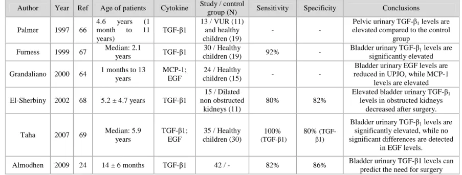
Table 1 resumes the principal studies about UPJO and cytokines.
Vesicoureteral reflux
VUR is a congenital anomaly that increases the risk of repeated pyelonephritis and, consequently, can result in renal scarring, renin-mediated hypertension, and, in some cases, renal insufficiency 70, 71. VUR is a heterogeneous condition that can be primary or associated with multicystic kidney, hypodysplasic kidneys, renal agenesia and renal or ureteral ectopia.
Kidneys with reflux nephropathy have disjointed glomeruli from proximal tubules, interstitial infiltration with chronic inflammatory cells and periglomerular fibrosis. Dysplasic feature is one of the characteristics of congenital reflux nephropathy. The main findings are areas of mesenchymal tissue containing primitive tubules 73.
There were found associations between gene polymorphisms of TNF-α and of TGF-β
and VUR 74, 75, 76, 77. Some of these polymorphisms were also associated to reflux nephropathy and progressive renal damage 76, 77. These associations could help in understanding the mechanisms of reflux nephropathy and could allow the detection of patients at risk of severe renal damage.
TNF-α and TGF-β are abundant in the smooth muscle cell of the ureter of VUR patients
78
. On the other hand, patients without VUR have higher expression of growth promoting factors like insulin-growth factor-1 (IGF-1), nerve growth factor (NGF) and vascular endothelial growth factor (VEGF) than those with VUR 78. In this regard, Chertin et al 73 have showed that the reduced production of EGF associated with high expression of MCP-1 might cause an over production of proinflammatory and profibrotic cytokines that trigger apoptosis, ultimately leading to tubular atrophy and renal dysfunction in reflux nephropathy
73
.
for the diagnostic of VUR. A cutt-off concentration of 5 pg of IL-8/ mol of creatinine has a sensitivity of 88% and a specificity of 69% 79.
The IL-6 may also be involved in the pathogenesis of reflux nephropathy. IL-6 induces B and T cells activation and differentiation during inflammation 29. Ninan et al 81 have detected a significant elevation of urinary IL-6 levels in patients with VUR. In addition, Wang et al 82 have found that urinary IL-6 was significantly higher in children with severe bilateral renal scarring than in those with mild scarring and normal controls. Gokce et al 83 have related high urinary levels of IL-6 with the presence of VUR and increased IL-8 concentrations with renal scarring. Concerning serum measurements of cytokines, Jutley et al 84 have detected significant elevation of IL-6 and TNF-α in patients with reflux nephropathy when compared to those without reflux nephropathy or to healthy controls.
Since the main histological alteration in reflux nephropathy is renal fibrosis, Sabasiñska et al 85 have measured urinary levels of TGF-β1 in patients with VUR. These authors have found that urinary concentrations of TGF-β1 were increased in high-grade reflux and in bilateral cases 85.
Table 2 shows studies about VUR and cytokines.
Concluding Remarks
Congenital obstructive nephropathy accounts for a great fraction of chronic kidney disease in children. Genetic and nongenetic factors responsible for the lesions are largely unidentified, and attention has been focused on minimizing obstructive renal injury and optimizing long-term outcomes. The renal response to urinary tract obstruction is complex and involves a wide array of interacting molecules. Elucidation of these interactions will
lead to the identification of biomarkers that will allow a more precise prediction to the response to surgical intervention and, hopefully, to novel therapies to prevent renal deterioration.
Acknowledgements. This study was partially supported by CNPq (Brazilian National Research Council) and FAPEMIG.
REFERENCES
1. Estrada CR, Jr. Prenatal hydronephrosis: early evaluation. Curr Opin Urol 2008;18:401-3.
2. Lee RS, Cendron M, Kinnamon DD, Nguyen HT. Antenatal hydronephrosis as a predictor of postnatal outcome: a meta-analysis. Pediatrics 2006;118:586-93.
3. Mallik M, Watson AR. Antenatally detected urinary tract abnormalities: more detection but less action. Pediatr Nephrol 2008;23:897-904.
4. Cohen-Overbeek TE, Wijngaard-Boom P, Ursem NT, Hop WC, Wladimiroff JW, Wolffenbuttel KP. Mild renal pyelectasis in the second trimester: determination of cut-off levels for postnatal referral. Ultrasound Obstet Gynecol 2005;25:378-83.
5. Bouzada MC, Oliveira EA, Pereira AK, et al. Diagnostic accuracy of fetal renal pelvis anteroposterior diameter as a predictor of uropathy: a prospective study. Ultrasound Obstet Gynecol 2004;24:745-9.
6. Coplen DE, Austin PF, Yan Y, Blanco VM, Dicke JM. The Magnitude of Fetal Renal Pelvic Dilatation can Identify Obstructive Postnatal Hydronephrosis, and Direct Postnatal Evaluation and Management. J Urol 2006;176:724-7.
7. Gramellini D, Fieni S, Caforio E, et al. Diagnostic accuracy of fetal renal pelvis anteroposterior diameter as a predictor of significant postnatal nephrouropathy: second versus third trimester of pregnancy. Am J Obstet Gynecol 2006;194:167-73.
9. Dias CS, Bouzada MC, Pereira AK, et al. Predictive factors for vesicoureteral reflux and prenatally diagnosed renal pelvic dilatation. J Urol 2009;182:2440-5.
10. Phan V, Traubici J, Hershenfield B, Stephens D, Rosenblum ND, Geary DF. Vesicoureteral reflux in infants with isolated antenatal hydronephrosis. Pediatr Nephrol 2003;18:1224-8.
11. Coelho GM, Bouzada MC, Pereira AK, et al. Outcome of isolated antenatal hydronephrosis: a prospective cohort study. Pediatr Nephrol 2007;22:1727-34.
12. Coelho GM, Bouzada MC, Lemos GS, Pereira AK, Lima BP, Oliveira EA. Risk factors for urinary tract infection in children with prenatal renal pelvic dilatation. J Urol 2008;179:284-9.
13. Farhat W, McLorie G, Geary D, et al. The natural history of neonatal vesicoureteral reflux associated with antenatal hydronephrosis. J Urol 2000;164:1057-60.
14. Ismaili K, Hall M, Donner C, Thomas D, Vermeylen D, Avni FE. Results of systematic screening for minor degrees of fetal renal pelvis dilatation in an unselected population. Am J Obstet Gynecol 2003;188:242-6.
15. Moorthy I, Joshi N, Cook JV, Warren M. Antenatal hydronephrosis: negative predictive value of normal postnatal ultrasound--a 5-year study. Clin Radiol 2003;58:964-70.
16. Tibballs JM, De Bruyn R. Primary vesicoureteric reflux--how useful is postnatal ultrasound? Arch Dis Child 1996;75:444-7.
18. Leung VY, Chu WC, Metreweli C. Hydronephrosis index: a better physiological reference in antenatal ultrasound for assessment of fetal hydronephrosis. J Pediatr 2009;154:116-20.
19. Ismaili K, Hall M, Piepsz A, Alexander M, Schulman C, Avni FE. Insights into the pathogenesis and natural history of fetuses with renal pelvis dilatation. Eur Urol 2005;48:207-14.
20. Hubert KC, Palmer JS. Current diagnosis and management of fetal genitourinary abnormalities. Urol Clin North Am 2007;34:89-101.
21. Piepsz A. Antenatally detected hydronephrosis. Semin Nucl Med 2007;37:249-60. 22. Lee RS. Biomarkers for pediatric urological disease. Curr Opin Urol 2009;19:397-401.
23. LaBaer J. So, you want to look for biomarkers (introduction to the special biomarkers issue). J Proteome Res 2005;4:1053-9.
24. Almodhen F, Loutochin O, Capolicchio JP, Jednak R, El-Sherbiny M. The role of bladder urine transforming growth factor-beta1 concentrations in diagnosis and management of unilateral prenatal hydronephrosis. J Urol 2009;182:292-8; discussion 8. 25. Wen JG, Frøkiær J, Jørgensen TM, Djurhuus JC. Obstructive nephropathy: an update of the experimental research. Urol Res. 1999; 27:29-39.
26. Klahr S, Morrisey. Obstructive nephropathy and renal fibrosis. Am J Renal Physiol. 2002; 283: F861-F875.
27. Matsell DG, Tarantal AF. Experimental models of fetal obstructive nephropathy. Pediatric Nephrology. 2002; 17: 470-476.
29. Borish LC, Steinke JW. Cytokines and chemokines. J Allergy Clin Immunol. 2003; 111(2): S460-S465.
30. Sereger S, Alpers CE. Chemokines and chemokine receptors in renal pathology. Current Opinion in Nephrology and Hypertension. 2003; 12:243-249.
31. Sereger S, Nelson PJ, Schlondorff D. Chemokines, chemokine receptors, and renal disease: from basic science to pathophysiologic and therapeutic studies. J Am Soc Nephrol 2000; 11:152-176.
32. Viedt C, Dechend R, Fei J, et al. MCP-1 induces inflammatory activation of human tubular epithelial cells: involvement on transcription factors, nuclear factor-kappaB and activating protein-1. J Am Soc Nephrol 2002; 13:1534-1547.
33. Gröne, HJ, Cohen CD, Gröne E, Schimidt C, Kretzler M, Schlöndorff D, Nelson PJ. Spatial and temporally restricted expression of chemokines and chemokine receptors in the developing human kidney. J Am Nephrol. 2002; 13: 957-967.
34. Sheu JN, Chen MC, Cheng SL, Lee IC, Chen SM, Tsay GJ. Urine interleukin-1β in children with acute pyelonephritis and renal scarring. Nephrology. 2007; 12: 487-493. 35. Sheu JN, Chen MC, Lue KH, Cheng SL, Lee IC, Chen SM, Tsay GJ. Serum and urine of interleukin-6 and interleukin-8 in children with acute pyelonephritis. Cytokine. 2006; 36:276-282.
36. Souto MFO, Teixeira AL, Russo RC, Penido MGMG, Silveira KD, Teixeira MM, Simões e Silva AC. Immune mediators in idiopathic nephrotic syndrome: Evidence for a relationship between interleukin 8 and proteinuria. Pediatr Res. 2008;64(6):637-42.
38. Artifoni L, Negrisolo S, Montini G, Zuchetta P, Molinari PP, Cassar W, Destro R, Anglani F, Rigamonti W, Zacchello G, Murer L. Interleukin-8 and CXCR1 receptor functional polymorphisms and susceptibility to acute pyelonephritis. The Journal of Urology.2007; 177: 1102-1106.
39. Monga M, Gabal-Shehab LL, Stein P. Urinary transforming growth factor-β1 levels correlate with bladder outlet obstruction. International Journal of Urology. 2001; 8:487-489.
40. Klahr S, Morrisey JJ. Comparative study of ACE inhibitors and angiotensin II receptor antagonists in international scarring. Kidney International. 1997; 52(63): S111-S114.
41. Miayjima A, Chen J, Lawrence C, Ledbetter S, Soslow RA, Stern J, Jha S, Pigato J, Lemer ML, Poppas DP, Vaughan Jr ED, Felsen D. Antibody to transforming growth
factor-β ameliorates tubular apoptosis in unilateral obstruction. Kidney International. 2000; 58:2301-2313.
42. Trachtman H, Weiser AC, Valderrama E, Morgado M, Palmer LS. Prevention of renal fibrosis by spironolactona in mice with complete unilateral ureteral obstruction. The Journal of Urology. 2004; 172:1590-1594.
43. Seseke F, Thelen P, Heuser M, Zöller G, Ringert RH. Impaired nephrogenesis in rats with congenital obstrutive uropathy. The Journal of Urology. 2001; 165:2289-2292. 44. Chevalier RL, Thornhill BA, Chang AY, Cachat F, Lackey A.Recovery from release of ureteral obstruction in the rat: Relationship to nephrogenesis. Kidney International. 2002; 61: 2033-2043.
as an indicator of the degree of hydronephrotic atrophy in partial ureteral obstruction. The Journal of Urology. 2002; 167:1497-1502.
46. Seseke F, Thelen P, Hemmerlein B, Kliese D, Zöller G, Ringert RH. Histologic and molecular evidence of obstructive uropathy in rats with hereditary congenital hydronephrosis. Urol Res. 2000; 28: 104-109.
47. Chevalier RL, Thornhill BA, Wolstenholme JT, Kim A. Unilateral ureteral obstruction in early development alters renal growth: dependence on the duration of obstruction. The Journal of Urology. 1999; 161: 309-313.
48. Zhou, Y, Takahashi G, Shinagawa T, Okuhara T, Yonamine K, Aida Y, Tadokoro M. Increased transforming growth factor-β1 and tubulointersticial fibrosis in rats with congenital hydronephrosis. International Journal of Urology. 2002; 9: 491-500.
49. Truong LD, Petrusevska G, Yang G, Gurpinar T, Shappell S, Lechago J, Rouse D, Suki WN. Cell apoptosis and proliferation in experimental chronic obstructive uropathy. Kidney International. 1996; 50: 200-207.
50. Truong LD, Choi YJ, Tsao CC, Ayala G, Sheikh-Hamad D, Nassar G Suki WN. Renal cell apoptosis in chronic obstructive uropathy: the role of caspases. Kidney International. 2001; 60:924-934.
51. Chevalier RL, Goyal S, Wolstenholme JT, Thornhill BA. Obstructive nephropathy in the neonatal rat is attenuated by epidermal growth factor. Kidney International. 1998; 54: 38-47.
53. Mizuno S, Matsumoto K, Nakamura T. Hepatocyte growth factor suppresses interstitial fibrosis in a mouse model of obstructive nephropathy. Kidney International. 2001; 59: 1304-1314.
54. Isaka Y, Tsujie M, Ando T, Nakamura H, Kaneda Y, Imai E, Hori M. Transforming growth factor-β1 antisense oligodeoxynucleotides block interstitial fibrosis in unilateral ureteral obstruction. Kidney International. 2000; 1885-1892.
55. Sato M, Muragaki Y, Saika S, Roberts AB, Ooshima A. Targeted disruption of TGF-β1/ Smad3 signaling protects against renal tubulointerstitial fibrosis induced by unilateral ureteral obstruction. Journal of Clinical Investigation, 2003; 112(10): 1486-1494. 56. Derynck R, Zhang Y, Feng XH. Smads: transcriptional activators of TGF-β
responses. Cell. 1998; 95: 737-740.
57. Misseri R, Meldrum DR, Dagher P, Hile K, Rink RC, Meldrum KK. Unilateral ureteral obstruction induces renal tubular cell production of tumor necrosis factor-α
independent of inflamatory cell infiltration. The Journal of Urology. 2004; 172; 1595:1599. 58. Vielhauer V, Anders HJ, Mack M, Cihak J, Strutz F, Sangasswinger M, Luckow B, Gröne HJ, Schlöndorff. Obstructive nephropathy in the mouse: progressive fibrosis correlates with tubulointerstitial chemokine expression and accumulation of CC chemokine receptor 2- and 5-positive leukocytes. J Am Soc Nephrol. 2001; 12:1173-1187.
59. Crisman JM, Richards LL Valach DP, Valach DF, Franzoni DF, Diamond JR. Chemokine expression in the obstructed kidney. Exp Nephrol. 2001; 9: 241-248.
61. Hochberg D, Johnson CW, Chen J, Cohen D, Stern, Vaughan Jr ED, Poppas D, Felsen D. Intertitial fibrosis of unilateral ureteral obstruction is exacerbated in kidneys of mice lacking the gene for inducible nitric oxide synthase. Laboratory Investigation. 2000; 11: 1721-1728.
62. Bulas DI, Fonda JS. Prenatal evaluation of fetal anomalies. Pediatric Radiology. 1997; 44:537-553.
63. Zhang PL, Peters CA, Rosen S. Ureteropelvic junction obstruction: morphological and clinical studies. 2000; 14:820-826.
64. Grandaliano G, Gesualdo L, Bartoli F, Ranieri E, Monno Rafaela, Leggio A, Paradies G, Caldarulo E, Infante B, Schena FP. MCP-1 and EGF renal expression and urine excretion in human congenital obstructive nephropathy. Kidney International. 2000; 58:182-192
65. Bartoli F, Gesualdo L, Paradies G, Caldarulo E, Infante B, Grandaliano G, Monno R, Leggio S, Salzillo F, Schena FP, Leggio A. Renal expression of monocyte chemotactic protein-1 and epidermal growth factor in children with obstructive hydronephrosis. Journal of Pediatric Surgery. 2000; 35:569-572.
66. Palmer LS, Maizels M, Kaplan WE, Firlit CF, Cheng EY. Urine levels of transform growth factor-beta 1 in children with ureteropelvic junction obstruction. Pediatric Urology. 1997; 50(5): 769-773.
68. El-Sherbiny MT, Mousa OM, Shokier AA, Ghoneim MA. Role of urinary transforming growth factor-β1 concentration in the diagnosis of upper urinary tract obstruction in children. The Journal of Urology. 2002; 168: 1798-1800.
69. Taha MA, Shokeir AA, Osman HG, El-Aziz AEAFA, Farahat SE. Pelvi-ureteric obstruction in children: the role of urinary transform growth factor-β1 and epidermal growth factor. Pediatric Urology. 2007; 99:899-903.
70. Elder, J. S., C. A. Peters, et al. Pediatric vesicoureteral reflux Guidelines Panel summary report on the management of primary vesicoureteral reflux in children. J Urol. 1997; 157: 1846-1851.
71. Silva, J. M., J. S. Santos Diniz, et al. Clinical course of 735 children and adolescents with primary vesicoureteral reflux. Pediatr Nephrol. 2006;21: 981-988.
73. Chertin B, Farkas A, Puri P. Epidermal growth factor and monocyte chemotactic peptide-1 expression in reflux nephropathy. European Urology. 2003; 44:144-149.
74. Yim HE, Bae IS, Yoo KH, Hong YS, Lee JW. Genetic control of VEGF and
TGF-β1 gene polymorphisms in childhood urinary tract infection and vesicoureteral reflux. Pediatric Research. 2007; 62 (2):183-187.
75. Kuronda S, Solari V, Puri P. Association of Transform growth factor-β1 gene polymorphism with familial vesicoureteral reflux. The Journal of Urology. 2007; 178:1650-1653.
77. Solari V, Ennis S, Cascio S, Puri P. Tumor necrosis factor-α gene polymorphism in reflux nephropathy. The Journal of Urology. 2004; 172:1604-1606.
78. Schwentner C, Oswald J, Lunacek A, Pelzer AE, Fritsch H, Schlenck B, Karatzas A, Bartsch G, Radmayr C. Extracellular Microenvironment and cytokine profile of the ureterovesical junction in children with vesicoureteral reflux. 2008; 180: 674-700.
79. Galanakis E, Bitsori M, Dimitriou H, Giannakopoulou C, Karkavitsas NS, Kalmanti M. Urine Interleukin-8 as a Marker of Vesicoureteral Reflux in Infants. Pediatrics. 2006 Apr 3.
80. Haraoka M, Senoh K, Ogata N, Furukawa M, Matsumoto T, Kumazawa J. Elevated interleukin-8 levels in the urine of children with renal scarring and/or vesicoureteral reflux. The Journal of Urology. 1996; 155:678-680.
81. Ninan GK, Juntley RS, Eremin O. Urinary Cytokines as markers of reflux nephropathy. The Journal of Urology. 1999 November; 162:1739-1742.
82. Wang J, Konda R, Sato H, Sakai K, Ito S, Orikasa S. Clinical significance of urinary interleukin-6 in children with reflux nephropathy. The Journal of Urology. 2001 January; 165:210-214.
83. Gokce I, Alpay H, Biyikli, Unluguzel G, Dede F, Topuzoglu A. Urinary levels of interleukin-6 and interleukin-8 in patients with vesicoureteral reflux and renal parenchymal scar. Pediatric Nephrology.2010.
84. Jutley RS, Youngson GG, Eremin O, Ninan GK. Serum Profile in reflux nephropathy. Pediatr Surg Int. 2000; 16:64-68.
FIGURE 1
Figure 1: Main mechanisms involved in obstructive models
OBSTRUCTIVE UROPATHY
Mechanical Stretch
MCP-1 TNF-α
TGF-β
Apoptosis Fibrosis Monocytes
Chronic interstitial inflammation
RANTES
Fibroblasts Angiotensin II
Collagen
Table 1: Recent studies on urinary cytokines in patients with ureteropelvic junction obstruction
Ref: reference number
Author Year Ref Age of patients Cytokine Study / control
group (N) Sensitivity Specificity Conclusions Palmer 1997 66
4.6 years (1 month to 11 years)
TGF-β1
13 / VUR (11) and healthy children (19)
- -
Pelvic urinary TGF-β1 levels are
elevated compared to the control group
Furness 1999 67 Median: 2.1
years TGF-β1
30 / Healthy
children (19) 92% -
Bladder urinary TGF-β1 levels are
significantly elevated Grandaliano 2000 64 1 months to 13
years
MCP-1; EGF
24 / Healthy
children (15) - -
Bladder urinary EGF levels are reduced in UPJO, while MCP-1
levels are elevated El-Sherbiny 2002 68 5.2 ± 4.7 years TGF-β1
15 / Dilated non obstructed
kidneys (11)
80% 82%
Elevated bladder urinary TGF-β1
levels in obstructed kidneys decreased after surgery.
Taha 2007 69 Median: 5.9 years
TGF-β1; EGF
35 / Healthy children (30)
100%
(TGF-β1)
80%
(TGF-β1)
Bladder urinary TGF-β1 levels are
significantly elevated, while no significant differences are detected
in EGF levels.
Ref: reference number
Author Year Ref Age of patients Cytokine Study/ control
group (N) Sensitivity Specificity Conclusions Haraoka 1996 80 Mean age 6.7
years IL-8 32 / - - -
Levels of IL-8 are elevated in patients with VUR or renal scarring Ninan 1999 81 5 months to
13.33 years
IL-6; TNF-α
17 / Healthy
children (15) - -
Levels of IL-6 and TNF-α receptor-1 are elevated in reflux associated
with renal damage Wang 2001 82 Mean age 14.6
years IL-6
66 / Healthy children (28)
Levels of IL-6 are elevated in severe bilateral renal scarring
Galanakis 2007 79 1 month to 2
years IL-8
24 / History ITU but no VUR (14); No
ITU, no VUR (21)
88% 69% Levels of IL-8 are elevated in VUR patients
Sabasiñska 2008 85 6.23+4.15 years TGF-β1
54 / Healthy children
(27)
- -
Highest urinay concentrations of TGF-β1 are detected in grade IV
and V reflux Gokce 2010 83 1 month 16 years 6;
IL-8
87 / Healthy
children (27) - -
3.
OBJETIVOS
________________________________________________
Objetivo Geral:
Avaliação dos níveis urinários de citocinas pró-inflamatórias e do fator de
crescimento e transformação do tipo beta em crianças e adolescentes com hidronefrose diagnosticada intraútero.
Objetivos específicos:
Os objetivos específicos desse projeto foram:
1.Determinar as concentrações urinárias de IL-6, TNF-α (fator de necrose tumaoral alfa, tumor necrosis factor-α) e TGF-β1, expressas em valores absolutos e relativos à creatinina urinária, em crianças e adolescentes portadores de anomalias do trato urinário, subdivididos em portadores de hidronefrose idiopática, de uropatia e de displasia/ hipoplasia renal;
2.Comparar esses parâmetros obtidos nos pacientes portadores de hidronefrose idiopática e portadores de uropatias clinicamente significativas;
3. Comparar esses marcadores nos pacientes com captação normal versus captação reduzida à cintilografia renal estática (DMSA);
4.
PACIENTES E MÉTODOS
_________________________________________________
4.1 DELINEAMENTO, POPULAÇAO E LOCAL DE ESTUDO
Trata-se de um estudo transversal de uma coorte de crianças e adolescentes que tiveram o diagnóstico de dilatação da pelve renal intraútero e que foram acompanhados desde o nascimento no Ambulatório Bias Fortes de atendimento terciário da Unidade de Nefrologia Pediátrica do Departamento de Pediatria da Faculdade de Medicina da Universidade Federal de Minas Gerais.
4.1.1 PERÍODO DE ESTUDO
Foram coletados exames do período de janeiro de 2008 a janeiro de 2009.
4.1.2 CRITÉRIOS DE INCLUSÃO
Foram incluídos pacientes que apresentavam o diâmetro ântero-posterior da pelve renal medido na seção transversa do hilo renal fetal ≥ 5mm no terceiro trimestre da gestação 2-4.
4.1.3 CRITÉRIOS DE EXCLUSÃO
Foram excluídos pacientes com aneuploidia ou malformações múltiplas.
Foram excluídos os pacientes que se apresentarem agudamente doentes no momento da coleta, tais como, aqueles com infecção não controlada, insuficiência renal crônica (IRC) estágios maior que 3, desequilíbrio hemodinâmico e /ou metabólico agudo.
Os pacientes e os responsáveis por eles foram devidamente esclarecidos sobre a natureza do estudo e o que seria feito, tendo resguardado seu direito, em caso de recusa, de receberem a avaliação e o tratamento indicados (para maiores detalhes, ver termo de consentimento em anexo). Além disso, as amostras de urina dos pacientes, que aceitaram participar da pesquisa, foram colhidas simultaneamente a outros exames de sangue e urina que fazem parte da avaliação clínica rotineira dessas crianças e adolescentes portadores de hidronefrose fetal.
Este projeto de pesquisa foi aprovado pela Câmara Departamental do Departamento de Pediatria da Faculdade de Medicina da UFMG (vide parecer em anexo) e pelo cômite de ética do Hospital das Clínicas e da UFMG (COEP) – Protocolo número ETIC 0487/06 e DEP 143/06, respectivamente (vide parecer em anexo).
4.2 DEFINIÇÕES E CLASSIFICAÇÃO DA DILATAÇÃO DA PELVE
RENAL
As seguintes definições foram adotadas no presente estudo:
Uropatia significativa – diagnóstico de uma entidade nosológica bem definida através da
dividindo-se os pacientes em dois grupos: uropatas (com uropatia significativa e rins displásicos) e não-uropatas (com hidronefrose idiopática).
Obstrução de junção ureteropélvica - foi considerada quando houve dilatação da pelve
renal associada a padrão de excreção intermediário ou obstrutivo, independentemente da captação renal relativa pelo 99tm-DMSA.
Obstrução de junção ureteropélvica cirúrgica - quando houver dilatação da pelve renal
associada a padrão de excreção intermediário ou obstrutivo, com captação renal relativa pelo 99tm-DMSA < 40%.
Lesão renal – presença de qualquer alteração da morfologia e/ou crescimento dos rins
e também presença de captação renal relativa pelo 99tm-DMSA < 45%. Para avaliação do crescimento renal foram utilizados os gráficos de Han & Bancock 5.
Hidronefrose – é o termo que descreve a dilatação do sistema coletor renal, não
implicando na presença de obstrução 6. Hidronefrose idiopática – dilatação da pelve renal sem causa definida.
Classificações da hidronefrose - para avaliação da dilatação renal serão vários
pontos de corte do diâmetro ântero-posterior (DAP): 1) dilatação leve - ≥ 5 mm e < 10 mm 2) dilatação moderada - ≥ 10 mm e < 15mm 3) dilatação acentuada - ≥ 15 mm para construir intervalos para análises. As unidades renais também foram classificadas de acordo com a classificação da SFU (Society of Fetal Urology) 7. Quando bilateral, foi considerada a unidade renal com dilatação maior e/ou com classificação em grau maior pela SFU.
Infecção do trato urinário – sinais e sintomas como febre, disúria e hematúria