Re gulato ry ro le s o f m icro tubule
-asso ciate d pro te ins in ne uro nal
m o rpho ge ne sis. Invo lve m e nt o f
the e xtrace llular m atrix
1Laboratorio de Biología Celular y Molecular, Facultad de Ciencias,
Universidad de Chile, Santiago, Chile
2Laboratório de Morfogênese Celular, Departamento de Anatomia,
Universidade Federal do Rio de Janeiro, Rio de Janeiro, RJ, Brasil G. Ramírez1, A. Alvarez1,
J. Garcia-Abreu2,
F.C.A. Gomes2,
V. Moura-Neto2 and
R.B. Maccioni1
Abstract
As a result of recent investigations, the cytoskeleton can be viewed as a cytoplasmic system of interconnected filaments with three major integrative levels: self-assembling macromolecules, filamentous poly-mers, e.g., microtubules, intermediate filaments and actin filaments, and supramolecular structures formed by bundles of these filaments or networks resulting from cross-bridges between these major cytoskeletal polymers. The organization of this biological structure appears to be sensitive to fine spatially and temporally dependent regulatory signals. In differentiating neurons, regulation of cytoskeleton organization is particularly relevant, and the microtubule-associated protein (MAP) tau appears to play roles in the extension of large neuritic processes and axons as well as in the stabilization of microtubular polymers along these processes. Within this context, tau is directly involved in defining neuronal polarity as well as in the generation of neuronal growth cones. There is increasing evidence that elements of the extracellular matrix contribute to the control of cytoskeleton organiza-tion in differentiating neurons, and that these regulaorganiza-tions could be mediated by changes in MAP activity. In this brief review, we discuss the possible roles of tau in mediating the effects of extracellular matrix components on the internal cytoskeletal arrays and its organization in growing neurons.
Co rre spo nde nce
R.B. Maccioni
Laboratorio de Biología Celular y Molecular
Facultad de Ciencias Universidad de Chile Las Palmeras 3425, Ñuñoa Santiago
Chile
Fax: + 56-2-218-6245
E-mail: rmaccion@ abello.dic.uchile.cl
Presented at the 5th Brazilian Symposium on Extracellular Matrix - SIMEC, Angra dos Reis, RJ, Brasil, September 7-10, 1998.
Research partially supported by the Presidential Chair on Science (Chile), and by an international CNPq-Conicyt cooperation project.
Received November 17, 1998 Accepted December 21, 1998
Ke y wo rds
·Extracellular matrix
·Tau functions
·Regulatory patterns
·Neuronal morphogenesis
Intro ductio n
The molecular factors that affect neu-ronal morphogenesis and its differentiation constitute a central paradigm in modern cell biology. The structure and integrity of the cytoplasm of eukaryotic cells depend on the assembly and organization of the network of interlinked filaments that form the
centrosomes, lysosomes, and the cell nucleus and membrane.
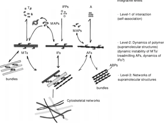
The structural and functional organiza-tion of living cells, and therefore the spatial array of their components, are generated through a process of growing complexity. Cell morphology is determinant for specific cell functions, and therefore the architecture of cells appears to be finely regulated in response to structural demands of cells within the context of their functions. The cytoskel-eton is a system of interconnected filaments, in which three major integrative levels can be distinguished: self-assembling macromol-ecules, filamentous polymers, e.g. microtu-bules, intermediate filaments and actin fila-ments, and supramolecular structures formed by bundles of these filaments or cross-bridges between these major cytoskeletal polymers (Figure 1). When cytoplasmic macromol-ecules assemble into supramolecular struc-tures that shape the cytoskeleton, a set of
properties of living cells emerge, such as their capacity of dividing, growing, moving and changing their morphology during dif-ferentiation. The architecture of cells is pro-vided to a large extent by the cytoskeletal organization (3). However, in addition to the internal components and regulatory signals, extracellular events can also contribute to the complex regulatory patterns of cytoskel-etal organization (4).
In the analysis of the different associa-tions between cytoskeletal components, a major question emerges: how can cytoplas-mic macromolecules or extracellular signals contribute to cytoplasmic organization and neuronal shape? On the other hand, how is this structure modulated as a consequence of the multiple functional demands of neuronal cells? The study of protein-to-protein inter-actions that play a key role in the structure of the cytoskeletal network has been one of the main aspects of our recent investigations (5).
Figure 1 - Simplified scheme show ing the three integrative levels in the process by w hich macromolecular units assemble into cytoskeletal polymers, and their association to form su-pramolecular structures, mainly bundles of these polymers, and netw orks involving microtubules (M T), intermediate filaments (IF) (neurofilaments) and actin fila-ments (AF). M icrotubule-associ-ated proteins (M APs) appear to play a major modulatory role at the level of microtubule assem-bly and in regulating the linkages betw een the different filaments of the cytoskeleton. T, Tubulin; A, actin; ABPs, actin binding pro-teins; IFPs, interm ediate-fila-ments proteins.
· Level-2: Dynamics of polymers (supramolecular structures) (dynamic instability of M Ts/ treadmilling AFs, dynamics of IFs?)
Integrative levels
· Level-1 of interaction (self-association)
· Level-3: Netw orks of supramolecular structures
Cytoskeletal netw orks bundles
ABPs AFs
M APs A IFPs
M APs
M Ts IFs
bundles
Cytoplasmic dynamics
These interactions include: i) homologous associations like those occurring in the oli-gomerization of actin in the assembly of microfilaments or self-assembly of tubulin leading to microtubule formation, and ii) heterologous associations like the interac-tions between cytoskeletal polymers and microtubule-associated proteins (MAPs). However, there is another branch in this field of study, which is the involvement of the extracellular matrix (ECM) in the deter-mination of modulatory signals that affect the neuronal cytoskeleton in processes like neuritogenesis, and the possible role of MAPs in mediating these cellular changes. This is the main focus of the present analysis.
Facto rs influe ncing m icro tubule o rganizatio n and ne urite stability and o utgro wth
Neuronal microtubules constitute major structures directly involved in neuronal mor-phogenesis, with different subsets of tubulin specifically expressed in neuronal and glial cells (6). Their assembly and dynamics ap-pear to be modulated by MAPs. Different animal tissues contain variable sets of MAPs, and the cell type-specificity of these proteins may account for their differential expression in each type of cell (7). In experiments in which cDNAs of tau and MAP-1B were transfected into HeLa cells and fibroblasts, microtubules were stabilized against depo-lymerizing agents (8). Post-translational modifications of microtubules are directly involved in the determination of morpho-genesis in neurons (9). The fact that these microtubules are enriched in acetylated tu-bulin suggests that this post-translational modification of tubulin renders microtubules more resistant to depolymerization. In N2A neuroblastoma cells, studies using depoly-merizing agents demonstrated that tau colo-calized in a discrete fashion along stable microtubular polymers, while a more con-tinuous association of tau along less stable
microtubules containing tyrosinated tubulin was also observed (10). The experiments indicate that there is a differential associa-tion of tau isoforms into stable and weak microtubules, and that tau association into more dynamic polymers containing tyrosin-ated tubulin contributes to their stabiliza-tion.
Further evidence of the roles of MAPs in neuritogenesis comes from studies on N18 neuroblastoma cells, which grow as loosely attached and morphologically undifferenti-ated spherical cells. However, removal of serum leads to greater adherence to a sub-stratum and to the initiation of neurite out-growth (11). Neurite differentiation begins within minutes and differentiated morpholo-gy is attained within 3 days in the low-serum environment. Even though the nature of ex-tracellular matrix components used as sub-strate contributes to neuronal outgrowth, ini-tiation of neuritogenesis is not simply a mat-ter of adhesion, since N-18 cells grown over poly-D-lysine but in the presence of 10% serum do not extend neurites. Studies have indicated a role of MAPs in regulating mi-crotubule assembly during nerve fiber outgrowth (12). High speed supernatants from N-18 differentiated cells were active in inducing in vitro microtubule assembly from
purified brain tubulin, but extracts of undif-ferentiated cells were unable to promote mi-crotubule polymerization.
The ro le o f tau in ne uro nal gro wth
to regulate microtubule assembly and dy-namics. This is supported by a substantial increase in tau expression concomitant with neurite extension (16). In essence, the changes in the expression of tau in trans-fected cells appear to play a critical role in the ability of tau to promote neuritic stability in cultured cells (17).
Early microinjection studies have pointed out that the in vivo actions of neuronal tau to
induce and stabilize microtubules, which are necessary for neuronal activity, may not be sufficient to explain neuronal morphogen-esis (18). Therefore, an important role re-vealed for tau is the directing of the neuronal outgrowth process from the cell soma to the distal ends of axon-like neurites (19). Within this context it is proposed that critical targets for the action of tau on neuronal growth are precisely the distal axons and growth cones (19). In fact, in polarized hippocampal neu-rons, the enzyme cdk-5 becomes concen-trated at the distal tip of growing axons, associating with the subcortical cytoskel-eton (20). Also, it was shown that specific phosphorylations of MAP-1b occur during neuronal outgrowth in neuroblastoma cells, and this modification is related to an en-hancement of microtubule assembly (21). Therefore, besides tau the high-molecular weight MAPs (MAP-1b, MAP-3) also ap-pear to be involved in neuronal growth (22). A major question to address is how tau function is regulated. At this level it is worth mentioning that tau proteins are produced by a single gene by alternative splicing, which yields several protein products. Thus, splic-ing of a common RNA transcript, as well as post-translational modification of the tau mol-ecule are the main regulatory mechanisms for tau activity on neurons. The single tau gene contains 14 exons encoding for the six different isoforms of this protein (for a re-view, see 5). Expression of tau isoforms is developmentally regulated, with some dif-ferences in the central and peripheral ner-vous system. In the fetal brain, only the short
tau isoforms containing only three repetitive binding motifs are expressed, but after post-natal development neuronal cells can ex-press all six major isotypes of tau. A marked decrease in fetal tau expression occurs in rats after postnatal day 8. At the level of the peripheral nervous system, only the high-molecular weight (110 kDa) isoform of tau containing two additional exons is expressed (23).
Within this context, another aspect to emphasize in the analysis of cellular factors affecting the extension of neurites by neu-ronal cells is the role of tau in mediating microtubule-actin filament interactions (5,10,24), an activity that could be involved in regulating the actin cortex and association with plasma membrane in the establishment of architectural patterns during neuronal growth (25).
Extrace llular m atrix cyto ske le to n and MAPs in the co nte xt o f ne uro nal gro wth
which is a neuronal substrate of cdk-5, is involved in mediating the laminin effects on neuronal growth. Previous observations have suggested that cdk-5 may serve as a regula-tory linker between environmental signals of ECM elements, and MAPs involved in the axonal formation process (26). The effects of laminin appear to be linked to specific phosphorylations of either MAP-1b or tau, and a reorganization of these MAPs within the neuronal cytoskeleton. These findings are strongly supported by additional obser-vations that inhibition of certain protein ki-nases abolishes the ECM-mediated promo-tion of neuronal polarity (28). A schematic representation of the effects of the ECM is illustrated in Figure 2.
Extracellular matrix components appear to affect the direction of neurite outgrowth by interacting with the growth cone;
how-ever, the way they function has not been elucidated. Elements of the ECM, such as laminin, stimulate neuronal growth (29), but are also involved in modulating cell contacts and the spreading phenomenon (30). The concept of a differential cell adhesion pro-vided by ECM elements is restricted to sub-strate conditions, and the molecular nature of these elements (4). When growth cones are experimentally detached, they show a greater level of contact with collagen and fibronectin substrates as compared with la-minin. However, there is evidence that lami-nin is a significantly better substrate than fibronectin and collagen (31). Evidence against differential adhesion as a mechanism for laminin function is that adhesion is not a determinant for growth cone preference for substrates. Outgrowth of neurons in culture is faster on a laminin substrate than on a
Hippocampal neuron
ECM
M AP-P* M APs
Protein kinases
axonal grow th
· mobilization of protein kinases tow ard distal tips · redistribution of M AP-P
Figure 2 - Schematic representation of a hippocampal neuron, and the effects of the extracellular matrix (ECM ) in stimulating axonal extension in grow ing neurons. The scheme indicates that ECM effects (laminin, others) can be transduced in a sequence of molecular events (1 and 2) leading to phosphorylation of neuronal M APs and incorporation of M AP-P (phosphorylated M APs) into grow ing microtubules, and/or activation and redistribution of protein kinases that could be critical for neuronal grow th. In the upper part of the figure, closed circles denote differentially phosphorylated M APs at specific sites, and open circles the normally phosphorylated M APs.
control substrate, but eventually neurons achieve polarity on either substrate (32). On the other hand, besides laminin, neuronal proteoglycans are directly involved in the outgrowth of fetal hippocampal neurons (33,34). However, the precise mechanisms by which tau protein and other MAPs di-rectly involved in neuronal polarity partici-pate in the action of ECM components are not clear at all.
Extrace llular m atrix and the glial cyto ske le to n
Elements of the ECM are also known to
affect the cytoskeleton and morphological changes in cultured astrocytes (35). It was reported that transforming growth factor (TGF)-beta1 stimulates the production of laminin and fibronectin and their incorpora-tion into the ECM of astrocytes. Further-more, it is possible that a TGF-beta1-like growth factor is secreted by neuronal cells and induces the glial fibrillary acidic protein (GFAP) gene (36). This factor also promoted formation of stress fibers and increased the content of actin. These changes in cell shape, cytoskeleton and ECM promoted by TGF beta 1 may be of relevance for understanding the connections between ECM and cell cyto-skeleton in the context of brain development (37). On the other hand, T3 hormone and conditioned media from cerebellar T3-treated astrocytes enriched with EGF and TNF-beta (36) promoted alterations in the GFAP cyto-skeleton (38) and fibronectin. Fibronectin, which usually exhibits a discontinuous dis-tribution of control, on the cell surface be-came diffuse in T3-treated astrocytes (39). Furthermore, this conditioned medium has been recently reported to induce cerebellar neuronal proliferation due to the presence of EGF and TNF-beta.
Tau pro te ins co uld be se nsitive to the m o dulato ry e ffe cts o f ECM o n ne uro nal o utgro wth
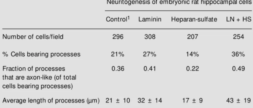
An interesting observation comes from studies indicating that subsets of tau may act differentially in tau interactions with micro-tubules, actin and neurofilaments (24). It is also possible that certain isoforms of tau may respond with different sensitivity to the effects of ECM components on neuritogen-esis. However, there is no clear evidence of the specific roles of tau isoforms due to limitations in generating probes to examine their specific roles. Tau function appears to be modulated by ECM and this regulation seems to depend on the nature of ECM ele-ments. In a study of neurite extension in Table 1 - M orphometric analysis of hippocampal cells.
1Hippocampal cells in primary cultures w ere plated onto culture dishes and incubated
under substrate control conditions, or in the presence of either ECM element. LN, Laminin, HS, heparan sulfate.
Neuritogenesis of embryonic rat hippocampal cells
Control1 Laminin Heparan-sulfate LN + HS
Number of cells/field 296 308 207 254
% Cells bearing processes 21% 27% 14% 36%
Fraction of processes 0.36 0.41 0.22 0.49
that are axon-like (of total cells bearing processes)
Average length of processes (µm) 21 ± 10 32 ± 14 17 ± 9 43 ± 19
Table 2 - In vitro polymerization of tubulin in the presence of brain extracts of primary cultures of hippocampal cells.
1Net assembly obtained after subtraction of the value of control for tubulin
self-aggregation in the absence of any cell extract. Final concentration of proteins in the assembly system: 1.5 mg/ml tubulin; 0.14 mg/ml brain extracts. 2Hippocampal cells in primary cultures w ere incubated under substrate control conditions, or in the presence of either ECM element. LN, Laminin; HS, heparan sulfate. Tubulin assembly w as monitored by the sedimentation assay (40).
Net assembly1 (µg microtubules)
Tubulin control (no factors added) 0.0
Tubulin + brain tau 49.9 ± 5.1
Tubulin + control brain extract2 10.4 ± 2.2
Tubulin + extract incubated w ith LN 16.4 ± 4.6
Tubulin + extract incubated w ith HS 14.8 ± 3.0
hippocampal cells in primary cultures, lami-nin stimulated the number of neurons bear-ing processes, an effect potentiated by the presence of heparan sulfate. An increase was also observed in the fraction of pro-cesses that are axon-like as well as in the average length of processes; however, hepa-ran sulfate by itself failed to produce this effect (Table 1). Therefore, an experiment was carried out in which tubulin polymeriza-tion was analyzed in the presence of hippo-campal brain extracts obtained from cells cultured under different ECM experimental conditions. Cell that were incubated with laminin and heparan sulfate showed the high-est activity of factors inducing microtubule assembly as compared with controls
incu-bated with purified brain tau (Table 2). Cosedimentation studies revealed that a higher fraction of tau was associated with polymers obtained from pure tubulin and extracts of the laminin-heparan sulfate-treated cells. These studies shed further light on the molecular analysis of the roles of MAPs, and particularly tau proteins, in the sequence of events by which ECM affects neuritogenesis.
Ackno wle dgm e nts
We thank Dr. Alfredo Cáceres, Lorena Saragoni and Rodrigo Toro for helpful dis-cussions.
Re fe re nce s
1. Porter K (1984). The cytomatrix: a short story of its study. Journal of Cell Biology, 99 (Suppl): 3s-12s.
2. M itchison T (1992). Compare and con-trast actin filaments and microtubules. M olecular Biology of the Cell, 3: 1309-1315.
3. M accioni RB & Arechaga J (1987). The Cytoskeleton in Cell Differentiation and Development. IRL Press, Oxford. 4. Luckenbill-Edds L (1997). Laminin and the
mechanism of neuronal grow th. Brain Re-search Review s, 23: 1-27.
5. M accioni RB & Cambiazo V (1995). Role of microtubule-associated proteins in the control of microtubule assembly. Physi-ologicalReview s, 75: 835-863.
6. M oura Neto V, M allat M , Jeantet C & Prochiantz A (1986). M icroheterogeneity of tubulin proteins in neuronal and glial cells from the mouse brains in culture. EM BO Journal, 2: 1241-1248.
7. M accioni (1986). M olecular Cytology of M icrotubules. Basque Country University Press-Springer-Verlag, Leioa.
8. Takemura R, Okabe S, Umeyama T, Kanai Y, Cow an NJ & Hirokaw a N (1992). In-creased microtubule stability and alpha tubulin acetylation in cells transfected w it h m icrot ubule-associat ed prot eins M AP1B, M AP-2 or tau. Journal of Cell Science, 103: 445-456.
9. Bulinski JC & Gundersen GG (1991). Sta-bilization of post-translational modification
of microtubules during cellular morpho-genesis. Bioessays, 13: 285-293. 10. M accioni RB, Tapia L & Cam biazo V
(1995). Functional organization of tau pro-teins during neuronal differentiation and development. Brazilian Journal of M edical and Biological Research, 28: 827-841. 11. Seeds NW & M accioni RB (1978).
Pro-teins from morphologically differentiated neuroblastoma cells promote tubulin po-lymerization. Journal of Cell Biology, 78: 547-555.
12. Seeds NW & M accioni RB (1987). Charac-terization of a microtubule-assembly pro-moting factor in differentiated neuroblas-toma cells. In: M accioni RB & Arechaga J (Editors), The Cytoskeleton in Cell Differ-entiation and Development. IRL Press, Oxford, 187-192.
13. Cáceres A & Kosik K (1990). Inhibition of neurite polarity by tau anti-sense oligo-nucleotides in primary cerebellar neurons. Nature, 343: 461-463.
14. Gordon-Weeks P (1991). Grow th cones: the mechanism of neurite advance. Bio-essays, 13: 235-239.
15. Trinczek B, Biernat J, Baum an K, M andelkow EM & M andelkow E (1995). Domains of tau protein, differential phos-phorylation, and dynamic instability of mi-crotubules. M olecular Biology of the Cell, 6: 1887-1902.
16. Ferreira A, Busciglio J & Cáceres A (1989). M icrotubule formation and neurite grow th
in cerebellar macroneurons w hich de-velop in vitro: evidence for the involve-ment of the microtubule associated pro-teins M AP-1a, M AP2 and tau. Develop-mental Brain Research, 49: 215-228. 17. Esmaeli-Azad B, M cCarty JH & Feinstein
SC (1994). Sense and antisense transfec-tion analysis of tau functransfec-tion: tau influ-ences net microtubule assembly, neurite outgrow th and neuritic stability. Journal of Cell Science, 107: 869-879.
18. Drubin D & Kirschner M W (1986). Tau protein function in living cells. Journal of Cell Biology, 103: 2739-2745.
19. Black M M , Slaughter T, M oschiach S, Obrocka M & Fisher I (1996). Tau is en-riched on dynamic microtubules in the distal region of grow ing axons. Journal of Neuroscience, 16: 3601-3619.
20. Pigino G, Paglini G, Ulloa L, Avila J & Cáceres A (1997). Analysis of the expres-sion, distribution and function of cyclin dependent kinase cdk-5 in developing cer-ebellar macroneurons. Journal of Cell Sci-ence, 110: 257-270.
21. Diaz-Nido J, Serrano L, M éndez E & Avila J (1988). A casein kinase II-related activity is involved in phosphorylation of microtu-bule-associated protein M AP-1b during neuroblastoma cell differentiation. Jour-nal of Cell Biology, 106: 2057-2065. 22. M atus A (1991). M icrotubule-associated
23. Couchie D, M avilia C, Georgieff IS, Liem RK & Nunez J (1992). Primary structure of high-molecular w eight tau present in the peripheral nervous system. Proceedings of the National Academy of Sciences, USA, 89: 4378-4381.
24. Henriquez JP, Vial C & M accioni RB (1995). Subpopulations of tau interacts w ith microtubules and actin filaments in different cell types. Cell Biochemistry and Function, 13: 239-250.
25. Brandt R (1996). The tau proteins in neu-ronal grow th and development. Frontiers in Biosciences, 1: 118-130.
26. DiTella M , Feiguin F, Carri N & Cáceres A (1996). M AP-1b/tau functional redundancy during laminin-enhanced axonal grow th. Journal of Cell Science, 109: 467-477. 27. Harada A, Ogushi K, Okabe S, Kuno J,
Terada S, Oshima T, Sao Y, Takei T & Hirokaw a N (1994). Altered microtubule organization in small caliber axons of mice lacking tau protein. Nature, 369: 488-491. 28. Lochter A & Schachner M (1997). Inhibi-tors of protein kinases abolish ECM -medi-ated promotion of neuronal polarity. Ex-perimental Cell Research, 235: 124-129. 29. Garcia-Abreu J, Cavalcante LA & M oura
Neto V (1995). Differential patterns of la-minin expression in lateral and medial
midbrain glia. NeuroReport, 6: 761-764. 30. M ooney DJ, Langer R & Ingber DE (1995).
Cytoskeletal filament assembly and the control of cell spreading and function by extracellular matrix. Journal of Cell Sci-ence, 108: 2311-2320.
31. Gundersen RW (1988). Interference re-flection microscopic study of dorsal root grow th cones on different substrates: as-sessment of grow th cone-substrate con-tacts. Journal of Neuroscience Research, 21: 298-306.
32. Lochter A, Taylor J, Fuss B & Schachner M (1994). The extracellular matrix mole-cule janusin regulates neuronal morphol-ogy in a substrate and culture time-de-pendent manner. European Journal of Neuroscience, 6: 597-606.
33. Wang W & Dow KE (1997). Effects of neuronal proteoglycans on activity-de-pendent grow th responses of fetal hippo-cam pal neurons. M olecular Brain Re-search, 48: 355-366.
34. Fernaud I, Niet o-Sam pedro M & Bovolenta P (1994). Differential effects of glycosaminoglycans on neurite outgrow th from hippocampal and thalamic neurons. Journal of Cell Science, 107: 1437-1448. 35. M at t hiessen HP, Schm alenbach C &
M uller HW (1989). Astroglial-released
neurite grow th inducing activity for em-bryonic hippocampal neurons is associ-ated w ith laminin bound in sulfassoci-ated com-plex and free fibronectin. Glia, 2: 177-188. 36. Gomes FCA, M aia CG, M enezes JRL & M oura Neto V (1999). Cerebellar astro-cytes treated by thyroid hormone modu-late neuronal proliferation. Glia (in press). 37. Baghdassarian D, Toru-Delbauf f e D, Gavaret J & Piere M (1993). Effects of transforming grow th factor beta 1 on the ECM and cytoskeleton of cultured astro-cytes. Glia, 7: 193-202.
38. Lima FRS, Trentin A, Rosenthal D, Chagas C & M oura Neto V (1997). Thyroid hor-mone induces protein secretion and mor-phological changes in astroglial cells w ith an increase in expression of glial fibrillary acidic protein. Journal of Endocrinology, 154: 167-175.
39. Trentin AG & M oura Neto V (1995). T3 affects cerebellar astrocyte proliferation, GFAP and fibronectin organization. Neu-roReport, 6: 293-296.