Antegrade pressure measurement of urinary tract in children
with persistent hydronephrosis
Patrícia Traballi de Carvalho Pegolo, Marcio Lopes Miranda, Sheila Kim, Antonio Gonçalves de Oliveira
Filho, Leonardo Oliveira Reis, Joaquim Murray Bustorff Silva
Division of Pediatric Surgery, Genitourinary Division (PTCP, MLM, SK, AGO, JMBS) and Urologic Division (LOR), University of Campinas - UNICAMP, Campinas, SP, Brazil and Faculty of Medicine, (LOR) Pontifical University of Campinas, PUC-Campinas, SP, Brazil
ABSTRACT ARTICLE INFO
_________________________________________________________ ___________________
Key words:
Hydronephrosis; Kidney; Pyonephrosis; Urinary Tract; Whitaker test; obstruction; renal pelvis; percutaneous; functional deficit
Int Braz J Urol. 2012; 38: 448-55
__________________
Submitted for publication: January 31, 2012
__________________
Accepted after revision: July 19, 2012
Introduction: Dilation of urinary tract occurs without the presence of obstruction. Diag-nostic methods that depend on renal function may elicit mistaken diagnosis. Whitaker (1973) proposed the evaluation of urinary tract pressure submitted to constant flow. Other investigators proposed perfusion of renal pelvis under controlled pressure, mak-ing the method more physiological and reproducible. The objective of the present study was to evaluate the results of the anterograde pressure measurement (APM) of the uri-nary tract of children with persistent hydronephrosis after surgery suspected to present persistent obstruction.
Materials and Methods: Along 12 years, 26 renal units with persistent hydronephrosis after surgery (12 PUJ and 14 VUJ) were submitted to evaluation of the renal tract pres-sure in order to decide the form of treatment. Previous radionuclide scans with DTPA, intravenous pyelographies and ultrasounds were considered undetermined in relation to obstruction in 10 occasions and obstructive in 16. APM was performed under ra-dioscopy through renal pelvis puncture or previous stoma. Saline with methylene blue + iodine contrast was infused under constant pressure of 40 cm H2O to fill the urinary system. The ureteral opening pressure was measured following the opening of the sys-tem and stabilization of the water column.
Results: Among the 10 cases with undetermined previous diagnosis, APM was consid-ered non-obstructive in two and those were treated clinically and eight were considconsid-ered obstructive and were submitted to surgery. Among the 16 cases previously classified as obstructive, nine confirmed obstruction and were submitted to surgery. Seven cases were considered non-obstructive, and were treated clinically, with stable DMSA and hydronephrosis.
Conclusions: APM avoided unnecessary surgery in one third of the cases and was im-portant to treatment decision in 100%. We believe that this simple test is an excellent diagnostic tool when selectively applied mainly in the presence of functional deficit.
INTRODUCTION
Dilation of the upper urinary tract is a frequent finding in all pediatric ages. The dis-tinction between obstructive and non-obstructive
the presence of functional deficit, they may elicit mistaken diagnosis (1).
In 1973, Whitaker (2,3) described the eval-uation of the urinary tract pressure submitted to a constant flow, but with low reproducibility (4) and with many criticism (5,6). In order to obtain a more physiological evaluation, other investiga-tors proposed the perfusion of the renal pelvis un-der controlled pressure, obtaining a more precise and sensitive method (7); they defined the ureteral opening pressure the one in which the contrast surpass the suspected point of obstruction (8).
After approximately 40 years of study, there is still difficulty in ascertain precisely the existence of obstruction of the upper urinary tract in children with hydronephrosis and renal func-tion deficit, making obligatory the need of other exams as urography and radionuclide renogra-phy (dynamic scintigrarenogra-phy with DTPA - 99mTC diethylene-triamin-pentacetic acid), the last one considered the gold standard test for obstruction (9,10). The urological magnetic resonance im-age combines anatomical and functional aspects without the use of radiation and many research-es advocate that in a short period of time it will supersede radionuclide renography; however, its higher cost and the need of a collaborative child limit its use (11).
The antegrate pressure measurement of the upper urinary tract (APM), although in-vasive, is an important diagnostic tool for the decision-making of forms of treatment of chil-dren with persistent post-surgical hydronephro-sis (12). This method is widely used in adults, but rarely in children. In our service, it has been used in the last decade.
The present study aims to evaluate the role of APM of the renal tract of children with persistent post-surgical hydronephrosis suspected of the presence of obstruction. We used a modi-fied protocol of the originally proposed by Whita-ker (2,3) in order to make it more physiological and reproducible.
MATERIALS AND METHODS
This retrospective study was approved by the Local Ethical Committee (241/2010) and
in-volved the analysis of the charts of the exams per-formed in a period of 12 years (1997-2008). Twenty and four children (3 months to 15 years old), with 26 renal units with persistent post-surgical hydro-nephrosis, 22 of which with low tubular function (12 anomalies of the pelviureteric junction – PUJ and 14 with anomalies of the vesicoureteric junc-tion – VUJ) were submitted to 26 APM tests for evaluation of obstruction (2 children with bilateral anomalies of VUJ). All children were previously submitted to surgery for the correction of the de-tected anomalies, with a median period of time of 23.5 months and with post-operatory exams (intra-venous pyelogram, renal ultrasound and 99mTC-DTPA) considered obstructive or undetermined (Table-1). The undetermined pattern included cases with low excretion and gross hydronephrosis at the pyelogram although with passage of the dye beyond the suspected point of obstruction and/or functional deficit and low excretion, without ex-clusion of obstructive pattern at DTPA.
Children with persistent hydronephrosis without obstruction at 99mTC-DTPA were ex-cluded as well as those who missed ambulatory follow-up. Among the selected children, 8 had been previously submitted to urinary diversion due to distal obstruction. Previous intravenous pyelograms, renal ultrasounds and 99mTc-DTPA scans showed undetermined pattern in 10 cases and obstructive in 16 (Table-1). Hydronephrosis was described according to the classification of the Society for Fetal Urology (13).
Technique
Figure 1 - Measurement of the Antegrade Pressure of the Upper Urinary Tract.
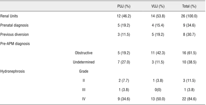
Table 1 - Number of children with persistent hydronephrosis.
PUJ (%) VUJ (%) Total (%)
Renal Units 12 (46.2) 14 (53.8) 26 (100.0)
Prenatal diagnosis 5 (19.2) 4 (15.4) 9 (34.6)
Previous diversion 3 (11.5) 5 (19.2) 8 (30.7)
Pre-APM diagnosis
Obstructive 5 (19.2) 11 (42.3) 16 (61.5)
Undetermined 7 (27.0) 3 (11.5) 10 (38.5)
Hydronephrosis Grade
II 2 (7.7) 1 (3.8) 3 (11.5)
III 1 (3.8) 0(0) 1 (3.8)
IV 9 (34.6) 13 (50.0) 22 (84.6)
to reach the collecting system, avoiding the renal hilum lowering the risks of bleeding and leakage of urine, below the 12th rib, to avoid pneumothorax.
Intrarenal pelvis with a lower degree of hy-dronephrosis was accessed using radioscopy, after cystoscopy and ascending pyelogram. The pubic symphysis was considered the zero marker for the water column and the permeability of the system was tested by an increase of the pressure provoked
by manual compression of the abdominal wall. For the infusion, it was used an Y system, using saline with methylen blue + iodine contrast (50%), under a constant pressure of 40 cm H2O, until the com-plete filling of the renal tract under fluoroscopy. From that moment on, the infusion was stopped and the pressure of the renal tract was measured after stabilization of the water column.
was not considered since the measurement of the urinary tract pressure was done intermittently and after interruption of the infusion (14).
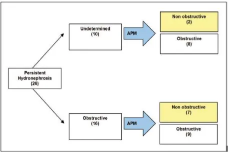
RESULTS
From a total of 26 exams, 10 were per-formed in children with pre-operatory exams with undetermined results (Figure-2). In all patients the exam was able to discriminate obstructive and non-obstructive cases, guiding the choice of treat-ment. Among the 8 patients with obstruction, with median values of 30 cm H2O (10-50 cm H2O), 7 were reoperated (one ureterostomy, two new
py-Among the 16 exams performed in patients with previous diagnosis of obstruction (ultrasound/ intravenous pyelogram/99mTC-DTPA), in 9 the obstruction was confirmed and they were submit-ted to surgery (1 ureterostomy; 1 pyelostomy; 1 ureteral meatotomy; 1 ureteral reimplantation; 2 uretero-uretero-anastomosis; 1 pyelo-pyelic anas-tastomosis; 1 nephrostomy; 1 new pyeloplasty).
Seven tests were considered non-obstruc-tive and clinically treated, without prejudice to function (according to DMSA) in a median fol-low-up of 7 years (Figure-2). Only one renal unit (anomaly of PUJ) presented pressure of 16 cm H2O and was considered non-obstructive since this was
Figure 2 - Flowchart of cases and test results.
eloplasties, two uretero-calix-anastomosis and two endopyelotomies).
Only one was submitted to ureteral cathe-terization due to end-stage renal disease (posterior urethral valve). In two patients of this group, the test revealed non-obstructive hydronephrosis (10-14 cm H2O), avoiding unnecessary new surgeries. For those patients the treatment of choice was ex-pectant and they were stable on the follow-up of 3 and 6 years. The grade of ultrasonographic dilation and tubular function (static renal scintigraphy with DMSA – dimercaptossuccinic acid) were stable.
a patient with Prune-Belly Syndrome with high grade bilateral ureteral reflux. No patient showed worsening of renal function during follow-up (10 months – 12 years, median 63.4 months). When studying the nine renal units considered non-ob-structive after the test and conservatively treated (median 77 months), tubular function remained stable even in the persistence of the hydronephro-sis (Table-2).
Those with urinary diversion without proved obstruction at APM were submitted to reversion of diversion (n = 3 nephrostomy withdrawal / n = 1 closure of ureterostomy / n = 1 closure of pyelosto-my) and only two maintained prophylaxis with an-tibiotics due to the presence of voiding disturbances.
DISCUSSION
The test proposed by Whitaker, that com-bines urodynamic and radiological aspects, was in-tended to differentiate residual obstructive hydrone-phrosis or recurrence due to dilation secondary to permanent changes, with five precise indications: persistent dilation of the upper urinary tract fol-lowing surgical correction of obstruction, possible
obstruction of pelviureteric and vesicoureteric junc-tions, safe withdrawal of urinary diversions, evalu-ation of primary defects of ureteral muscle and evaluation of the influence of bladder volume and pressure on the ureteral dynamics (16).
Just like him, several other authors tried to define the process of obstruction of the upper urinary tract however not correlating the grade of obstruc-tion with the risk of renal damage (17). Our study was able to benefit children submitted to at least one surgical procedure that remained with hydronephro-sis, or those with urinary diversion due to obstruc-tion that needed surgery to withdraw the diversion.
In this study the test was safe and able to guide the treatment in all evaluated cases. In those previously classified as undetermined (n- = 10), the
Table 2 - Patients with test results considered non obstructive treated clinically.
Patient Diagnosis/ Surgery USG DMSA(%) RK/LK
DTPA (RK/LK)
Test (cmH2O)
DMSA(%) RK/LK
Follow-up US Post surgical
AFO L PUJ /Pyeloplasty G IV 81 /19 NO/I 10 80/20 3 years GIV
EBS R PUJ /Pyeloplasty GIII 23 /77 O /NO 10 24/76 7 years GIII
IAS L PUJ (Prunne Belly)/L Pyelostomy
G II 4 /96 I/O 16 0/100 10 years GII
KCRL L PUJ/L Pyeloplasty G IV 62 /38 NO/O 11 62/38 5 years GIV
LSR R PUJ/R Pyeloplas-ty + R nephrostomy
GII 92 /8 O/I 10 91/9 12 years GI
JMM L VUJ/L reimplan-tation
G IV 79 /21 NO/I 14 78/22 6 years GIV
LAT Bilateral VUJ / Vesicostomy +R Pyelostomy + L
Ureterostomy
Exclusion/ G IV
Exclusion RK
I/O 9 6/94 2 years GIV
LMSC Bilateral VUR / Bila-teral reimplantation
GIV /GII 15 /85 O /NO 10 21/79 3 years GIV
TCB Bilateral PUJ /Pye-loplasty +bilateral
nephrostomy
test was able to propose two different groups, those with obstruction (n = 8) and those without obstruc-tion (n = 2), that were clinically treated without the need of surgical intervention in a median follow up period of 4.5 years.
In cases with previous diagnosis of ob-struction (n = 16), it was observed a disparity between 99m Tc-DTPA and the APM test (18,19); the obstruction was confirmed in only nine renal units (56.3%).
Wang et al. agreed that the renogram with only diuretics is not a reliable parameter for the di-agnosis of obstructive hydronephrosis after surgery of renal units with functional deficit and they sug-gested that the measurement of the antero-posteri-or diameter and the evaluation of the renal func-tion must be considered for the confirmafunc-tion of the presence of obstruction (20).
None renal unit was lost during follow up, that ranged from 10 months to 12 years (median = 63.4 months) and only one child presented signifi-cant loss of renal function following pyeloplasty (pre-test). In this child, the dynamic radionuclide renogram after surgery was considered undeter-mined due to a gross functional deficit and the APM was considered obstructive, resulting in a ure-tero-calyx-anastomosis. After this procedure, the child is asymptomatic with stable renal function after one year of follow-up. Four patients presented end-stage renal disease at the moment of the test: one due to posterior urethral valve, two with Prune Belly Syndrome and high grade bilateral ureteral reflux and one patient with a solitary kidney with obstruction of the vesicoureteric junction.
All children maintained DMSA and se-rum creatinine levels stable, and were attended by a multidisciplinary team of pediatricians and nephrologists, and were non-dependent of dialy-sis. In spite of the chosen treatment (surgical or clinical), all children maintained their hydrone-phrosis during follow-up, although with stability of tubular function (DMSA) and absence of renal infections.
The main criticism of the test proposed initially by Whitaker was the low reproducibility mainly due to the high grade flow of infusion (10 mL/min.), leading to a non-real rise of the upper urinary tract pressure (17).
Ripley (1982) and Woodbury (1989) pro-posed the infusion of saline under constant pres-sure and presented high reproducibility with simple equipment. These tests were initially per-formed in animals without ureteral dilation, and in this situation, ureteral contraction in response to constant flow may have been more evident than in a chronically dilated ureter with atrophic ureteral musculature (4,7).
The study of the ureteral opening pres-sure (by constant flow or prespres-sure) has a high positive predictive value when above 14 cm H2O (8). Otherwise, Veenboer and Jong showed a high negative predictive value (12). In the present study, all patients treated clinically had no progression of the obstruction (asymptom-atic and without renal damage) in a follow-up that varied from 2 to 12 years.
In 1981, Newhouse et al. already advocat-ed the proposal of a superior normal limit of the renal perfusion pressure of dilated patients (21). After that, several experimental studies showed that pressures on the renal system above 14 cm H2O determined adverse alterations of the renal blood flow, glomerular filtration rate and tubular function; with pressures above 20 cm H2O there was apoptosis and lowering of the vascular endo-thelial growth factor (22).
This study did not require urodynamic studies or complex mathematical formulae and avoided unnecessary surgeries in nine cases (34.6%), guiding treatment in 100% of the cas-es, in agreement with the study of Lupton and George (23).
There were three modifications of the originally proposed test by Whitaker, without changing the results: 1. Infusion with constant pressure without the need of an infusion pump (simplifying the test, with easily reproducible re-sults); 2. Patient position (oblique lateral recum-bency), and 3. Applying the tests in cases initially considered undetermined (15-22 cm H2O) as if ob-structive, due to the maintenance of the dilation grade and persistence of urinary infection, with-out any prejudice to function or behavior (24).
can be reproducible in any hospital dedicated to pediatric patients.
CONCLUSIONS
Antegrate Pressure Measure is a safe di-agnostic tool with minimal morbidity. The test guided the treatment of all cases and avoided un-necessary new surgeries in one third of the stud-ied cases and helped understand the dynamic and radiological behavior of the upper urinary tract, with a considerable clinical value in the follow-up of children suspected with urinary obstruction. The test is an important tool to evaluate persistent post-surgical dilation of the urinary tract, when used selectively and mainly in the presence of functional deficit.
“That intangible art of clinical judgment must still dominate practice of medicine”
ABBREVIATIONS
DTPA: Diethylene triamin pentacetic acid
APM: Antegrade pressure measurement
USG: Ultrasonography
PUV: Posterior urethral valve
PUJ: Pelviureteric junction
VUJ: Vesicoureteric junction
DMSA: Dimercaptossuccinic acid
CONFLICT OF INTEREST
None declared.
REFERENCES
1. Whitaker RH, Buxton-Thomas MS: A comparison of pres-sure flow studies and renography in equivocal upper urinary tract obstruction. J Urol. 1984; 131: 446-9.
2. Whitaker RH: Methods of assessing obstruction in dilated ureters. Br J Urol. 1973; 45: 15-22.
3. Tchetgen MB, Bloom DA: Robert H. Whitaker and the Whita-ker test: a pressure-flow study of the upper urinary tract. Urology. 2003; 61: 253-6.
4. Ripley SH, Somerville JJ: Whitaker revisited. Br J Urol. 1982; 54: 594-8.
5. Ellis JH, Campo RP, Marx MV, Cohan RH, Platt JF, Sonda LP, et al.: Positional variation in the Whitaker test. Radiology. 1995; 197: 253-5.
6. Wentzell PG, Arnold AJ, Carty H, Rickwood AM: Two-needle modification of the Whitaker test. Br J Urol. 1988; 62: 388. 7. Woodbury PW, Mitchell ME, Scheidler DM, Adams MC, Rink
RC, McNulty A: Constant pressure perfusion: a method to determine obstruction in the upper urinary tract. J Urol. 1989; 142: 632-5; discussion 667-8.
8. Fung LC, Churchill BM, McLorie GA, Chait PG, Khoury AE: Ureteral opening pressure: a novel parameter for the evalua-tion of pediatric hydronephrosis. J Urol. 1998; 159: 1326-30. 9. Senac MO Jr, Miller JH, Stanley P: Evaluation of obstructive
uropathy in children: radionuclide renography vs. the Whita-ker test. AJR Am J Roentgenol. 1984; 143: 11-5.
10. Lupton EW, Richards D, Testa HJ, Gilpin SA, Gosling JA, Bar-nard RJ: A comparison of diuresis renography, the Whitaker test and renal pelvic morphology in idiopathic hydronephro-sis. Br J Urol. 1985; 57: 119-23.
11. Jones RA, Grattan-Smith JD, Little S: Pediatric magnetic reso-nance urography. J Magn Reson Imaging. 2011; 33: 510-26. 12. Veenboer PW, de Jong TP: Antegrade pressure
measure-ment as a diagnostic tool in modern pediatric urology. World J Urol. 2011; 29: 737-41.
13. Fernbach SK, Maizels M, Conway JJ: Ultrasound grading of hydronephrosis: introduction to the system used by the Society for Fetal Urology. Pediatr Radiol. 1993; 23: 478-80. 14. Gill B, Levitt S, Kogan S, Reda E, Weiner S, Donner K: The
dilated urinary tract in children. Prospective analysis with correlation of radiological, isotope, pressure perfusion and surgical findings. Br J Urol. 1988; 61: 413-9.
15. Pfister RC, Papanicolaou N, Yoder IC: Diagnostic morpho-logic and urodynamic antegrade pyelography. Radiol Clin North Am. 1986; 24: 561-71.
16. Jaffe RB, Middleton AW Jr.: Whitaker test: differentiation of obstructive from nonobstructive uropathy. AJR Am J Roent-genol. 1980; 134: 9-15.
17. Wåhlin N, Magnusson A, Persson AE, Läckgren G, Sten-berg A: Pressure flow measurement of hydronephrosis in children: a new approach to definition and quantification of obstruction. J Urol. 2001; 166: 1842-7.
18. Gonzalez R, Chiou R: The diagnosis of upper urinary tract obstruction in children: comparison of diuresis renography and pressure flow studies. J Urol. 1985; 133: 646-9. 19. Dacher J, Pfister C, Thoumas D, Véra P, Liard-Zmuda A,
Chomant J, et al.: Shortcomings of diuresis scintigraphy in evaluating urinary obstruction: comparison with pressure flow studies. Pediatr Radiol. 1999; 29: 742-7.
21. Newhouse JH, Pfister RC, Hendren WH, Yoder IC: Whitaker test after pyeloplasty: establishment of normal ureteral perfu-sion pressures. AJR Am J Roentgenol. 1981; 137: 223-6. 22. Fung LC, Khoury AE, McLorie GA, Chait PG, Churchill BM:
Evaluation of pediatric hydronephrosis using individualized pressure flow criteria. J Urol. 1995; 154(2 Pt 2): 671-6.
23. Lupton EW, George NJ: The Whitaker test: 35 years on. BJU Int. 2010; 105: 94-100.
24. Vela-Navarrete R: Constant pressure flow-controlled antegrade pyelography. Eur Urol. 1982; 8: 265-8.
Correspondence address:
Dr. Marcio Lopes Miranda University of Campinas - UNICAMP, Campinas Division of Pediatric Surgery, Genitourinary Division Cidade Universitária “Zeferino Vaz” Barão Geraldo, Campinas, 13083-970, SP, Brazil Fax: +55 19 3521-2121 E-mail: [email protected]
EDITORIAL COMMENT
The authors should be congratulated for revisiting the results with a modification of the Whitaker test, in which has had loose popularity in the past decades. The authors described how many patients who maintain the hydronephrosis after up-per urinary tract surgery will have a pelvic pressure higher than 14 cmH20. This is important data.
Despite being an interesting study, it fails to validate Whitaker test or its modifications as a standard diagnosis test of obstruction after surgery. The endpoint used by the authors was the neces-sity of reoperation in cases of pyelic pressure above 14 cm H20. However, because we do not know the specificity of this method in humans we cannot rule out that in some cases the operation may be unnecessary. The best answer to the research ques-tion would be the improvement of renal dilataques-tion in the long term.
In significant renal dilatations, there is fail-ure of pelvic and calicial muscles because of the high intrarenal pressure before surgery. After cor-rective surgery, many children persist with the same renal dilatation as before (1). Methods to evaluate diagnosis that may better interpret theses cases are welcome. Renal scintigraphy with DTPA or MAG3 has a lot of limitations, and most of the time does not differentiate between obstruction and significant dilatation without obstruction. This exam commonly gives a false diagnosis of obstruction in very dilated kidneys. When the diuretic renogram is equivocal, usually there is no kidney obstruction. In this study,
surgery was indicated for patients with an equivo-cal curve of obstruction. Furthermore, the authors reported that the test was performed in Grade II and III hydronephrosis. Usually, less than Grade IV hy-dronephrosis are not obstructed. Therefore, all these considerations suggest that, in this series, some pa-tients could have been treated conservatively.
Whitaker test or its modifications might be useful sometime, however it cannot be considered the gold standard. It does not reproduce the py-elic physiology. It is not well demonstrated in hu-mans to which pyelic pressure is associated to loss of kidney function. Therefore, the cutoff point of 14 cmH20 for surgery indication is only a theory and should not be used as the only indication of reoperation. In my view, the best way to diagnosis urinary tract obstruction after surgery is by scin-tigraphic loss of kidney function and increase of hydronephrosis on ultrasound. The place of APA in the clinical practice is still obscure.
REFERENCES
1. Barroso U,Jr., Barroso VA, Calado A, Zerati M: Renal ultra-sonographic findings before and after pyeloplasty. Int Braz J Urol 2000; 26: 190-5.
Dr. Ubirajara Barroso Jr.