Cells Independent of Foxp3 Induction
Takatoku Oida¤, Howard L. Weiner*
Center for Neurologic Diseases, Brigham and Women’s Hospital, Harvard Medical School, Boston, Massachusetts, United States of America
Abstract
Background: It has been reported that human FOXP3+ CD4 Tregs express GARP-anchored surface latency-associated peptide (LAP) after activation, based on the use of an anti-human LAP mAb. Murine CD4 Foxp3+Tregs have also been reported to express surface LAP, but these studies have been hampered by the lack of suitable anti-mouse LAP mAbs.
Methodology/Principal Findings:We generated anti-mouse LAP mAbs by immunizing TGF-b2/2 animals with a mouse
Tgfb1-transduced P3U1 cell line. Using these antibodies, we demonstrated that murine Foxp3+CD4 Tregs express LAP on their surface. In addition, retroviral transduction of Foxp3 into mouse CD4+CD252T cells induced surface LAP expression. We then examined surface LAP expression after treating CD4+CD252 T cells with TGF-b and found that TGF-b induced surface LAP not only on T cells that became Foxp3+but also on T cells that remained Foxp32after TGF-btreatment. GARP expression correlated with the surface LAP expression, suggesting that surface LAP is GARP-anchored also in murine T cells.
Conclusions/Significance:Unlike human CD4 T cells, surface LAP expression on mouse CD4 T cells is controlled by Foxp3 and TGF-b. Our newly described anti-mouse LAP mAbs will provide a useful tool for the investigation and functional analysis of T cells that express LAP on their surface.
Citation:Oida T, Weiner HL (2010) TGF-bInduces Surface LAP Expression on Murine CD4 T Cells Independent of Foxp3 Induction. PLoS ONE 5(11): e15523. doi:10.1371/journal.pone.0015523
Editor:Derya Unutmaz, New York University, United States of America
ReceivedAugust 8, 2010;AcceptedOctober 6, 2010;PublishedNovember 24, 2010
Copyright:ß2010 Oida, Weiner. This is an open-access article distributed under the terms of the Creative Commons Attribution License, which permits unrestricted use, distribution, and reproduction in any medium, provided the original author and source are credited.
Funding:This work was supported by the National Institutes of Health Grants AI435801 and NS38037. The funders had no role in study design, data collection and analysis, decision to publish, or preparation of the manuscript.
Competing Interests:The authors have declared that no competing interests exist.
* E-mail: [email protected]
¤ Current address: BioLegend Inc., San Diego, California, United States of America
Introduction
TGF-b controls immune responses by multiple mechanisms including the suppression of Th1 and cytotoxic lymphocytes, and the induction of Th17 cells depending on the context [1]. TGF-bis first synthesized as pro-TGF-band is then intracellularly processed by furin proprotein convertase to form a latent TGF-b complex which consists of non-covalently associated dimmers of the N-terminal region of pro-TGF-b (latency-associated peptide, LAP) and of the C-terminal region of pro-TGF-b(mature TGF-b) [2]. Expression of pro-TGF-b, LAP, latent TGF-b and/or mature TGF-b(hereafter referred as LAP/TGF-b) on mouse CD4 T cells was first reported in 2001 by Nakamura et al. [3]. They proposed that CD4+
CD25+
regulatory T cells (Tregs) mediated their suppressive function by presenting active TGF-b to effector cells in a cell-cell contact manner. They used a polyclonal chicken anti-TGF-bantibody and a monoclonal anti-human LAP mAb (clone 27232) for FACS staining of mouse CD4 T cells. Our laboratory has also reported the presence of surface LAP+
on mouse T cells using a polyclonal goat anti-human LAP antibody [4,5]. However, use of a polyclonal antibody is problematical due to the inherent variance between different polyclonal preparations. The anti-LAP mAb (clone 27232) used by Nakamura, et al., was raised against recombinant human LAP (R&D Systems). Although Nakamura et al. used this antibody to stain mouse CD4 T cells [3], in our hands,
we did not find that this anti-human LAP mAb cross-reacted with mouse LAP. Thus, although clone 27232 stained humanTGFB1 -transduced cells [6], it did not stain mouseTgfb1-tranduced cells at all (Figure S1). To overcome these problems, a fully characterized anti-mouse LAP mAb would be required for staining mouse T cells. Recently, by using the anti-human LAP mAb 27232 [7,8], it was reported that human FOXP3+
Tregs express surface LAP after activation and that the surface LAP is anchored by GARP/ LRRC32 [8,9].
We raised anti-mouse LAP mAbs by immunizing TGF-b2/2mice with mouseTgfb1-transduced cells, and used them to stain mouse CD4 T cells. We found that the majority of mouse Foxp3+CD4 T cells expressed surface LAP after activation. Surface LAP was induced by Foxp3-transduction into mouse CD4+
CD252 T cells and by addition of TGF-bto mouse CD4+
CD25-T cell cultures. In contrast to human T cells [8], TGF-binduced surface LAP not only on T cells that converted to Foxp3+but also on T cells in which Foxp3 was not expressed. GARP expression correlated with the surface LAP expression suggesting that surface LAP is anchored by GARP.
Results
Generation of anti-mouse LAP mAbs
TGFB1-transduced P3U1 (P3U1-huTGF-b) cells express LAP/ TGF-bon the surface [6]. Surface expression of murine LAP was also expected on P3U1-muTGF-bcells since we found that anti-TGF-b(clone 9016) surface stained P3U1-muTGF-bcells as well as P3U1-huTGF-b cells (Figure S1). We elected to immunize TGF-b-deficient mice. TGF-b2/2mice manifest an autoimmune syndrome and die at 3–4 wks after birth [10,11]. We attempted to prolong their life by injecting galectin-1, which has been reported to suppress other autoimmune diseases [12], starting at day 7 of birth. P3U1-muTGF-bcells were injected i.p. every other day 5 times beginning at day 8 after birth, and spleen cells were taken at day 22 after birth and fused with P3U1 myeloma cells. The hybridoma cells were grown in hypoxanthine-aminopterin-thymi-dine (HAT)-supplemented methylcellulose medium. Approximate-ly 2,800 clones were picked from the plates, and transferred to hypoxanthine-thymidine (HT)-supplemented DMEM in 96-well plates. The culture supernatants were screened by surface staining of P3U1-muTGF-bcells by FACS. Thirty-six positive clones were selected and recovered (TW7 series) (Figure S2). Of the 36 clones, 32 clones were IgG and 4 clones were IgM. To check their specificity, we tested the ability of the antibodies to immunopre-cipitate Flag-tagged mouse LAP (Flag-mLAP) produced by retrovirally Flag-mLAP-transduced P3U1 cells. Of the 32 IgG clones, 26 clones, including TW7-16B4 and TW7-20B9, immu-noprecipitated Flag-mLAP (Figure S3, underlined) and thus were true anti-mouse LAP mAbs. Several clones, including 28G11, did not immunoprecipitate Flag-mLAP (Figure S3). TW7-28G11, however, stained human latent TGF-b-coated beads, but not human LAP- or human active TGF-b-coated beads (Figure S4A). TW7-28G11 immunoprecipiated Flag-mLAP only when active TGF-b was exogenously added to Flag-mLAP solution (Figure S4B), and immunoprecipiated pro-TGF-b and latent TGF-b from the culture supernatant of P3U1-muTGF-b cells (Figure S5A). These results indicate that TW7-28G11 is a conformation specific anti-mouse/human latent TGF-b /pro-TGF-b mAb which recognize LAP and TGF-bin combination. The specificity of some clones, including TW7-16B4, TW7-20B9 and TW7-28G11, were further confirmed by testing their ability to detect mouse pro-TGF-b and/or LAP by Western blot (Figure S5B), and by their ability to immunoprecipiate pro-TGF-b and latent TGF-b from culture supernatant of P3U1-muTGF-bcells (Figure S5A).
Surface LAP expression on mouse Foxp3+CD4 T cells
It has been reported that human FOXP3 Tregs express surface LAP after activation [7,8] by a GARP-mediated anchoring mechanism [8,9]. We tested our anti-LAP/TGF-bmAbs for their ability to stain pre-activated mouse CD4 T cells. CD4 T cells were stimulated with plate-bound anti-CD3/anti-CD28 for 2 days and rested for 1 day. Following this, they were surface stained with anti-LAP/TGF-b mAbs using PE-labeled anti-mouse IgG1 (for
IgG1subtype clones) or anti-mouse Igk secondary antibody (for
non-IgG1clones), then fixed and intracellularly stained with
anti-Foxp3-Alexa Fluor647. Of the 36 potential anti-LAP/TGF-b candidate clones, 31 clones surface stained Foxp3+
cells. Three representative clones (TW7-16B4, TW7-20B9, and TW7-28G11) are shown in Figure 1A and all 36 clones are shown in Figure S6. It should be noted that 24 of the 26 clones which immunopre-cipitated Flag-mLAP as described above stained Foxp3+
CD4 T cells with a similar pattern. Among them, TW7-16B4 produced the highest staining signal followed by TW7-20B9. An anti-pro-TGF-b/latent TGF-b clone, TW7-28G11, also stained Foxp3+ CD4 T cells (Figure 1A and Figure S6), suggesting that surface LAP exists as pro-TGF-b and/or latent TGF-b rather than free
LAP without mature TGF-b. Surface LAP staining strongly correlated with GARP expression (Figure 1B), indicating that surface LAP on mouse Foxp3+
CD4 Tregs is also anchored by GARP as on human FOXP3+CD4 Tregs.
For further analysis we selected TW7-16B4 (IgG1,k) and
TW7-20B9 (IgG1,k) as the highest staining anti-LAP clones, and
TW7-28G11 (IgG2b, k) as an anti-pro-TGF-b/latent TGF-b clone.
These clones were used with secondary antibodies or as antibodies directly labeled with PE or Allophycocyanin (APC). We tested whether unstimulated CD4 T cells also express surface LAP using the direct conjugates. We found that freshly prepared mouse CD4+
25+
T cells also weakly expressed surface LAP (Figure 2A). We also investigated the time course of surface LAP expression. We found that surface LAP expression on Foxp3+cells peaked on days 1 and 2, and then gradually decreased when the cells were rested (days 3 and 5) (Figure 2B, upper panels). We found that GARP was co-expressed with LAP in all time points (Figure 2B, lower panels).
Foxp3-induced surface LAP expression
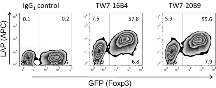
We then asked whether surface LAP expression is controlled by Foxp3. We found that retroviral Foxp3 transduction into mouse CD4+
CD252 T cells induced surface LAP (GFP+
population vs. GFP2 population in Figure 3). This result demonstrates that surface LAP is under control of Foxp3.
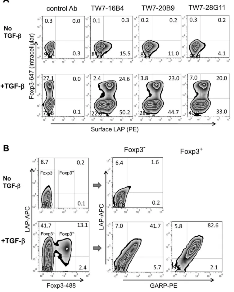
TGF-b-induced surface LAP expression
TGF-b converts Foxp32 CD4 T cells into induced Foxp3+ Tregs (iTregs) [1]. To determine whether iTregs also express surface LAP, we stimulated mouse CD4+
CD252 T cells in the presence or absence of recombinant TGF-b and checked for surface LAP expression. As expected ,25% of CD4+CD25- T cells were converted to Foxp3+
iTregs in presence of TGF-b (Figure 4A). We found that these iTregs expressed surface LAP. Interestingly, the Foxp32-remaining cells also became surface LAP+
cells following culture in the presence of TGF-b. GARP expression correlated with surface LAP expression on both Foxp3+ cells and Foxp32cells (Figure 4B), suggesting that surface LAP is GARP-dependent not only on natural Tregs and iTregs cells but also on non-Tregs.
It is possible that surface LAP expression on natural Foxp3+ Tregs might also be maintained by TGF-b produced by Tregs themselves. We found, however, that the ALK5 inhibitor or anti-TGF-b 1D11 did not affect surface LAP expression or GARP expression on Foxp3+ Tregs (Figures S7 and S8). Thus, these results suggest that surface LAP expression on Foxp3+
Tregs is independent of TGF-b.
Discussion
The existence and function of surface LAP on Tregs has been a matter of debate. Contrary to the first report by Nakamura et al. [3], Shevach’s group questioned the function of TGF-bin Treg-mediated suppression [13], and their staining of mouse T cells was quite faint, if at all present [14]. As a part of our investigation of TGF-b, we found that the anti-human LAP mAb 27232 used by Nakamura et al. does not cross-react with mouse LAP (Figure S1). In this report, we raised anti-mouse LAP mAbs by immunization of TGF-b2/2mice and revisited the existence of surface LAP on mouse CD4 T cells. We found that anti-mouse LAP mAbs stained majority of Foxp3+
Tregs, but not Foxp32T cells after activation (Figure 1A). Fresh CD4+CD25+T cells also expressed surface LAP at a weak level (Figure 2A). Thus our results establish that mouse Foxp3+
Tregs do express surface LAP. It should be mentioned,
however, that it is not yet determined whether surface LAP/TGF-bhas a functional contribution to Treg-mediated suppression.
Using the anti-LAP mAb 27232 [7,8] it was recently reported that human FOXP3+
Tregs express surface LAP and that the surface LAP is anchored by GARP [8,9]. It appears that this is also the case with mouse CD4 T cells since GARP expression strongly correlated with surface LAP expression (Figure 1B and Figure 2B).
We recently reported the occurrence of GARP-independent, GRP78-associated surface LAP onTGFB1-transduced cells [6]. It is unknown at this time whether GARP-independent surface LAP also can be seen on T cells.
In humans, TGF-b-induced FOXP3+ CD4 T cells do not express surface LAP or GARP [8]. On the contrary, in mice, not only did TGF-b-induced Foxp3+
CD4 T cells express surface LAP
Figure 1. Surface LAP/TGF-bexpression on mouse activated CD4 T cells.(A) BALB/c CD4 T cells were stimulated with plate-bound anti-CD3/ anti-CD28 for 2 days and rested for 1 day. The cells were surface stained with anti-LAP mAbs using PE-labeled secondary antibodies, then intracellularly stained with anti-Foxp3-Alexa Fluor647. Staining with representative clones, anti-LAP mAbs TW7-16B4 and TW7-20B9, and anti-latent TGF-b/pro-TGF-bmAb TW7-28G11, are shown. (B) Activated BALB/c CD4 T cells were stained with anti-LAP TW7-20B9 (surface) and anti-Foxp3 (intracellular) (left), with anti-GARP (surface) and anti-Foxp3 (intracellular), or anti-LAP TW7-20B9 (surface) and GARP (surface) (right).
and GARP, but TGF-b-exposed CD4+
CD252T cells that did not become Foxp3+
CD4 T cells also expressed surface LAP and GARP (Figure 4B). Some Foxp32 CD4 T cells also expressed surface LAP/TGF-b without exogenous of TGF-b. We do not know whether this LAP/TGF-bexpression was induced by TGF-b in an autocrine fashion or occurred independent of TGF-b. However, TGF-b signaling seems not absolutely required for surface LAP expression since natural Foxp3+
Tregs maintained surface LAP expression even when TGF-bsignaling was blocked
(Figure S7). Thus, surface LAP expression may be controlled independently by Foxp3 and TGF-bsignaling.
In summary, we raised anti-mouse LAP mAbs and revisited surface LAP expression on mouse CD4 T cells. We found that Foxp3+
Tregs expressed surface LAP and that surface LAP is induced by forced expression of Foxp3 or by TGF-birrespective of Foxp3 induction. Furthermore, surface LAP expression strongly correlated with GARP, suggesting that surface LAP is GARP-mediated. These newly described anti-mouse LAP mAbs will
Figure 2. Surface LAP/TGF-bexpression on mouse unstimulated CD4 T cells and time course analysis.(A) Freshly prepared BALB/c CD4 T cells were surface stained with PE-conjugated anti-LAP/TGF-bmAbs (TW16B4, TW20B9, or TW28G11), anti-CD25-FITC, anti-CD4-APC, and 7-AAD. CD4+
7-AAD2cells were gated. (B) BALB/c CD4 T cells were stimulated with plate-bound anti-CD3/anti-CD28 for two days, and then split in 10%
FBS-IMDM containing 100 U/ml IL-2. The cells were surface stained with PE-conjugated anti-LAP TW7-20B9 followed by anti-Foxp3-Alexa Fluor647 (intracellular staining) (upper panels), or with APC-conjugated anti-LAP TW7-20B9 and GARP-PE (lower panels).
doi:10.1371/journal.pone.0015523.g002
provide a useful tool for functional analysis of T cells that express LAP on their surface.
Materials and Methods
Generation of anti-mouse LAP mAbs (TW7 series)
Mice were housed in a pathogen-free environment and the animal protocols were approved according to the guidelines of the Committee on Animals of Harvard Medical School (Protocol No. 02683). TGF-b2/2 mice [10] were injected i.p. with 20mg galectin-1 (Sigma-Aldrich) [12] every other day starting at 7 day after birth to prevent the fatal autoimmunity seen in TGF-b2/2 mice [11]. MouseTgfb1-transduced P3U1 (P3U1-muTGF-b) cells (clone#11) were injected i.p. at 1-46106cells (in 10–25ml PBS) every other day 5 times starting at 8 days after birth. At age 22 days, the spleen cells were fused with P3U1 myeloma cells, and the hybridoma cells were plated in methylcellulose medium (ClonaCell-HY, Stemcell Technologies). Screening was conducted by surface staining of P3U1-muTGF-b cells by FACS. Anti-mouse LAP specificity was confirmed by immunoprecipitation of recombinant Flag-tagged mouse LAP (lacking C-terminal mature TGF-b sequence) (Flag-mLAP) [15], immunoprecipitation of pro-TGF-b and latent TGF-b, staining recombinant human latent TGF-b (R&D Systems)-coated polystyrene beads, and/or staining recom-binant human TGF-b(R&D Systems) coated polystyrene beads.
Other antibodies and reagents
Anti-human LAP mAb clone 27232, anti-TGF-bmAb clone 9016, and biotinylated goat anti-LAP (BAF246) were obtained from R&D Systems. Anti-mouse CD3 (145-2C11), anti-mouse CD28 (37.51), Allophycocyanin (APC)-labeled goat anti-mouse Ig, PE- or APC-labeled anti-mouse IgG1(A85-1), and PE-labeled
anti-mouse Igk (187.1) were from BD Biosciences. PE-labeled mouse GARP (YGIC86), and Alexa Fluor647-labeled anti-Foxp3 (FJK-16s) were from eBioscience. Alexa Fluor488-labeled anti-Foxp3 (150D) was from Biolegend. TGF-breceptor I kinase inhibitor (ALK5 inhibitor II) was from EMD/Calbiochem. Anti-Flag mAb (M2) was from Sigma-Aldrich. (caga)12-MLP-Luc
TGF-breporter plasmid [16,17] was kindly provided by Dr. D. Vivien (the Universite’ de Caen, Daix, France). Mv1Lu cells (ATCC) were stably transfected with (caga)12-MLP-Luc plasmid
and used for testing dose-response of ALK5 inhibitor II in TGF-b bioassay.
CD4 T cell stimulation and FACS staining
CD4 T cells were separated from BALB/c mice (The Jackson Laboratories) or C57BL/6 background Foxp3-GFP knock-in (Foxp3-KI) mice [18] using MACS CD4 purification kit (Miltenyi Biotec). When CD4+
CD252 T cells were prepared, biotinylated anti-CD25 antibody was additionally mixed to the MCAS antibody cocktail. T cells were stimulated with plate-bound anti-CD3 and anti-CD28 for 2 days. The cells were split into non-coated wells and rested for 1 day, then stained by FACS. Surface LAP staining was conducted by either PE- or APC-directly conjugated anti-mouse LAP mAbs, or unconjugated anti-mouse LAP mAbs followed by PE- or APC-conjugated goat anti-mouse Ig, monoclonal anti-mouse IgG1 or monoclonal anti-mouse Igk
secondary antibody. Intracellular Foxp3 staining was done with Alexa Fluor647- or Alexa Fluor488-labeled anti-Foxp3 using Foxp3 Staining Buffer Set (eBioscience).
Retroviral transduction
Retroviral vector pMCs-IRES-GFP [19], ecotropic retroviral packaging cell line Plat-E [20] were kindly provided by Dr. Kitamura (Tokyo Univ., Tokyo, Japan). Foxp3 was cloned into pMCs-IRES-GFP vector, and the retroviral supernatant was produced by Plat-E. Mouse CD4+252 T cells from BALB/c mice pre-activated with plate-bound anti-CD3 and anti-CD28 for 30 hrs were infected with Foxp3 ecotropic retrovirus by centrifugation at 3,000 rpm for 1 hr. 1 day after infection, the cells were split onto a non-coated wells, and rested. The transduced cells were re-stimulated with plate-bound anti-CD3/anti-CD28 for 14 hrs, rested for 2 days, and surface stained with LAP mAbs and then intracellularly with anti-Foxp3.
Figure 3. Induction of surface LAP by Foxp3 transduction.BALB/c CD4+CD252T cells were stimulated with plate-bound anti-CD3/anti-CD28
and retrovirally transduced with pMCs-Foxp3-IRES-GFP vector. The cells were re-stimulated with plate-bound anti-CD3/anti-CD28 for 14 hrs and transferred to uncoated wells. 2 days after re-stimulation, the cells were stained with anti-LAP TW7-16B4 or TW7-20B9 using anti-mouse IgG1-APC
secondary antibody.
Figure 4. Induction of surface LAP by TGF-b.(A) BALB/c CD4+
CD252T cells were stimulated with plate-bound anti-CD3/anti-CD28 without
Supporting Information
Figure S1 Negative staining of mouse TGF-b-transduced cells with anti-human LAP mAb 27232. Non-transduced P3U1 cells (green), human TGF-b gene (TGFB1)-transduced P3U1 cells (clone #32) (blue), or mouse TGF-b gene (Tgfb1 )-transduced P3U1 (clone#11) cells (red) were surface stained with anti-TGF-bmAb 9016 (left) or with anti-human LAP mAb 27232 (right). Note that mouse TGF-b-transduced P3U1 cells were later found positive with anti-mouse LAP mAbs as shown in Figure S2. (PDF)
Figure S2 Staining of mouse TGF-b-transduced P3U1 cells with TW7 anti-LAP/TGF-bcandidate clones.Mouse TGF-b-transduced P3U1 (clone #11) cells (GFP+
) mixed with non-transduced P3U1 cells (GFP(-)) were surface stained with culture supernatants of anti-LAP/TGF-bcandidate clones (TW7 series) using goat anti-mouse Ig-APC after Fc receptor blocking. Immunoglobulin subtypes are also shown in the figures. Clones identified as anti-LAP in Fig. 3 are underlined.
(PDF)
Figure S3 Immunoprecipitation of Flag-tagged mouse LAP with TW7 anti-LAP/TGF-bcandidate clones.Culture supernatant of P3U1 cells transduced with retroviral pMCs vector carrying Flag-tagged mouse LAP lacking TGF-bsequence (Flag-mLAP) was immunoprecipitated with anti-LAP/TGF-bcandidate clones using anti-mosue IgG BioMag Plus (Polysciences). The immunoprecipitated samples were run on SDS-PAGE under reducing conditions and blotted with anti-Flag mAb M2. Ig H chain and Ig L chain were detected at 55 kDa and at 25 kDa, respectively, and Flag-mLAP migrated at 43 kDa. Clones that immunoprecipitaed Flag-mLAP were marked under the clone numbers. C, MOPC21 IgG1 control; 1, TW7-1C12 (IgG1); 2,
TW7-3G11 (IgM); 3, TW7-4G7 (IgG1); 4, TW7-5A1 (IgG1); 5,
TW7-5B2 (IgG1); 6, TW7-5B5 (IgG1); 7, TW7-5D4 (IgG1); 8,
TW7-5F5 (IgG1); 9, TW7-5G10 (IgG1); 10, TW7-6B3 (IgG1); 11,
TW7-7C7 (IgG1); 12, TW7-7G7 (IgG1); 13, TW7-7H4 (IgG1); 14,
TW7-8C11 (IgG1); 15, TW7-10C10 (IgG1); 16, TW7-11G5
(IgG1); 17, TW7-12E2 (IgG1); 18, TW7-13C5 (IgG1); 19,
TW7-13C8 (IgG1); 20, TW7-13D7 (IgG1); 21, TW7-13E12 (IgG1); 22,
TW7-16A2 (IgG1); 23, TW7-16B4 (IgG1); 24, TW7-17G8 (IgM);
25, TW7-18C4 (IgG2aor2b); 26, TW7-18C9 (IgG2aor2b); 27,
TW7-20B9 (IgG1); 28, TW7-22F7 (IgG1); 29, TW7-22F9 (IgG2a
or 2b); 30, TW7-22H5 (IgG1); 31, TW7-23D12 (IgG1); 32,
TW7-24B11 (IgG1); 33, TW7-24E3 (IgM); 34, TW7-24G5 (IgG1); 35,
TW7-26E10 (IgM); 36, TW7-28G11 (IgG2b).
(PDF)
Figure S4 Characterization of TW7-28G11 clone. (A) Recombinant human LAP- (left), human latent TGF-b- (middle), or human active TGF-b- (right) coated polystyrene beads were stained with TW7-28G11 mAb using goat anti-mouse Ig-APC. (B) Culture supernatant of Flag-mLAP-transduced P3U1 cells with/ without exogenously added recombinant human TGF-b was immunoprecipitated with TW7-28G11 or control Ab. The
samples were run on SDS-PAGE under reducing conditions and blotted with anti-Flag M2 antibody.
(PDF)
Figure S5 Western blotting and immunoprecipitation of LAP/TGF-bby TW7 mAbs.(A) Culture supernatant of P3U1-muTGF-b (clone #11) cells (lane 1), or immunoprecipitated samples from P3U1-muTGF-b culture supernatant with TW7-7H4 (lane 2), TW7-16B4 (lane 3), TW7-20B9 (lane 4), TW7-22F7 (lane 5), TW7-28G11 (lane 6), or or IgG1control MOPC21 (lane
7) were run on SDS-PAGE under non-reducing conditions, and blotted with biotinylated goat anti-LAP Ab. (B) Culture superna-tant of P3U1-muTGF-b(clone#11) cells were run on SDS-PAGE under non-reducing conditions and blotted with TW7-16B4 (lane 1), TW7-20B9 (lane 2), TW7-28G11 (lane 3), or biotinylated goat anti-LAP (lane 4).
(PDF)
Figure S6 Staining of pre-activated mouse CD4 T cells with TW7 anti-LAP/TGF-b mAb series. BALB/c CD4 T cells were stimulated with plate-bound anti-CD3/anti-CD28 for 2 days and rested 1 day. The cells were surface stained with TW7 anti-LAP/TGF-b mAbs using PE-labeled anti-mouse IgG1 or
anti-mouse Igksecondary antibodies, then intracellularly stained with anti-Foxp3-Alexa Fluor647 as Figure 2A. Staining with all 36 TW7 clones was shown.
(PDF)
Figure S7 Surface LAP expression under TGF-b block-ing conditions. B6 background Foxp3-GFP knock-in CD4 T cells were stimulated with plate-bound anti-CD3/anti-CD28 in presence of 10 ng/ml recombinant human TGF-b, 1mM ALK5 inhibitor II (Figure S8), or 50mg/ml anti-TGF-bmAb 1D11 for 2 days, and rested for 1 day. The cells were stained with anti-LAP TW7-16B4 using anti-mouse IgG1-APC secondary antibody and
anti-GARP-PE. The quadrants were set by isotype control staining.
(PDF)
Figure S8 Dose response curve of ALK5 inhibitor II.(A) Mv1Lu cells stably transfected with (caga)12-MLP-Luc vector were
cultured in the presence of recombinant human TGF-bfor 8 hrs, and luciferase was measured. (B) Mv1Lu-(caga)12-MLP-Luc cells
were cultured in presence of 100 pg/ml recombinant human TGF-bwith various concentrations of ALK5 inhibitor II for 8 hrs, and luciferease was measured.
(PDF)
Acknowledgments
We thank Thomas Koeglsperger for providingTgfb12/+mice. Author Contributions
Conceived and designed the experiments: TO HLW. Performed the experiments: TO. Analyzed the data: TO. Contributed reagents/ materials/analysis tools: TO. Wrote the paper: TO HLW.
References
1. Rubtsov YP, Rudensky AY (2007) TGFbsignalling in control of T-cell-mediated self-reactivity. Nat Rev Immunol 7: 443–453.
2. Miyazono K, Ichijo H, Heldin CH (1993) Transforming growth factor-b: latent forms, binding proteins and receptors. Growth Factors 8: 11–22.
with anti-Foxp3-Alexa Fluor647. (B) BALB/c CD4+
CD252T cells were stimulated with/without TGF-b, and then surface stained with ACP-conjugated
anti-LAP TW7-20B9 and GARP-PE, followed by intracellular staining with anti-Foxp3-Alexa Fluor488. Foxp32and Foxp3+cells populations were gated and plotted by LAP and GARP expression.
3. Nakamura K, Kitani A, Strober W (2001) Cell contact-dependent immunosup-pression by CD4+
CD25+
regulatory T cells is mediated by cell surface-bound transforming growth factorb. J Exp Med 194: 629–644.
4. Oida T, Zhang X, Goto M, Hachimura S, Totsuka M, et al. (2003) CD4+
CD252T cells that express latency-associated peptide on the surface suppress CD4+
CD45RBhigh-induced colitis by a TGF-b-dependent mechanism. J Immunol 170: 2516–2522.
5. Ochi H, Abraham M, Ishikawa H, Frenkel D, Yang K, et al. (2006) Oral CD3-specific antibody suppresses autoimmune encephalomyelitis by inducing CD4+
CD252LAP+
T cells. Nat Med 12: 627–635.
6. Oida T, Weiner HL (2010) Overexpression of TGF-b1gene induces cell surface
localized Glucose-Regulated Protein 78-associated Latency-associated peptide/ TGF-b. J Immunol 185: 3529–3535.
7. Tran DQ, Andersson J, Hardwick D, Bebris L, Illei GG, et al. (2009) Selective expression of latency-associated peptide (LAP) and IL-1 receptor type I/II (CD121a/CD121b) on activated human FOXP3+
regulatory T cells allows for their purification from expansion cultures. Blood 113: 5125–5133.
8. Tran DQ, Andersson J, Wang R, Ramsey H, Unutmaz D, et al. (2009) GARP (LRRC32) is essential for the surface expression of latent TGF-bon platelets and activated FOXP3+
regulatory T cells. Proc Natl Acad Sci U S A 106: 13445–13450.
9. Stockis J, Colau D, Coulie PG, Lucas S (2009) Membrane protein GARP is a receptor for latent TGF-b on the surface of activated human Treg. Eur J Immunol 39: 3315–3322.
10. Kulkarni AB, Huh CG, Becker D, Geiser A, Lyght M, et al. (1993) Transforming growth factor b1 null mutation in mice causes excessive inflammatory response and early death. Proc Natl Acad Sci U S A 90: 770–774.
11. Christ M, McCartney-Francis NL, Kulkarni AB, Ward JM, Mizel DE, et al. (1994) Immune dysregulation in TGF-b1-deficient mice. J Immunol 153: 1936–1946.
12. Liu FT, Rabinovich GA (2010) Galectins: regulators of acute and chronic inflammation. Ann N Y Acad Sci 1183: 158–182.
13. Piccirillo CA, Letterio JJ, Thornton AM, McHugh RS, Mamura M, et al. (2002) CD4+
CD25+
regulatory T cells can mediate suppressor function in the absence of transforming growth factorb1 production and responsiveness. J Exp Med 196: 237–246.
14. Andersson J, Tran DQ, Pesu M, Davidson TS, Ramsey H, et al. (2008) CD4+
FoxP3+
regulatory T cells confer infectious tolerance in a TGF-b -dependent manner. J Exp Med 205: 1975–1981.
15. Young GD, Murphy-Ullrich JE (2004) Molecular interactions that confer latency to transforming growth factor-b. J Biol Chem 279: 38032–38039.
16. Dennler S, Itoh S, Vivien D, ten Dijke P, Huet S, et al. (1998) Direct binding of Smad3 and Smad4 to critical TGFb-inducible elements in the promoter of human plasminogen activator inhibitor-type 1 gene. EMBO J 17: 3091–3100. 17. Docagne F, Colloc’h N, Bougueret V, Page M, Paput J, et al. (2001) A soluble
transforming growth factor-b(TGF-b) type I receptor mimics TGF-bresponses. J Biol Chem 276: 46243–46250.
18. Bettelli E, Carrier Y, Gao W, Korn T, Strom TB, et al. (2006) Reciprocal developmental pathways for the generation of pathogenic effector TH17 and regulatory T cells. Nature 441: 235–238.
19. Kitamura T, Koshino Y, Shibata F, Oki T, Nakajima H, et al. (2003) Retrovirus-mediated gene transfer and expression cloning: powerful tools in functional genomics. Exp Hematol 31: 1007–1014.
20. Morita S, Kojima T, Kitamura T (2000) Plat-E: an efficient and stable system for transient packaging of retroviruses. Gene Ther 7: 1063–1066.