CD4
+CD25
+T lymphocytes and regulation
of the immune system: perspectives for a
pathophysiological understanding of sepsis
Linfócitos T CD4+CD25+ e a regulação do sistema imunológico:
perspectivas para o entendimento fisiopatológico da sepse
INTRODUCTION
he incidence of sepsis has increased dramatically during the past two decades. It is estimated that 1.5 million people in the United States and 1.5 million in Europe develop severe sepsis and/or septic shock each
year, 35% to 50% of whom die.(1) his large number of fatal cases has
triggered a broad range of studies aimed at understanding the intricate pathogenic mechanisms of sepsis in association with the development of
immunomodulatory therapy.(1-3)
In patients with sepsis, the systemic inlammatory response is disorganized due to the disruption of the complex balance between pro-
and anti-inlammatory mechanisms.(4) Many components of the human
immune system are involved in anergy and reducing of the response to microorganisms, which characterize typical immunosuppression and may be designated as Compensatory Anti-inlammatory Response Syndrome
(CARS).(5) In this regard, some T lymphocyte populations, that have been
described with increasing details in the literature, seem to evade thymic selection or undergo a process of “thymic education” in which they acquire a status diferent from that of so-called “traditional” lymphocytes. For
example, self-reactive lymphocytes, which mature exclusively in the thymus,
play a prominent role in the regulation of autoimmunity. hese cells are
called CD4+CD25+ regulatory T lymphocytes (Treg).(6)
Treg (CD4+CD25+) cells play an important role in immune regulation.
Rodrigo Siqueira-Batista1, Andréia Patrícia
Gomes1, Sarah Fumian Milward Azevedo1,
Rodrigo Roger Vitorino2, Eduardo Gomes de
Mendonça3, Flávio Oliveira de Sousa4, Alcione de
Paiva Oliveira4, Fábio Ribeiro Cerqueira4, Sérgio
Oliveira de Paula5, Maria Goreti de Almeida
Oliveira3
1. Department of Medicine and Nursing, Universidade Federal de Viçosa - UFV - Viçosa (MG), Brazil.
2. Centro Universitário Serra dos Órgãos - UNIFESO - Viçosa (MG), Brazil.
3. Department of Biochemistry and Molecular Biology, Federal University of Viçosa - UFV - Viçosa (MG), Brazil.
4. Department of Informatics, Universidade Federal de Viçosa - UFV - Viçosa (MG), Brazil. 5. Department of General Biology, Universidade Federal de Viçosa - UFV - Viçosa (MG), Brazil.
ABSTRACT
he systemic inlammatory response represents the core pathogenic event of sepsis, underlying clinical manifestations and laboratory indings in patients. Numerous studies have shown that CD4+CD25+ T lymphocytes, also known as regulatory T lymphocytes (Treg), participate in the development of sepsis due to their ability to suppress the immune response. he present article discusses the role of Treg lymphocytes in sepsis based on a speciic search strategy
(Latin American and Caribbean Health Sciences / Literatura Latino-americana e do Caribe em Ciências da Saúde - LILACS, PubMed, and Scientiic Electronic Library Online - SciELO) focusing on two main topics: the participation of Treg cells in inlammation and immunity as well as perspectives in the computational physiological investigation of sepsis.
Keywords: Regulatory T lymphocytes; Sepsis/pathophysiology; Sepsis/therapy; Inlammation; Immunity; Computer simulation
Conflict of interest: None.
Submitted on July 24, 2012 Accepted on September 28, 2012
Corresponding author:
Notice that the control of the adaptive immune response is critical for the operation of immune system. Among the functions of the immune response that closely depend on such regulation, the responses signaled by self-reactivity are of great importance and are crucial to internal medicine with respect to
autoimmune(7) and infectious diseases. Several studies
have elucidated the role of this subset of T lymphocytes in the pathophysiology of sepsis to explain how this disease arises and to inform the development of new therapeutic strategies.(3,8-10)
With respect to these preliminary considerations, the aim of the present article is to present a succinct review of the role of Treg cells in sepsis, focusing on its pathophysiologic features, and to identify perspectives for scientiic research into the pathophysiology of
this disease via computational modeling/in silico
experimentation.
METHODS
The present article is a result of a specific search strategy of the following databases: LILACS (Literatura
Latino-americana e do Caribe em Ciências de Saúde/
Latin American and Caribbean Health Sciences), PubMed (National Library of Medicine), and SciELO (Scientific Electronic Library Online). The search terms followed DeCS (Descritores de Ciências da
Saúde/Health Sciences Descriptors), particularly T
lymphocyte subsets and sepsis. The term regulatory
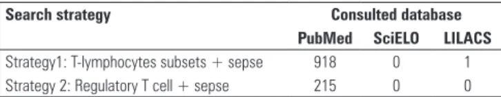
T cells was also used, although it is not listed in DeCs. The following strategies were utilized: strategy 1 - T lymphocyte subsets + sepsis; and strategy 2 - Regulatory T cells + sepsis.
he literature search resulted in 1134 citations, as described in table 1, out of which 50 articles were selected. he criteria for selection prioritized the focus on the regulatory role of Treg cells in the immune system and their participation in the pathophysiology of sepsis.
In addition to the selected articles, immunology and internal medicine textbooks were also used for the literature survey as well as important references previously known by the authors.
RESULTS
he data collected from the selected sources were organized under the following headings: “CD4+CD25+ regulatory lymphocytes” and “CD4+CD25+ T lymphocytes and pathogenesis of sepsis”. A inal short
section titled “Computational Frontiers: in silico
experiments with Treg lymphocytes in sepsis” was also included to present research perspectives in the ield of computational modeling, including experiments developed by the authors.
CD4+CD25+ regulatory T lymphocytes
he human organism has countless mechanisms to promote homeostasis, including the immune system, which comprises a complex combination of interacting elements to maintain the balance between immunity
and immune tolerance(11), still being responsible for
producing an efective response against infectious and non-infectious challenges (e.g., tumors) without triggering autoimmune events or processes that are harmful to the host.(11) In this regard, immune tolerance,
i.e., non-responsiveness to a previously recognized antigen, is crucial and occurs with a signiicant participation by Treg cells.(12)
Origin and suppressive function
Treg cells were initially identiied in the early 1970s in murine models and were later discovered in humans. Treg cells are a T lymphocyte subset characterized by the expression of CD4 and CD25 as well as the transcription factor FoxP3 (forkhead box P3) and are indispensable for the control of the immune response to self and non-self antigens by suppressing efector T cells.(13) Sakaguchi et al.(14) observed that adoptive
transfer of a T lymphocyte population depleted of CD25+ surface cells induced autoimmunity in several organs and systems, particularly in immunodeicient
individuals.(14) he increasing interest in the study of
Treg cells in recent years concerns their role in the maintenance of the mechanisms that ensure self-tolerance and the regulation of the immune response. When the T cell subsets that suppress the immune response were initially described, they were designated suppressor T cells because they were believed to be CD8+ T lymphocytes. However, recent studies have shown that suppressor cells participate in the regulation of the immune response but resemble CD4+ rather than CD8+ T cells. he population of CD4+CD25+ T cells includes the Treg lymphocytes, which may minimize Table 1 - Number of articles identified in literature searches
Search strategy Consulted database
PubMed SciELO LILACS
the proliferation of other T cell populations as it has been shown in vitro.(15) he suppressive efects of these
cells concern both the adaptive (T and B cells) and the innate (monocytes, macrophages, and dendritic cells) immune response. After activation of the T cell receptor (TCR), natural Treg cells hinder the in vivo and in vitro
immune response in a non-speciic antigen manner, by means of a mechanism independent of antigen-presenting cells (APCs) and related to non-restricted
major histocompatibility complex (MHC).(6)
Naïve CD4+CD25+ T cells are produced in the thymus in Hassall’s corpuscles and are activated upon reaching the peripheral blood and secondary lymphoid
organs, thereby acquiring the memory phenotype.(16)
These cells represent approximately 5-15% of the
CD4+ T lymphocytes in peripheral circulation.(17)
The interleukin 7 (IL7) receptor (CD127), which is negatively regulated by the FoxP3 nuclear factor, is currently considered to be the most specific marker for identifying Treg among T lymphocytes and allows the identification of those with greater suppressive capacity.(18)
he mechanisms employed by Treg cells to perform their suppressive function are quite complex and are still the subject of in vitro studies. At least three such
mechanisms are postulated:(19)
- physical contact (in time and space) between Treg
and CD4+ efector cells whereby CTLA-4 (cytotoxic T
lymphocyte antigen 4) releases inhibitory signals after binding to the CD80 receptor of dendritic cells or activated T cells;(20)
- participation of inhibitory cytokines, such as
IL-10 and tumor growth factor (TGF) β1, which have
been observed in an in vivo study;(21) IL-10 inhibits
the activation of APCs, is an antagonist of interferon (INF)-γ, and controls inlammatory processes;(21)
- competition with target cells for growth factors, particularly IL-2, possibly resulting in apoptosis triggered by cytokine deprivation.(19)
In addition to the mechanisms listed above, a fourth possible immunosuppressive mechanism has been described whereby regulatory T cells acquire cytotoxic activity and release granzymes and perforins, leading to the cytolysis of target cells.(22)
It is important to note that efector T cells resistant to suppression by Treg cells have been described.(23)
Groups of Treg lymphocytes
Currently, Treg cells are divided into two groups: natural and adaptive cells.(24)
Natural Treg cells constitutively express surface CD25+ and are therefore designated CD4+CD25+
T lymphocytes.(25) In addition to the CD25 marker,
natural Treg lymphocytes express other surface components that, although non-speciic, also contribute to the identiication of these cells. Some of the most important such components are: CTLA-4, GITR (Glucocorticoid-induced tumor necrosis factor receptor), TNFR-2 (tumor necrosis factor receptor
2), and HLA-DR (human leukocyte antigen).(19)
Natural Treg cells are further characterized as CD4+ T lymphocytes that express the alpha chain of the high-ainity IL-2 receptor (CD25) but do not express other markers typical of activated T cells. Indeed, genetic deiciency of the IL-2 receptor or IL-2 itself
results in the development of autoimmune diseases.(12)
Binding and the resulting paucity of IL-2 is one likely mechanism of suppression employed by Treg cells, as mentioned above. Signiicant evidence has been
found in vitro that the efector function of CD4+ T
cells is minimized by this mechanism; however, in vivo
experimental evidence is lacking. his same study also
demonstrated in vivo that the homeostasis of both Treg
and CD8+ T lymphocytes is susceptible to regulation by IL-2.(26)
Although the stimuli that trigger the production and development of natural Treg cells are not fully elucidated, it has been suggested that recognition of self antigens in the thymus mediated by high-ainity TCRs is the signal involved in this process.(12)
Some surface receptors of Treg cells, such as CD27, Fas, CD26L, and the chemokine receptors CCR6, CCR7, CCR8, and CD103, allow for their migration to the sites where inlammation occurs. Nevertheless, these markers relect the activated state of T lymphocytes and are not speciic to the Treg cell subset.(24)
Adaptive Treg cells are produced at peripheral sites under the inluence of a myriad of antigenic stimuli or under tolerogenic conditions and exert their suppressive function through the release of cytokines, such as
IL-10 and TGF-β.(27) hese cells include TR1 (type 1
regulatory cells), TR3, CD4+CD8+ T cells, natural killer cells (NK), suppressor CD8+ lymphocytes, and
gamma-delta T cells.(28) TR1 cells can control memory
T cells both in vitro and in vivo and suppress h1- and h2-mediated (T helper cells) immune responses to
microorganisms, allergens, and oncogenic processes,(29)
particularly through the production of IL-10.(30) In turn,
the suppressive efects of h3 cells are
a widely expressed factor that regulates the functional activity of several immune cell types. herefore,
TGF-β-producing h3 cells most likely play an important
role in several features of immune control and T cell homeostasis.(31)
Role of factor FoxP3
Treg cells may be produced in the peripheral blood by the induction of naïve CD4+ T lymphocytes, which are considered an important source of these cells. Diferent soluble substances, such as cytokines, retinoic acid, and neuropeptides, lead to increased FoxP3 expression, which facilitates the peripheral generation of Treg cells.(32,33) Hori et al. (2003)(34) showed that the
transcription factor FoxP3 is primarily expressed by Treg cells and that naïve T cells transfected with FOXP3
mRNA manifest the properties of regulatory cells.(34)
his study also found that the transfected cells assumed a phenotype similar to that of Treg cells, producing cytokines and other molecules characteristic of Treg cells, such as CD25, CTLA-4, CD103, and GIRT, in addition to suppressing the proliferation of other T cells and inhibiting the development of autoimmune disease in vitro.(34)
In humans, the FOXP3 gene is located on the short arm of the X chromosome and is primarily expressed by thymus, spleen, and lymph node cells, particularly CD4+CD25+ T cells. he FOXP3 gene synthesizes a transcription factor - the FoxP3 - that increases or inhibits the transcription of speciic genes.(24)
FOXP3 deficiency causes severe systemic autoimmune disorders. In addition, effects on the genesis and/or function of FOXP3-dependent Treg cells are associated with the development of rheumatoid arthritis in particular as well as collagenosis and vasculitis, mixed connective tissue disease, Kawasaki disease, Wegener’s granulomatosis, systemic
lupus erythematous, and Sjögren’s syndrome.(35)
In humans, mutations of this gene are associated with IPEX syndrome (Immunodysregulation polyendocrinopathy enteropathy X-linked syndrome), which is characterized by enteropathy, diabetes mellitus with destruction of pancreatic islet cells, thyroiditis, and eczema, with lethal outcomes occurring by the second year of life.(19,24,33)
Animal studies have shown that some types of CD4+CD25+ T cells hinder the development of autoimmune diseases, such as experimentally induced inflammatory bowel disease, experimental allergic encephalitis, and autoimmune diabetes mellitus. This
suppressive mechanism is activated via the TCR; therefore, it is antigen-specific and, consequently, involves contact between the suppressor cell and its target. The clinical implications arising from the suppression of the immune response by Treg cells are remarkable. For example, immunization following the reduction or limitation of Treg cells may increase the immune response to conventional vaccines. Allergic and autoimmune diseases and diseases involving tissue rejection and organ suppression may be treated by increasing the suppressive function of Treg cell subsets.(15,35)
With respect to mechanisms of FoxP3 nuclear signaling in Treg cells, several studies have shown that after antigens bind to the TCR, intracellular signals are attenuated due to the interaction between the
nuclear factors NF-κB (Nuclear factor kappa-B) and
NFAT (Nuclear factor of activated T cells) and FoxP3,
resulting in the inhibition of IL-2, IL-4, and INF-γ
transcription and increased expression of CD25 and CTLA-4.(36,37)
Current views
After several decades of doubt and questioning, regulatory cells have finally come to occupy a more central place in the current debates in immunology. Natural CD4+CD25+ Treg cells play an important role in the maintenance of tolerance to endogenous antigens and in the regulation of the immune response against external antigens, thereby protecting the host from injury. A better understanding of these cells may enable their use as adjuvant treatments for numerous conditions related to the immune system, such as
autoimmune and allergic illnesses,(38) cancer,(39)
primary immunodeficiencies,(24) dermatoses,(40)
transplant rejection,(17) rheumatic diseases,(35) and
infectious diseases,(41) particularly sepsis. This last
topic is discussed next.
CD4+CD25+ T lymphocytes and the pathogenesis of sepsis
he pathogenesis of sepsis is associated with the generalized dysfunction of the immune/inlammatory response, especially cellular immunity, which results in
increased morbidity among the afected patients.(42,43)
development of the condition, resulting in dysfunction of the innate and adaptive responses during the progression of the disease.(9) he main features corresponding to the
pathophysiological mechanisms currently characterized include the following: lymphopenia associated with apoptosis of B and T lymphocytes and dendritic cells;(44)
increased numbers of Treg cells at peripheral sites and increased suppressive activity;(8,45) and alteration of the
phenotype and function of monocytes, evidenced by the expression decrease of the HLA-DR receptor, the granulocyte-macrophage colony-stimulating factor
(GM-CSF), and pro-inlammatory cytokines.(46)
Increased serum levels of anti-inlammatory cytokines such as IL-10 reduce antigen presentation related to the decreased expression of MHCII molecules such as HLA-DR on the cell surface, in addition to apoptosis.(47)
Toll-like receptors (TLR), which recognize pathogen-associated molecular patterns (PAMPs), play an important role in the innate and acquired immune responses. Evidence indicates that TLR are also expressed by Treg cells and orchestrate speciic molecular
mechanisms that contribute to sepsis.(48) A controlled
study conducted with septic patients found increased levels of TLR-2, primarily during infections with
Gram-positive bacteria, suggesting that TLR tolerance
may afect the expression of Toll-like receptors of Gram
-negative bacteria involved in sepsis.(49) Several other
studies have shown that TLR-2 controls the synthesis and release of various cytokines during infection and thereby contributes to the immunopathogenesis of
sepsis through complex mechanisms.(50-52) In addition,
some alterations in FoxP3 expression caused by several TLR ligands are implicated in the complex immune mechanisms present in sepsis.(53)
Sepsis has been related to laws in the immune response. Indeed, defects in phagocytosis and the increased production of immunosuppressive cytokines such as IL-10 and CD25+FoxP3+ T cells have been
described.(54) One of the irst mechanisms causing such
alterations is the apoptosis of lymphoid and myeloid cells. A recent study elucidated one further mechanism contributing to this response in infections associated with stimulation by superantigens. Under these conditions, a greater number of efector cells express a regulatory phenotype, and T lymphocytes begin to exhibit a superantigen efect that is dependent on the
quantity of cytokines produced.(55) An in vivo study
that sought to demonstrate the regulation of CD8+ T cell diferentiation and expansion by Treg cells
mediated by the availability of IL-2 also described the efects of Treg on CD4+ cells. Treg cells were found to limit both the priming of these cells in the lymph nodes and their efector activity at sites of inlammation and to play an important role in cell homeostasis, priming,
and memory formation in CD8+ cells.(26) Such immune
alterations may contribute to fatal sepsis, particularly as a function of the state of lymphocytic anergy within
its pathophysiology.(56) Lymphocytic anergy has also
been associated with the development of late secondary infections.(57)
Some studies have shown that CD4+CD25+ T lymphocytes can suppress the adaptive immune
response involved in immune dysfunction in sepsis.(9,58)
One study of induced sepsis in rats found a significant increase in CD4+CD25+ T cells in the peripheral blood and the spleen, and the underlying molecular mechanism was associated with the expression of FoxP3 protein which amplified Treg
cells in the septic animals.(59) IL-6 was the mediator
implicated in the proliferation of CD4+CD25+
T cells, whereas the levels of IL-10 did not vary.(59)
Similar findings were described in a study in humans. The percentage of CD4+CD25+ T cells increased significantly in septic patients compared with healthy individuals. This observation was associated with lower expression of the FOXP3 gene and the consequent
impairment of lymphocyte proliferation.(9)
Animal models have provided evidence that an insuicient number of Treg cells may contribute to autoimmunity because adoptive transfer of these cells results in positive outcomes. Similarly, under favorable conditions, the production of Treg cells may be stimulated at peripheral sites and thereby protects against the development of autoimmune diseases. In humans, the efect of insuicient numbers of Treg cells is more evident in patients with IPEX syndrome, in whom Treg cells are completely lacking.
any quantiication of Treg cells by this method may also include recently activated efector T cells.(23)
Because Treg cells are usually implicated in
autoimmune diseases, in a recent study, Prots et al.(33)
demonstrated that reconstitution of these cells may ameliorate autoimmunity, inlammation, and graft rejection as observed in numerous animal models. hese indings represent an encouraging therapeutic perspective and point to the need for achieving a better understanding of the genesis, growth, and function of these cell subsets.(60,61)
COMPUTATIONAL FRONTIERS: IN SILICO
EXPERI-MENTS WITH TREG LYMPHOCYTES IN SEPSIS
Computational simulation, or in silico modeling,
of the immune system is a recently developed tool that is included in the range of methods available to researchers in immunology. Modeling of the immune system using computational devices serves to better characterize this system and to apply the acquired knowledge in other scientiic ields, such as computing and engineering, to the solution of complex problems.
Among the features that make the development of in
silico models relevant, Li et al.(62) observed that there
are many medical hypotheses on how the immune system reacts to infections, which must be tested.
Consequently, in silico models may aid researchers in
the understanding of the mechanisms involved in the immune response. In addition, the knowledge gained may be applied to the development of novel treatments, and their eicacy may be tested using the same model. Finally, these authors(62) also state that in addition to
being less expensive, in silico models take less time to complete than in vivo studies.(62)
Among the various approaches to immune system modeling, the system based on autonomous agents, also known as multi-agent systems (MAS), is promising. Some of the advantages of the use of agent-based models include the exploration of the “emergence of complex and deterministic macroscopic functions
from stochastic microscopic interactions”,(62) i.e.,
exploration in terms of complexity and the chance/
necessity debate.(63) For this reason, it is possible
to verify hypotheses on how cells interact and how behaviors emerge from such interactions.
BIS, also known as “he Basic Immune Simulator”,(64)
and AutoSimune(65,66) are some of the known
agent-based immune system simulators. BIS is an agent-agent-based model designed to investigate the interactions between
innate and adaptive immune cells. AutoSimune is an extension of BIS designed to test hypotheses of
autoimmune diseases.(67)
Based on the above succinct conjectures, simulation of Treg cell behavior might represent an important tool for testing hypotheses and demonstrating their role in sepsis. In this regard, the simulator developed by Possi et al.(65,66) is a natural candidate to perform such simulations.
For this purpose, an agent simulating the Treg cell, together with its regulatory behavior on efector T cells, must be included in the model. Moreover, the behavior of the cytokines involved, especially anti-inlammatory cytokines such as IL-10, must be simulated. Another important element to be modeled is the interaction between mast cells and Treg cells since, according to Lu et al.,(68) mast cells, which are already simulated in
AutoSimune(69), play a critical role in Treg
lymphocyte-dependent peripheral tolerance.(68) We strongly believe
that such in silico studies have a great potential to help elucidating the pathophysiology of sepsis.(70)
CLOSING REMARKS
he existence of regulatory T cells that speciically modulate the immune response bears signiicant clinical implications. here is great interest in demonstrating how the activity of populations of CD4+CD25+ T cells may be increased to minimize undesirable immune responses and how it may be reduced to promote desirable responses.
Because increased numbers of CD4+CD25+ T cells circulating in the peripheral blood of septic patients are associated with a reduced proliferative response, Treg cell counts may represent a simple and valuable biological marker of lymphocytic anergy, which requires further elucidation via in vivo, in vitro, and in silico experiments.Based on these considerations, the development of new markers to more easily identify Treg cells is of paramount importance because they may contribute to the diagnosis of patients with suspected sepsis.
ACKNOWLEDGMENTS
REFERENCES
1. Gogos C, Kotsaki A, Pelekanou A, Giannikopoulos G, Vaki I, Maravitsa P, et al. Early alterations of the innate and adaptive immune statuses in sepsis according to the type of underlying infection. Crit Care. 2010;14(3):R96. 2. Ulloa L, Brunner M, Ramos L, Deitch EA. Scientific and clinical challenges
in sepsis. Curr Pharm Des. 2009;15(16):1918-35.
3. Wang L, de Zoeten EF, Greene MI, Hancock WW. Immunomodulatory effects of deacetylase inhibitors: therapeutic targeting of FOXP3+ regulatory T cells. Nat RevDrugDiscov. 2009;8(12):969-81.
4. Ceccon ME, Vaz FA, Diniz EM, Okay TS. Interleucina 6 e proteína C reativa no diagnóstico de sepse tardia no recém-nascido. Rev Assoc Med Bras. 2006;52(2):79-85.
5. Siqueira-Batista R, Gomes AP, Calixto-Lima L, Vitorino RR, Perez MC, Mendonça EG, et al. Sepse: atualidades e perspectivas. Rev Bras Ter Intensiva. 2011;23(2):207-16.
6. Beissert S, Schwarz A, Schwarz T. Regulatory T cells. J Invest Dermatol. 2006;126(1):15-24. Review.
7. Haynes BF, Soderberg KA, Fauci AS. Introdução ao sistema imune e complexo gênico principal de histocompatibilidade. In: Fauci AS, Braunwald E, Kasper DL, Hauser SL, Longo DL, Jameson JL, Loscalzo J, editores. Harrison medicina interna. 17a ed. Rio de Janeiro: McGraw Hill; 2008. p. 2019-53.
8. Venet F, Chung CS, Kherouf H, Geeraert A, Malcus C, Poitevin F, et al. Increased circulating regulatory T cells (CD4(+)CD25 (+)CD127 (-)) contribute to lymphocyte anergy in septic shock patients. Intensive Care Med. 2009;35(4):678-86.
9. Venet F, Chung CS, Monneret G, Huang X, Horner B, Garber M, et al. Regulatory T cell populations in sepsis and trauma. J Leukoc Biol. 2008;83(3):523-35.
10. Huang LF, Yao YM, Dong N, Yu Y, He LX, Sheng ZY. Association between regulatory T cell activity and sepsis and outcome of severely burned patients: a prospective, observational study. Crit Care. 2010;14(1):R3. 11. Siqueira-Batista R, Geller M, Gomes AP, Antonio VE, Conceição-Silva F. O
sistema imunológico: atualidades e perspectivas para a prática clínica. J Bras Med. 2008;95(5/6):28-34.
12. Abbas AK, Lichtman AH, Pillai S. Imunologia celular e molecular. 6a ed. Rio de Janeiro: Elsevier; 2008.
13. Sojka DK, Huang YH, Fowell DJ. Mechanisms of regulatory T-cell suppression - a diverse arsenal for a moving target. Immunology. 2008;124(1):13-22.
14. Sakaguchi S, Sakaguchi N, Asano M, Itoh M, Toda M. Immunologic self-tolerance maintained by activated T cells expressing IL-2 receptor alpha-chains (CD25). Breakdown of a single mechanism of self-tolerance causes various autoimmune diseases. J Immunol. 1995;155(3):1151-64. 15. Kindt TJ, Goldsby RA, Osborne BA. Imunologia de Kuby. 6a ed. Porto
Alegre: Artmed; 2008.
16. Mesquita Júnior D, Araújo JA, CatelanTT, Souza AW, Cruvinel WM, Andrade LE, et al. Sistema imunitário - parte II: fundamentos da resposta imunológica mediada por linfócitos T e B. Rev Bras Reumatol. 2010;50(5):552-80.
17. Faria BA, Silva SM, Abreu MT, Napimoga MH. Ação dos linfócitos T regulatórios em transplantes. Rev Bras Hematol Hemoter. 2008;30(4):309-15.
18. Liu W, Putnam AL, Xu-Yu Z, Szot GL, Lee MR, Zhu S, et al. CD127 expression inversely correlates with FoxP3 and suppressive function of human CD4+ Treg cells. J Exp Med. 2006;203(7):1701-11.
19. Melo KM, Carvalho BT. Células T regulatórias: mecanismos de ação e função nas doenças humanas. Rev Bras Alergia Imunopatol. 2009;32(5):184-8. 20. Sakaguchi S. Naturally arising CD4+ regulatory t cells for immunologic
self-tolerance and negative control of immune responses. Annu Rev Immunol. 2004;22:531-62.
21. Piccirillo CA. Regulatory T cells in health and disease. Cytokine. 2008;43(3):395-401.
22. Braz-Silva PH. Recrutamento de células dendríticas imaturas e linfócitos Treguladores (Treg) em lesões associadas ao vírus Epstein-Barr (EBV): papel da citocina MIP3 [tese]. São Paulo: Faculdade de Odontologia da Universidade de São Paulo; 2009.
23. Buckner JH. Mechanisms of impaired regulation by CD4(+)CD25(+) FOXP3(+) regulatory T cells in human autoimmune diseases. Nat Rev Immunol. 2010;10(12):849-59.
24. Bacchetta R, Gambineri E, Roncarolo MG. Role of regulatory T cells and FOXP3 in human diseases. J Allergy Clin Immunol. 2007;120(2):227-35; quiz 236-7. Review.
25. Fontenot JD, Rasmussen JP, Williams LM, Dooley JL, Farr AG, Rudensky AY. Regulatory T cell lineage specification by the forkhead transcription factor Foxp3. Immunity. 2005;22(3):329-41.
26. McNally A, Hill GR, Sparwasser T, Thomas R, Steptoe RJ. CD4+CD25+ regulatory T cells control CD8+ T-cell effector differentiation by modulating IL-2 homeostasis. Proc Natl Acad Sci USA. 2011;108(18):7529-34. 27. Jonuleit H, Schmitt E. The regulatory T cell family: distinct subsets and
their interrelations. J Immunol. 2003;171(12):6323-7. Review.
28. Bach JF. Regulatory T cells under scrutiny. Nat Rev Immunol. 2003;3(3):189-98. Erratum in Nat Rev Immunol. 2003;3(6):509. François Bach J [corrected to Bach JF].
29. Groux H. Type 1 T-regulatory cells: their role in the control of immune responses. Transplantation. 2003;75(9 Suppl):8S-12S.
30. Levings MK, Bacchetta R, Schulz U, Roncarolo MG. The role of IL-10 and TGF-beta in the differentiation and effector function of T regulatory cells. Int Arch Allergy Immunol. 2002;129(4):263-76.
31. Weiner HL. The mucosal milieu creates tolerogenic dendritic cells and T(R)1 and T(H)3 regulatory cells. Nat Immunol. 2001;2(8):671-2. 32. Calich VL, Vaz CA, coordenadores. Imunologia. 2a ed. Rio de Janeiro:
Revinter; 2009. Tolerância imunológica. p.179-201.
33. Prots I, Skapenko A, Lipsky PE, Schulze-Koops H. Analysis of the transcriptional program of developing induced regulatory T cells. PLoS One. 2011;6(2):e16913.
34. Hori S, Nomura T, Sakaguchi S. Control of regulatory T cell development by the transcription factor Foxp3. Science. 2003;299(5609):1057-61. 35. Cruvinel WM, Mesquita Júnior D, Araújo JA, Salmazi KC, Kállas EG, Andrade
LE. Células T regulatórias naturais (Tregs) em doenças reumáticas. Rev Bras Reumatol. 2008;48(6):342-55.
RESUMO
A resposta inlamatória sistêmica representa o evento patogê-nico central da sepse, subjazendo às manifestações clínicas e aos achados laboratoriais presentes nos enfermos. Inúmeras pesquisas têm demonstrado que os linfócitos T CD4+CD25+ - também co-nhecidos como células T reguladoras (Treg) - participam dos pro-cessos de desenvolvimento da sepse, em virtude de sua capacidade de suprimir a resposta imune. Com base nessas ideias, propôs-se,
no presente artigo, a discussão do papel dos linfócitos Treg na sepse, com base na revisão da literatura com estratégia de bus-ca deinida (LILACS, PubMed e SciELO), tendo em vista duas abordagens principais: a participação dessas células nos processos de inlamação e imunidade, e as perspectivas de investigação isio-patológica computacional da condição mórbida.
36. Campbell DJ, Ziegler SF. FOXP3 modifies the phenotypic and functional properties of regulatory T cells. Nat Rev Immunol. 2007;7(4):305-10. 37. Siqueira-Batista R, Mendonça EG, Gomes AP, Vitorino RR, Miyadahira R,
Alvarez-Perez MC, et al. Atualidades proteômicas na sepse. Rev Assoc Med Bras. 2012;58(3):376-82.
38. Kearley J, Barker JE, Robinson DS, Lloyd CM. Resolution of airway inflammation and hyperreactivity after in vivo transfer of CD4+CD25+ regulatory T cells is interleukin 10 dependent. J Exp Med. 2005;202(11):1539-47.
39. Beyer M, Schultze JL. Regulatory T cells in cancer. Blood. 2006;108(3):804-11. Review.
40. Lima HC. Papel das células T reguladoras no desenvolvimento de dermatoses. An Bras Dermatol. 2006;81(3):269-81.
41. Maddur MS, Vani J, Lacroix-Desmazes S, Kaveri S, Bayry J. Autoimmunity as a predisposition for infectious diseases. PLoS Pathog. 2010;6(11):e1001077.
42. Rittirsch D, Flierl MA, Ward PA. Harmful molecular mechanisms in sepsis. Nat Rev Immunol. 2008;8(10):776-87.
43. Ayala A, Lomas JL, Grutkoski PS, Chung CS. Pathological aspects of apoptosis in severe sepsis and shock? Int J Biochem Cell Biol. 2003;35(1):7-15.
44. Le Tulzo Y, Pangault C, Gacouin A, Guilloux V, Tribut O, Amiot L, et al. Early circulating lymphocyte apoptosis in human septic shock is associated with poor outcome. Shock. 2002;18(6):487-94.
45. Venet F, Pachot A, Debard AL, Bohé J, Bienvenu J, Lepape A, et al. Increased percentage of CD4+CD25+ regulatory T cells during septic shock is due to the decrease of CD4+CD25- lymphocytes. Crit Care Med. 2004;32(11):2329-31.
46. Pangault C, Le Tulzo Y, Tattevin P, Guilloux V, Bescher N, Drénou B. Down-modulation of granulocyte macrophage-colony stimulating factor receptor on monocytes during human septic shock. Crit Care Med. 2006;34(4):1193-201.
47. Hotchkiss RS, Karl IE. The pathophysiology and treatment of sepsis. N Engl J Med. 2003;348(2):138-50.
48. Dai J, Liu B, Li Z. Regulatory T cells and Toll-like receptors: what is the missing link? Int Immunopharmacol. 2009;9(5):528-33.
49. Armstrong L, Medford AR, Hunter KJ, Uppington KM, Millar AB. Differential expression of Toll-like receptor (TLR)-2 and TLR-4 on monocytes in human sepsis. Clin Exp Immunol. 2004;136(2):312-9.
50. Kirschning CJ, Schumann RR. TLR2: cellular sensor for microbial and endogenous molecular patterns. Curr Top Microbiol Immunol. 2002;270:121-44.
51. Henneke P, Dramsi S, Mancuso G, Chraibi K, Pellegrini E, Theilacker C, et al. Lipoproteins are critical TLR2 activating toxins in group B streptococcal sepsis. J Immunol. 2008;180(9):6149-58. Erratum in J Immunol. 2009;182(4):2551.
52. Han SH, Kim JH, Martin M, Michalek SM, Nahm MH. Pneumococcal lipoteichoic acid (LTA) is not as potent as staphylococcal LTA in stimulating Toll-like receptor 2. Infect Immun. 2003;71(10):5541-8.
53. Fontenot JD, Gavin MA, Rudensky AY. Foxp3 programs the development and function of CD4+CD25+ regulatory T cells. Nat Immunol. 2003;4(4):330-6.
54. Annane D, Bellissant E, Cavaillon JM. Septic shock. Lancet. 2005;365(9453):63-78. Review.
55. Taylor AL, Lewelyn MJ. Superantigen-induced proliferation of human CD4+CD25- T cells is followed by a switch to a functional regulatory phenotype. J Immunol. 2010;185(11):6591-8.
56. Monneret G, Venet F, Pachot A, Lepape A. Monitoring immune dysfunctions in the septic patient: a new skin for the old ceremony. Mol Med. 2008;14(1-2):64-78.
57. Roth G, Moser B, Krenn C, Brunner M, Haisjackl M, Almer G, et al. Susceptibility to programmed cell death in T-lymphocytes from septic patients: a mechanism for lymphopenia and Th2 predominance. Biochem Biophys Res Commun. 2003;308(4):840-6.
58. Kalechman Y, Gafter U, Gal R, Rushkin G, Yan D, Albeck M, et al. Anti-IL-10 therapeutic strategy using the immunomodulator AS101 in protecting mice from sepsis-induced death: dependence on timing of immunomodulating intervention. J Immunol. 2002;169(1):384-92.
59. Wisnoski N, Chung CS, Chen Y, Huang X, Ayala A. The contribution of CD4+ CD25+ T-regulatory-cells to immune suppression in sepsis. Shock. 2007;27(3):251-7.
60. Pugin J. Immunostimulation is a rational therapeutic strategy in sepsis. Novartis Found Symp. 2007;280:21-7; discussion 27-36, 160-4. 61. Monneret G, Debard AL, Venet F, Bohe J, Hequet O, Bienvenu J, et al.
Marked elevation of human circulating CD4+ CD25+ regulatory T cells in sepsis-induced immunoparalysis. Crit Care Med. 2003;31(7):2068-71. 62. Li X, Wang Z, Lu T, Che X. Modelling immune system: Principles, models,
analysis and perspectives. J Bionic Eng. 2009;6(1):77-85.
63. Siqueira-Batista R, Helayel-Neto JA. The chance is necessary? The case of The Drunkard’s Walk: How Randomness Rules Our Lives. Rev Bras Ensino Fis. 2011;33(3):1-1.
64. Folcik VA, An GC, Orosz CG. The Basic Immune Simulator: an agent-based model to study the interactions between innate and adaptive immunity. Theor Biol Med Model. 2007;4:39.
65. Possi MA, Oliveira AP, Di Iório VO, Dias CM. A agent-based simulation tool of biological immune system: a case study of autoimmune diseases. In: Brazilian Symposium on Bioinformatics. BSB 2010 poster proceedings, August, 31 to September, 3, 2010, Búzios, Rio de Janeiro, Brasil. Búzios: Brazilian Computer Society;2010.
66. Possi MA, Oliveira AP, Chaves CM, Cerqueira FR, Arroyo JE. An in-silico
immune system model for investigating human autoimmune diseases. In: XXXVII Conferencia Latinoamericana de Informática (XXXVII CLEI). [S.l.: s.n.]; 2011.
67. Siqueira-Batista R, Gomes AP, Possi MA, Oliveira AP, Sousa FO, Silva CC, et al. Computational modeling of sepsis: perspectives for in silico
investigation of antimicrobial therapy. II International Conference on Antimicrobial Research - ICAR2012 Lisbon (Portugal); 2012.
68. Lu LF, Lind EF, Gondek DC, Bennett KA, Gleeson MW, Pino-Lagos K, et al. Mast cells are essential intermediaries in regulatory T-cell tolerance. Nature. 2006;442(7106):997-1002.
69. Silva CC, Oliveira AP, Possi MA, Cerqueira FR, Gomes AP, Santana LA, et al. Immune system simulation: modeling the mastcell. In: Proceeding of IEEE International Conference on Bioinformatics and Biomedicine (BIBM). Philadelphia, USA, October 4-7; 2012.