Curcumin Treatment Improves Motor
Behavior in
α
-Synuclein Transgenic Mice
Kateri J. Spinelli1,2*, Valerie R. Osterberg1,2, Charles K. Meshul3,4, Amala Soumyanath2, Vivek K. Unni1,2,5
1Jungers Center for Neurosciences Research, Oregon Health & Science University, Portland, Oregon, United States of America,2Department of Neurology, Oregon Health & Science University, Portland, Oregon, United States of America,3Research Services, Veterans Affairs Medical Center, Portland, Oregon, United States of America,4Department of Behavioral Neuroscience, Oregon Health & Science University, Portland, Oregon, United States of America,5Parkinson Center of Oregon, Oregon Health & Science University, Portland, Oregon, United States of America
Abstract
The curry spice curcumin plays a protective role in mouse models of neurodegenerative dis-eases, and can also directly modulate aggregation ofα-synuclein proteinin vitro, yet no
studies have described the interaction of curcumin andα-synuclein in genetic synucleinopa-thy mouse models. Here we examined the effect of chronic and acute curcumin treatment in the Syn-GFP mouse line, which overexpresses wild-type humanα-synuclein protein. We discovered that curcumin diet intervention significantly improved gait impairments and re-sulted in an increase in phosphorylated forms ofα-synuclein at cortical presynaptic termi-nals. Acute curcumin treatment also caused an increase in phosphorylatedα-synuclein in terminals, but had no direct effect onα-synuclein aggregation, as measured byin vivo multi-photon imaging and Proteinase-K digestion. Using LC-MS/MS, we detected ~5 ng/mL and ~12 ng/mL free curcumin in the plasma of chronic or acutely treated mice, with a glucuroni-dation rate of 94% and 97%, respectively. Despite the low plasma levels and extensive me-tabolism of curcumin, these results show that dietary curcumin intervention correlates with significant behavioral and molecular changes in a genetic synucleinopathy mouse model that mimics human disease.
Introduction
Curcumin, the main component of turmeric spice, is a natural compound found in the root of
theCurcuma longaplant. Of the three main curcuminoids found in the turmeric root,
curcu-min is the most abundant and the most biologically active. Much research has demonstrated that this polyphenolic compound has anti-oxidant, anti-inflammatory, and anti-amyloidogenic properties. These properties are particularly relevant for treatment of neurodegenerative dis-eases, and curcumin has been shown to alleviate cell death and neuronal loss in cellular and an-imal models of Alzheimer’s Disease (AD), Parkinson’s Disease (PD), Huntington’s Disease, and stroke [1,2]. Curcumin has been extensively studied in mouse models of AD, where it has
OPEN ACCESS
Citation:Spinelli KJ, Osterberg VR, Meshul CK, Soumyanath A, Unni VK (2015) Curcumin Treatment Improves Motor Behavior inα-Synuclein Transgenic
Mice. PLoS ONE 10(6): e0128510. doi:10.1371/ journal.pone.0128510
Academic Editor:Philipp J. Kahle, Hertie Institute for Clinical Brain Research and German Center for Neurodegenerative Diseases, GERMANY
Received:November 13, 2014
Accepted:April 29, 2015
Published:June 2, 2015
Copyright:This is an open access article, free of all copyright, and may be freely reproduced, distributed, transmitted, modified, built upon, or otherwise used by anyone for any lawful purpose. The work is made available under theCreative Commons CC0public domain dedication.
Data Availability Statement:All relevant data are within the paper and its Supporting Information files.
Funding:The work was supported by the following: National Institutes of Health—National Center for
Complementary & Alternative Medicine grant AT002688 to KJS (http://nccam.nih.gov/); National Institutes of Health—National Institute of Neurological
been shown to reduce A-beta plaque size, reduce soluble A-beta, suppress soluble tau dimers, and lower oxidative stress [3–6]. While studies in mouse models of PD and Dementia with Lewy Bodies (DLB) are more limited, experiments from a toxin-induced mouse model of PD show that curcumin reverses neurodegeneration by blocking mitochondrial dysfunction and apoptosis [7].
In addition to promoting mitochondrial health and reducing oxidative stress, curcumin may be beneficial in PD and DLB by directly modulating aggregation ofα-synuclein protein. α-synuclein is the most abundant protein found in Lewy Bodies (LBs), the pathological
hall-mark of PD and DLB, and is genetically and molecularly linked to neurodegeneration in both human patients and mouse models of disease [8,9]. Curcumin protects againstα
-synuclein-in-duced cell death in tissue culture cells by inhibiting mitochondrial toxicity and formation of re-active oxygen species [10,11]. Furthermore, curcumin directly binds toα-synucleinin vitro,
and can both inhibit and reverse the formation of toxicα-synuclein aggregate species [12].
Mo-lecular interaction studies reveal that curcumin binds to the non-amyloid-beta component (NAC) domain ofα-synuclein to shield hydrophobic residues that drive self-aggregation, thus
promoting and stabilizing non-aggregate forms of the proteinin vitro[13]. Ahmad and col-leagues also found that curcumin could dis-aggregate higher-order oligomeric and fibrillar forms of the protein, which are thought to be the toxic species. Because curcumin can dis-ag-gregate pre-formedα-synuclein aggregates, this suggests curcumin could potentially be used
therapeutically, not just preventatively, to halt or reverse the disease process after it has already caused clinical symptoms.
To date, the effect of curcumin in genetic mouse models of PD, DLB, and related synuclei-nopathies that closely mimic human disease, has not been examined. We use transgenic mice with 2-3-fold over-expression of human GFP-tagged wild-typeα-synuclein (Syn-GFP), which
mimics duplication and triplication mutations found in some human patients [14,15]. Using a combination ofin vivomulti-photon imaging through a cranial window and biochemical anal-ysis of fresh and fixed brain tissue, we have previously characterized a pool of phosphorylated Syn-GFP microaggregates in cortical presynaptic terminals from these mice [15]. In this study, we examined the effect of both chronic and acute curcumin treatment on motor behavior,α
-synuclein phosphorylation, and aggregation in these mice. We found that a moderate dose of dietary curcumin significantly improved the motor phenotype of Syn-GFP mice, and that this correlated with an increase in phosphorylatedα-synuclein protein at cortical presynaptic
ter-minals. We further found that acute treatment of curcumin for 2 weeks via i.p. injection did not significantly affect Syn-GFP aggregatesin vivoor in fixed and fresh brain tissue, but did have the same effect of increasing phosphorylatedα-synuclein. Combined these results indicate
that curcumin intervention promotes phosphorylation ofα-synuclein, which may contribute
to improved motor behavior over time.
Experimental Procedures
Mice
Mice were housed by Oregon Health and Science University’s Department of Comparative Medicine (DCM) or the Veterans Affairs Medical Center’s Veterinary Medical Unit, held in a light-dark cycle, temperature- and humidity-controlled animal vivarium, and maintained under ad libitum food and water diet supplied by the DCM. Syn-GFP (PDNG78 line; [14]) het-erozygous male mice were mated to BDF1 females from Charles River Laboratories. Male and female mice between 3 and 10 months old were used for experiments. All experiments were ap-proved by the OHSU Institutional Animal Care and Use Committee (Protocol number:
research.va.gov/services/shared_docs/merit_review. cfm); and National Institutes of Health—National
Institute of Neurological Disorders and Stroke P30 NS061800 Shared Resource Grant (http://www.ninds. nih.gov/). The contents do not represent the views of the U.S. Department of Veterans Affairs or the United States Government.
IS01185) and every effort was made to minimize the number of animals used and their suffering.
Chronic and acute curcumin treatments. Mouse diet containing 500 ppm dietary supple-ment grade curcumin (Chromadex, Irvine, CA), premixed in soybean oil prior to incorporation into the diet, or control diet containing an equal amount of oil without curcumin, was made by Harlan Teklad. For chronic treatment, age matched male and female mice (n = 2 per sex per treatment group) were placed on the modified diet at 3 months of age, and were maintained on the diet for 6 months until the time of sacrifice. Mice were single housed, and animals and diet were weighed every day for 5 days at the start of the diet intervention, then weekly thereafter to measure weight gain and consumption. For acute curcumin treatment, mice with previously installed cranial windows were treated with 15 mg/kg/day dietary supplement grade curcumin or 50% DMSO/D-PBS vehicle control i.p. daily for 15 days. Mice were given soft food and daily i.p. injections of D-PBS to maintain hydration for the duration of the treatment. Imaging ex-periments were performed one day prior to the start of treatment, and at 7 and 14 days of treat-ment. On day 15 of treatment, mice were sacrificed 5 hours after the last curcumin or DMSO injection.
Behavioral Analysis
1 day prior to behavioral testing, mice were acclimated to the testing room for 30 minutes. Gait analysis was performed on a DigiGait treadmill (Mouse Specifics, Quincy, MA), with a speed of 24 cm/sec, with 2–4 seconds of video analyzed for each mouse. In between animals, the treadmill was wiped down with Nolvasan and 70% ethanol. Mice were run on a declined plat-form (achieved by elevating the back end of the treadmill 8 inches off the ground) on day 1 of testing, rested for 4 days, then run on a flat platform. Muscle strength was tested using a Grip Strength Meter (Columbus Instruments, Columbus, OH); all limbs, then forelimbs only were tested.
Tissue Collection. Mice were anesthetized with ketamine/xylazine (100mg/kg/10mg/kg), and blood was collected from the heart into heparinized tubes using the cardiac puncture tech-nique. Plasma was isolated by centrifugation at 2,000 x g for 10 minutes, and plasma samples were immediately snap frozen in an ethanol/dry ice bath. Following blood removal, animals were sacrificed via cervical dislocation, and the brain was dissected out of the skull, cerebellum and olfactory bulbs were removed, and the remaining tissue was bisected along the midline. Half the brain was fixed in 4% paraformaldehyde in a circulating water bath for 1 h, 150 W, at 30°C using a BioWave (Pelco, Ted Pella, Inc, Redding, CA) microwave fixation system, then postfixed in 4% paraformaldehyde overnight at 4°C. The other half of the brain was homoge-nized in Syn-PER Synaptic Protein Extraction Reagent (Thermo Fisher), and synaptosome and cytosolic components were isolated according to the manufacturers protocol. Fixed brain tissue was mounted on a Leica VT1000 S Vibratome and sagittally sectioned into 50μm slices, which
were then stored in PBS/0.05% sodium azide at 4°C.
LC-MS/MS Analysis
LC-MS/MS analysis was based on a previously validated method for quantitation of curcumin in plasma [16]. Experimental plasma samples and blank mouse plasma spiked with known amounts of curcumin were treated as follows, in triplicate. Frozen samples were thawed on ice, and 50μL of sample was mixed with 150μL acetonitrile containing 0.15 ng/μL honokiol
inter-nal standard (Sigma). For enzyme treatment, curcumin spiked plasma standards and experi-mental plasma samples (50μL) were incubated with 5μL B-glucuronidase (20 units, Type H-2,
8,000 x g for 5 minutes. The remaining supernatant was filtered through 0.22μMillipore
Dura-por PVDF spin filter, and 10μL was injected onto a Zorbax Extend-C18 column (4.6x150mm,
5μm, Agilent, Palo Alto, CA). The mobile phase was a mixture of (A) water and (B)
acetoni-trile, both containing 0.02% formic acid. The flow rate was 0.8 mL/min, and the following gra-dient was applied: 50–95% B over 0–5 min, 95% B for 3 min, followed by a return to 50% B and equilibration. For MS/MS analysis the Shimadzu Prominence HPLC was interfaced to an Ap-plied BioSystems 4000 Q-TRAP hybrid triple-quadrupole, linear ion trap mass spectrometer with electrospray interface (ESI) used in negative ion mode, with the following source parame-ters: source voltage, -4.5 kV; source temperature, 500°C; Ion source gas 1, 20; ion source gas 2, 50l/h; curtain gas, 15; interface heater, on. Curcumin and honokiol were measured using multi-ple reaction monitoring (MRM) and optimal parameters were determined by direct infusion of each compound (Table 1). The collision gas was set to medium.
Immunohistochemistry
Human frontal cortex from a patient with Dementia with Lewy Bodies (DLB) and a normal healthy control were obtained from the OHSU Brain Bank, and 50μm sections were cut on a
Leica VT1000 S Vibratome. To detect phosphorylatedα-synuclein in both human and mouse
tissue, brain sections were blocked in 0.1% Triton-X, 10% goat serum for 1–2 hours, then stained with rabbit monoclonal S129-P-α-synuclein antibody (Abcam, catalogue number
ab51253, 1:1000 for mouse tissue, 1:500 for human tissue) overnight at 4°C, washed extensive-ly, then stained with goat anti-rabbit Alexa-647 (Invitrogen) at 1:2000 overnight at 4°C. Mouse brain tissue was mounted in CFMR2 (Citifluor, London, UK), which partially cleared the tissue to allow for deeper imaging depths, and imaged on a Zeiss LSM 780 confocal microscope. IMARIS software (Bitplane, Zurich, Switzerland) was used to quantify GFP and S129-P fluo-rescence at individual synapses, and the CoLoc function was used to analyze S129-P/GFP colo-calization. For curcumin or Thioflavin-S (ThS) co-staining in human brain tissue, sections were first stained with antibodies, then washed in 70% ethanol, incubated with 10μM
curcu-min or 0.05% ThS in 50% ethanol for 10–15 minutes, washed again in 70% ethanol, and rehy-drated in water. Tissue was mounted in Vectashield (Vector Labs, Burlingame, CA) and imaged on a Zeiss LSM 780 confocal microscope equipped with the spectral imaging with line-ar unmixing module. Spectral imaging was performed in lambda mode using a 9.7nm gradient width and 34 PMT detectors for a 419–691 nm wavelength range.
Proteinase-K and Western Blot analysis
Proteinase-K-resistant aggregates were analyzed by incubating synaptosome and cytosolic brain fractions with 10μg/mL Proteinase-K (Thermo Scientific) for 30 minutes at 37°C,
fol-lowed by SDS-PAGE western blot using the Li-cor Odyssey CLx quantitative western blot im-aging system. Syn-GFP blot signal was detected using a rabbit polyclonal antibody against GFP (Abcam, 1:2000), and data was quantified using LiCor Image Studio software.
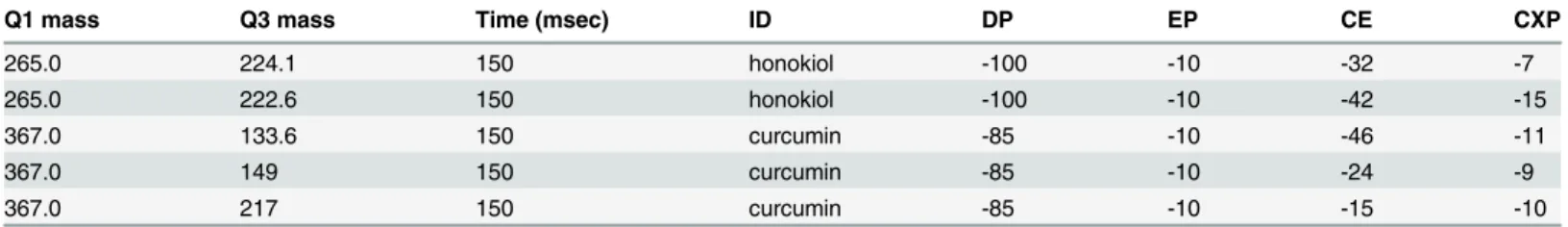
Table 1. LC-MS/MS parameters.
Q1 mass Q3 mass Time (msec) ID DP EP CE CXP
265.0 224.1 150 honokiol -100 -10 -32 -7
265.0 222.6 150 honokiol -100 -10 -42 -15
367.0 133.6 150 curcumin -85 -10 -46 -11
367.0 149 150 curcumin -85 -10 -24 -9
367.0 217 150 curcumin -85 -10 -15 -10
Cranial window surgery and
in vivo
imaging
Cranial window surgery,in vivoimaging and photobleaching experiments were performed as previously described [15]. Briefly, mice were anesthetized with isoflurane (1–2%) and a cranial window was surgically installed on the skull. Following surgery, animals were monitored for pain and given daily i.p. injections of buprenorphine for 1–3 days, as needed. Imaging experi-ments were begun at least 3 weeks following placement of the cranial window. For multiphoton imaging, animals were anesthetized with isoflurane, mounted to a custom stereotaxic frame, and imaged using a 20x/1.0NA water immersion objection and a Zeiss LSM 7MP multiphoton microscope, outfitted with dual channel BiG (binary GaAsP) detectors and the Coherent Cha-meleon titanium-sapphire femtosecond pulsed laser source, tuned to 860 nm. Baseline images of Syn-GFP-labeled cell bodies and presynaptic terminals in cortical layers 2/3 were obtained using 1–3 mW power (measured at objective exit). Large square regions of interest (ROI, 53x53μm2) encompassing 50–100 terminals were bleached with ~100–150 mW power over
<10 ms, and recovery images were acquired at 1–5 minute intervals, for 15 minutes. Zeiss Zen
2011 image acquisition software was used for image acquisition, and data was analyzed using Fiji image analysis software (http://fiji.sc/). The background-subtracted fluorescence intensity of the ROI was calculated for each time point and graphed as normalized fluorescence intensity vs. time using Prism software (GraphPad, La Jolla, CA). The data was fit as a one-phase associ-ation to calculate the tau recovery rate and the immobile fraction.
Statistics
All statistical analyses were carried out using Prism software (Graphpad, La Jolla, CA), and all data are presented as mean ± standard deviation. For behavioral analysis, right and left paw data was averaged for the fore paws or hind paws for each animal, then data for each animal in a group were averaged, to yield an n = 4 animals per group. Unless otherwise noted, unpaired, two-tailed t-tests were used to determine significance between groups. Since no significant dif-ferences were detected between male and female mice, results from both sexes were combined.
Results
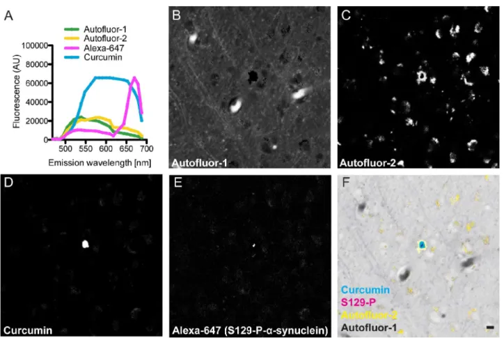
Curcumin binds to Lewy Bodies in diseased human brain tissue
To eliminate confounding effects due to variations in purification methods or the presence of other curcuminoids, we used synthetic curcumin, as opposed to turmeric root or extracted cur-cumin. We found that curcumin from Chromadex was>99% pure by HPLC, stable in 100%
DMSO solution over 1 week, and stable as a powder for over 1 year.
found that curcumin binds to Serine-129-phosphorylated (S129-P)-α-synuclein-positive
corti-cal LBs in human brain tissue from patients with DLB (Fig 1B–1G). While it appeared as though the LBs may contain a smaller central core of S129-P-α-synuclein, with a larger region
of curcumin staining outside this core, we believe that this is likely a result of the software at-tempting to clearly assign only one spectral component to the LB core. In normal control brain tissue, no staining for S129-P-α-synuclein was seen (S1 Fig). To our knowledge, this is the first
report of curcumin binding to LBs in human brain tissue.
Free and glucuronidated curcumin are present in plasma from mice on a
curcumin diet
Given this data that curcumin can bind to, and therefore potentially modulate, aggregatedα
-synuclein in human tissue, we next set out to test its effects in -synucleinopathy mouse models. To examine the long-term effects of curcumin, we treated Syn-GFP mice with 500 ppm curcu-min diet or control diet for 6 months. Food consumption and body weight were monitored weekly; no significant differences between curcumin and control diet mice were found at any time point (Fig 2A and 2B). Motor behavior was assessed at 23 weeks, and brain tissue and plasma was harvested at 24 weeks.
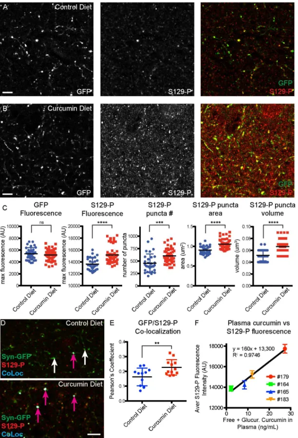
Fig 1. Curcumin stains Lewy Bodies.(A) Individual fluorescence spectra of curcumin, Alexa-647 secondary antibody, and two autofluorescent
components, defined by spectral imaging and linear unmixing. Autofluorescence components (B, C) were clearly separated from adjacent curcumin-positive (D) structures, which were defined as Lewy Bodies because they co-labeled with S129-P-α-synuclein-A647 antibodies (E, G). Minimal unassigned residual
light (F) demonstrates a high efficiency of the unmixing technique for the defined spectra. (Scale bar = 10μm)
We used a previously validated liquid chromatography tandem mass spectrometry (LC–
MS/MS) method to measure curcumin levels in plasma samples after dietary treatment [16]. In both spiked control plasma matrix and experimental samples, we detected a single peak corre-sponding to curcumin (molecular weight 368) by following the 367/133 and 367/217 MRM transitions (Fig 2C). The 367/133 transition values were used for quantification of curcumin. The lower limit of quantitation (LLOQ) was calculated as 5 ng/mL due to this being the lowest value on the standard curve, although curcumin peaks were clearly detectable with signal to noise ratios greater than 10:1 at lower concentrations, and the lower limit of detection (LLOD) was 0.033 ng/mL. Standard curves were linear in the range of 0–500 ng/mL curcumin
(r = 0.998±0.002). We found an average plasma curcumin concentration of<5 ng/mL (4.40±
5.72 ng/mL) in 3 out of 4 mice on a curcumin diet (Table 2). One sample did not contain tectable curcumin, likely because of rapid metabolism of free curcumin (see below) and a de-layed period of time between the last food consumption time point (at the end of the dark cycle in the early morning) and sacrifice (early afternoon). We did not detect curcumin in plasma from control diet mice.
Curcumin is rapidly metabolizedin vivothrough two pathways—reduction of the com-pound to dihydrocurcumin, tetrahydro-curcumin, and hexahydrocurcumin, and addition of a sugar group to yield curcumin-glucuronide versions of the parent compound and each of the three metabolites. Previous research has shown that up to 99% of curcumin is rapidly glucuro-nidatedin vivo[19]. To examine the levels of curcumin-glucuronide in mice on a curcumin diet, we treated plasma samples with B-glucuronidase prior to LC-MS/MS, to remove the sugar and reduce the compound back to free curcumin. After enzyme treatment, we found an average curcumin concentration of 12.52±10.68 ng/mL, indicating that 94.1±7.3% of curcumin was
Fig 2. LC-MS/MS detection of curcumin in plasma from mice on a curcumin diet.Food consumption (A) and body weight (B) for mice on a 500 ppm curcumin diet or control diet, recorded weekly for 6 months. Extracted-ion chromatogram for plasma matrix spiked with 100 ng/mL curcumin (C), showing distinct peaks for curcumin and honokiol internal standards.
glucuronidated in plasma from curcumin diet mice (Table 2). Enzyme treatment did allow us to detect curcumin in the mouse sample that had no free curcumin (mouse #183), indicating that 100% of the curcumin in this mouse was glucuronidated. Surprisingly, mouse #164 had a lower concentration of curcumin in plasma after enzyme treatment, perhaps indicating that the curcumin in this sample had degraded during freeze/thaw cycles between the initial mass spec run and the enzyme treatment experiment.
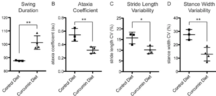
Curcumin diet intervention improves motor behavior
Automated gait analysis is a highly sensitive tool for measuring changes in multiple gait param-eters in mouse models of PD, Huntington’s Disease, and Amyotrophic Lateral Sclerosis [20]. To examine the effects of curcumin intervention on motor behavior, we tested curcumin and control diet mice using automated gait analysis on the DigiGait machine, and measured changes in several parameters. On a declined platform, there was a significant increase in hind paw angle in curcumin diet mice (5.0±2.5 degrees for control diet, 14.1±5.6 degrees for curcu-min diet, p<0.05). On a flat platform, mice on a curcumin diet showed a significant increase in
fore paw swing duration (87.7±0.3 ms for control diet, 101.3±5.1 ms for curcumin diet, p<0.01;Fig 3A) and a non-significant trend towards a decrease in hind paw stride frequency
(3.9±0.1 steps/sec for control diet, 3.6±0.2 steps/sec for curcumin diet, p = 0.0671), compared to control diet mice. Combined, these two parameters indicate that control diet mice took a greater number of shorter steps in a given time period, which is consistent with gait changes re-ported in a toxin-induced mouse model of PD [21]. These altered gait dynamics in our mouse model are due to expression of the Syn-GFP transgene, and curcumin diet intervention was able to partially normalize this deficit.
Strikingly, the ataxia coefficient, an index of step-to-step variability that is increased in human PD patients, was significantly decreased in the fore paws for curcumin diet mice com-pared to control (0.55±0.10 for control diet, 0.32±0.05 for curcumin diet, p<0.01;Fig 3B). Two
other PD-associated parameters, the variability in stride length and stance width in the fore paws, were also decreased in curcumin diet mice (stride length variability = 15.76±2.72% for control diet, 10.18±1.74% for curcumin diet, p<0.05; stance width variability = 27.55±3.85%
for control diet, 12.95±5.05% for curcumin diet, p<0.01;Fig 3C and 3D). Stride length
variabil-ity is higher in patients with PD [22,23]. In mice, the normal range is 10–12% and is increased
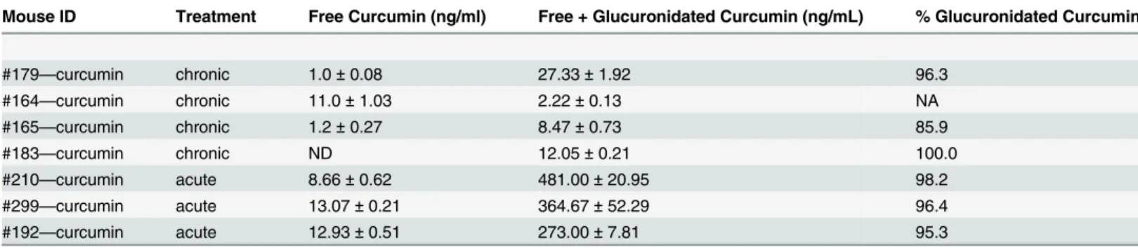
Table 2. Levels of curcumin in plasma from chronic and acutely treated mice.
Mouse ID Treatment Free Curcumin (ng/ml) Free + Glucuronidated Curcumin (ng/mL) % Glucuronidated Curcumin
#179—curcumin chronic 1.0±0.08 27.33±1.92 96.3
#164—curcumin chronic 11.0±1.03 2.22±0.13 NA
#165—curcumin chronic 1.2±0.27 8.47±0.73 85.9
#183—curcumin chronic ND 12.05±0.21 100.0
#210—curcumin acute 8.66±0.62 481.00±20.95 98.2
#299—curcumin acute 13.07±0.21 364.67±52.29 96.4
#192—curcumin acute 12.93±0.51 273.00±7.81 95.3
For mice treated chronically with a curcumin diet for 6 months, average free curcumin in plasma was 4.40±5.72 ng/mL and average free plus
glucuronidated curcumin was 12.52±10.68 ng/mL, indicating that 94.1±7.3% of curcumin was glucuronidated. For mice treated acutely with 15 mg/kg/day
curcumin i.p. for 15 days and sacrificed 5 hours after thefinal treatment, average free curcumin was 11.55±2.51 ng/mL and average free plus
glucuronidated curcumin was 372.89±104.24 ng/mL, indicating that 96.6±1.5% of curcumin was glucuronidated (samples from individual mice were run in
triplicate; mean±SD; ND, not detectable; NA, not applicable).
in a toxin-induced mouse model of PD [21]. Increased stance width variability is associated with PD in human patients, and is normally in the range of 10–15% in mice [21,24]. While control diet mice fall outside of the normal range for both of these parameters, curcumin diet intervention normalized the effect of over-expressing the Syn-GFP transgene to return these mice to the normal range.
We also tested muscle strength in curcumin and control diet mice, using a Grip Strength Meter, and found no significant difference between groups (control diet = 2.65±0.31 Newtons, curcumin diet = 2.62±0.29 Newtons; p = 0.8723). This result is consistent with a separate study that showed no change in grip strength in a Pink1 genetic model of PD [25]. This supports the idea that the motor phenotype we detect is similar to PD-like phenotypes, and is not a result of changes in muscle strength.
Curcumin diet intervention results in an increase in phosphorylated
α
-synuclein
We next examined molecular changes inα-synuclein as a result of curcumin diet intervention,
using immunohistochemical and biochemical assays on fixed and fresh brain tissue. We previ-ously found that presynaptic Syn-GFP terminal microaggregates contain S129-P forms ofα
-synuclein, and are resistant to digestion by Proteinase-K [15]. Here we find that staining for S129-P-α-synuclein was increased in presynaptic terminals in mice that were on a curcumin diet
(Fig 4A and 4B). We found that there was no change in GFP fluorescence in presynaptic termi-nals, but there was an increase in the number, area, volume, and fluorescence of presynaptic S129-P puncta (control diet GFP fluorescence = 5373±1039 AU, curcumin diet GFP fluores-cence = 5171±955 AU, p = 0.3767; control diet S129-P fluoresfluores-cence = 13641±1006 AU, curcumin diet S129-P fluorescence = 15170±1471 AU, p<0.0001; control diet S129-P number = 454±186,
curcumin diet S129-P number = 604±146; p<0.0005; control diet S129-P area = 0.9±0.1μm2,
curcumin diet S129-P area = 1.1±0.1μm2, p<0.0001; control diet S129-P volume = 0.052±
0.009μm3, curcumin diet S129-P volume = 0.066±0.011μm3, p<0.0001;Fig 4C). When we
Fig 3. Curcumin diet mice show improved gait.Syn-GFP mice on a 6 month curcumin diet intervention had significant changes in gait compare to control diet mice, including increased fore paw swing duration (A), decreased fore paw ataxia coefficient (B), decreased for paw stride length variability (C), and decreased fore paw stance width variability (D). The ataxia coefficient, and variability in stride length and stance width, are all associated with a PD-like phenotype in both humans and mice (flat platform run, n = 4 animals per group,*p<0.05,**p<0.01).
looked exclusively at Syn-GFP-positive terminals, we also detected a significant increase in S129-P levels, as measured by analyzing the amount of colocalization between S129-P and Syn-GFP (control diet Pearson’s coefficient = 0.16±0.06, curcumin diet Pearson’s coefficient = 0.23± 0.5, p<0.01,Fig 4D and 4E). Combined, these IHC findings indicate that chronic exposure to
curcumin results in a global increase inα-synuclein phosphorylation state, at both synapses that
contain endogenous mouseα-synuclein and synapses that over-express the human Syn-GFP
protein. We also found a strong correlation between plasma levels of curcumin and S129-P fluo-rescence intensity, in individual mice (Fig 4F).
To biochemically examine aggregates, we isolated synaptosome and cytosolic proteins from whole brain extracts of mice that were on a curcumin or control diet. Our previous work dem-onstrated that Syn-GFP mice contain a single species of Syn-GFP, identified by molecular weight under denaturing SDS-PAGE western blot conditions, and do not contain multiple SDS-resistant aggregate species of different molecular weights [15]. We further found that ~35% of Syn-GFP in synaptosomes is resistant to digestion by Proteinase-K, compared to ~2% of cytosolic Syn-GFP [15]. In curcumin diet mice, we did not detect any significant changes in the Syn-GFP Proteinase-K-resistant fraction in synaptosomes or cytosolic compartments, indi-cating that chronic curcumin treatment has no effect on Proteinase-K-resistant aggregates in these mice (control diet synaptosome fraction = 0.30±0.14, curcumin diet synaptosomes frac-tion = 0.40±0.09, p = 0.2654; control diet cytosolic fracfrac-tion = 0.03±0.02, curcumin diet cytosol-ic fraction = 0.03±0.03, p = 0.9762;Fig 5).
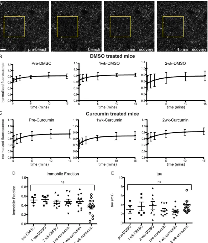
Acute curcumin treatment modulates phosphorylation of
α
-synuclein, but
has no effect on
in vivo
aggregation
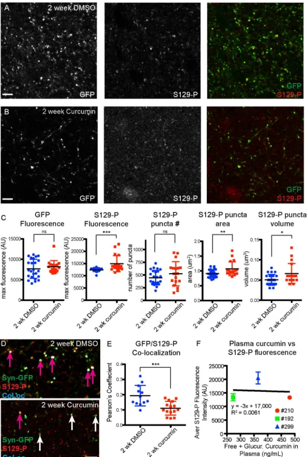
To look more directly at the effect of curcumin on terminal microaggregatesin vivo, we adopted an acute curcumin treatment paradigm that we could apply to Syn-GFP mice that had surgically-installed cranial windows over the cortex. We have shown previously that Syn-GFP terminal microaggregates have slower recovery kinetics and a significant immobile fraction in Fluorescence Recovery After Photobleaching (FRAP) experiments, compared to soluble Syn-GFP in the cell body [15]. We treated mice with 15 mg/kg/day curcumin or DMSO vehicle con-trol i.p. for 15 days, and examined FRAP kinetics at pre-injection, 7 day, and 14 day time points. We found no significant change in the immobile fraction (IF) or the tau recovery rate due to curcumin treatment (pretreatment DMSO IF = 0.52±0.11, 1 week DMSO IF = 0.53± 0.10, 2 week DMSO IF = 0.48±0.11, pretreatment curcumin IF = 0.49±0.15, 1 week curcumin IF = 0.48±0.14, 2 week curcumin IF = 0.36±0.16; pretreatment DMSO tau = 3.00±1.66 ms, 1 week DMSO tau = 3.66±2.17 ms, 2 week DMSO tau = 3.90±2.38 ms, pretreatment curcumin tau = 2.83±1.16 ms, 1 week curcumin tau = 2.58±0.96 ms, 2 week curcumin tau = 2.56±5.68 ms;Fig 6). A 2-way repeated measures ANOVA showed that there no effect of time or treat-ment, and no interaction effect (interaction p = 0.6927, time p = 0.1846, treatment p = 0.1471), although there was a trend towards a decrease in the immobile fraction over time in some cur-cumin regions analyzed.
In agreement with thein vivoFRAP data, we also found no significant change in the Pro-teinase-K-resistant fraction in fresh brain tissue from acutely-treated mice (S2 Fig). Similar to chronic curcumin treatment, we detected an increase in S129-P-α-synuclein in cortical
demonstrates an increase in S129-P at GFP-positive terminals. (F) Strong correlation between plasma curcumin levels and terminal S129-P fluorescence in individual mice. (n = 4 mice for control diet, n = 4 mice for curcumin diet; each data point represents the average value for a 3μm z-stack, with 7–15 z-stacks analyzed per mouse; pink arrows indicate colocalized synapses, white arrows indicate lack of colocalization;**p<0.01,***p<0.0005,****p<0.0001, Scale bar = 5μm).
synapses in acutely-treated curcumin mice, compared to DMSO treated controls (DMSO GFP fluorescence = 7659±2327 AU, curcumin GFP fluorescence = 8185±1483 AU, p = 0.4065; DMSO S129-P fluorescence = 12419±690 AU, curcumin S129-P fluorescence = 14931±3302 AU, p<0.001; DMSO S129-P number = 447±143, curcumin S129-P number = 523±238;
p = 0.2023; DMSO S129-P area = 0.9±0.1μm2, curcumin S129-P area = 1.1±0.2μm2, p<0.01;
DMSO S129-P volume = 0.050±0.013μm3, curcumin S129-P volume = 0.066±0.025μm3,
p<0.05;Fig 7A–7C). However, when we looked specifically at Syn-GFP-positive synapses, we
detected a significant decrease in S129-P levels after 2 weeks of curcumin treatment, as
Fig 5. No change in Proteinase-K-resistant aggregates in curcumin diet mice.(A) Western blot detection of Syn-GFP microaggregates, following Proteinase-K digestion of synaptosome and cytosolic protein fractions from control and curcumin diet mice. (B) Quantification of Syn-GFP band intensity shows no change in the resistant fraction between control and curcumin diet mice.
Fig 6. No change in in vivo FRAP kinetics in acutely treated curcumin mice.(A) Photobleaching and recovery imaging of presynaptic Syn-GFP protein in cortical brain tissue, through a cranial window in the skull. (B) FRAP recovery curves from DMSO and curcumin (C) treated animals, at pre-treatment, 1 week, and 2 week time points. (D) Immobile Fraction and recovery time constant tau values (E) do not change over time or with treatment (n = 3 animals curcumin, n = 2 animals DMSO, 2–5 regions per animal per time point; yellow box indicates bleach ROI; Scale bar = 10μm).
indicated by a smaller Pearson’s colocalization coefficient between GFP and S129-P (DMSO Pearson’s coefficient = 0.19±0.07, curcumin Pearson’s coefficient = 0.11±0.05, p<0.001;Fig 7D
and 7E).
Finally, we detected an average of 11.55±2.51 ng/mL free curcumin in plasma from 15-day curcumin treated mice, which were sacrificed 5 hours after the last curcumin treatment (Table 2). Following B-glucuronidase treatment of plasma, we detected an average of 372.89± 104.24 ng/mL curcumin, indicating that 96.6±1.5% of curcumin in the blood is glucuronidated 5 hours after i.p. injection (Table 2). Unlike chronic curcumin treatment, there was no correla-tion between plasma levels of curcumin and S129-P fluorescence intensity, in individual mice that received acute curcumin treatment (Fig 7F).
Discussion
We have found that transgenicα-synuclein mice show improvements in gait after chronic
treatment with a moderate dose of dietary curcumin. For a 500 ppm curcumin diet, daily con-sumption rates of ~ 4 g of food would result in 2 mg of curcumin consumed per day over a 6 month period. Despite well-established problems of low bioavailability [16,26], many of curcu-min’s cellular mechanisms of action occur in the nM toμM range. Indeed, 0.1–1.0μM
concen-trations of curcumin can protect againstα-synuclein-induced cell death in tissue culture cells,
and curcumin was shown to disaggregateα-synuclein aggregatesin vitroat concentrations of
0.5–5.0μM [10,12]. Here we extend thesein vitrofindings to show, for the first time in a
genet-ic synucleinopathy mouse model, that a moderate dose of dietary curcumin is suffgenet-icient to im-prove motor phenotype.
Using LC-MS/MS, we measured mean values of ~4.5 ng/mL free curcumin (range 0–11 ng/mL) and ~12.5 ng/mL total curcumin (range 2–27 ng/mL; free and glucuronidated) in plas-ma from curcumin diet mice. However, these values are probably lower than peak plasplas-ma levels actually attained. Plasma was collected in the early afternoon, leaving at least 6 hours between the end of the dark cycle and thus, likely the last significant food consumption time point. Be-cause the half-life of oral curcumin in mouse plasma is 3.5 hours [19], it is likely that curcumin plasma levels significantly declined due to the lag in collection time. The lag between the last feeding and collection may also contribute to some of the variability between samples. A previ-ous group reported 35 ng/mL curcumin in plasma from mice on a 500 ppm diet for 4 months [3], but these authors did not report timing of the plasma collection relative to the dark cycle or last feeding time point. With acute curcumin dosing, we detected mean values of ~12 ng/mL free curcumin (range 9–13 ng/mL) and ~373 ng/mL total curcumin (range 273–481 ng/mL; free and glucuronidated) in plasma from mice that were treated with 15 mg/kg/day (450μg)
i.p. curcumin for 15 days, and sacrificed 5 hours after the last curcumin treatment. This is also lower than previous reports, which found 127 ng/mL curcumin in plasma 4 hours after 140μg
was injected i.p. into an AD mouse model [3]. These differences could be due to variable me-tabolism in the different mouse strains used.
In this study, in order to measure curcumin’s molecular effects onα-synuclein, half of each
mouse brain was fixed for IHC and the other half processed for synaptosome isolation and sub-sequent biochemistry; therefore, there was insufficient tissue to attempt to measure brain
shows significant changes in S129-P puncta, but no change in GFP. (D) Analysis of GFP/S129-P colocalization (CoLoc channel), and quantification of the Pearson’s Colocalization Coefficient (E), shows a decrease in S129-P at GFP-positive terminals. (F) No correlation between plasma curcumin levels and terminal S129-P fluorescence in individual mice. (n = 2 mice for DMSO treatment, n = 3 mice for curcumin treatment; each data point represents the average value for a 3μm z-stack, with 4–12 z-stacks analyzed per mouse; pink arrows indicate colocalized synapses, white arrows indicate lack of colocalization; *p<0.05,**p<0.01,***p<0.001, Scale bar = 5μm).
curcumin levels by LC-MS/MS. Curcumin does cross the blood-brain barrier [3,19], and Begum et al. found 0.47μg/g (1.28μM) and 0.74μg/g (2.01μM) in mouse brain tissue
follow-ing chronic dietary and i.p. delivery methods, respectively. These authors showed that with 4 months of chronic dosing with a curcumin diet identical to ours (500 ppm curcumin diet), cur-cumin was highly enriched in mouse brain tissue, with a brain-to-plasma ratio of 13.4 [3]. With acute dosing, using an i.p. dose of curcumin that is half of the dosage used in our study, they found a brain-to-plasma ratio of 5.85, 4 hours after treatment [3]. These results highlight that curcumin, a lipophilic compound, is more stable in brain than in plasma, and can reach concentrations that are within the range of what has been shown to affectα-synuclein cellular
functionin vitro(see above). Interestingly, we found a strong positive correlation between in-creased levels of S129-P at individual cortical terminals and plasma levels of curcumin, in indi-vidual mice that were on a curcumin diet (Fig 4). This underscores the accumulation and stability of curcumin in lipid-rich brain tissue, particularly with chronic treatment, and lends further support to the brain-specific effects of dietary curcumin in our system. Because curcu-min is intrinsically auto-fluorescent, we also attempted to directly measure global curcucurcu-min fluorescence or curcumin localization to individual presynaptic terminals usingin vivocranial window imaging, but did not detect any significant changes in fluorescence at either 1–3 hours or 7–14 days after acute curcumin treatment.
The Syn-GFP mouse mimics human duplication and triplication mutations to model cellu-lar changes that occur due to 2-3-fold over-expression ofα-synuclein. The presence ofα
-synu-clein microaggregates in presynaptic terminals, but not cell bodies, makes this mouse an excellentin vivomodel of early cellular aggregation of alpha-synuclein, which occurs in many diseases including PD, DLB, and related synucleinopathies [15]. Syn-GFP mice do not show extensive neurodegeneration, severe motor decline, or early mortality rates, compared to more severe mouse models which more strongly overexpressα-synuclein bearing point mutations
[14]. Despite this mild phenotype, we were able to measure significant changes in motor behav-ior due to curcumin diet intervention. Of these gait changes, 3 parameters that are associated with PD-like symptoms were found to be abnormally elevated in control diet mice, and 6 months of curcumin diet intervention was sufficient to bring these parameters back into the normal range. In control diet mice, expression of Syn-GFP increased the variability in both stride length and stance width, which are the same gait parameters reported to increase in a toxin-induced mouse model of PD [21]. It would be interesting to examine curcumin diet in-tervention in the A53T and E46Kα-synuclein point mutation mouse models that show more
severe motor dysfunction and early mortality [27,28], at varying time points of disease progres-sion, to test whether curcumin treatment could halt or reverse these more severe neurodegen-erative phenotypes.
We measured a global increase in S129-P-α-synuclein in cortical presynaptic terminals in
mice that were chronically and acutely treated with curcumin. With chronic curcumin treat-ment, this increase in phosphorylation was also present at Syn-GFP-positive terminals, while in acutely-treated mice we measured a decrease in S129-P at Syn-GFP-positive terminals. Combined, these data indicate that the effects of curcumin on phosphorylation of human Syn-GFP are differentially modulated over acute and chronic treatment time points, despite there being an overall increase in endogenous mouse S129-P for both acute and chronic curcumin exposure. With acute treatment, where S129-P-α-synuclein is decreased in terminals
contain-ing human Syn-GFP but increased in terminals containcontain-ing endogenous mouseα-synuclein,
this could represent an effect of short-term curcumin treatment specifically on humanα
-synu-clein that is reversed with long-term exposure to curcumin. Human and mouseα-synuclein
increased aggregation and toxicity in human patients [9]. It has been proposed that curcumin binds to amino acids 89–91 ofα-synuclein [13]; thus if threonine-53 present in mouse protein
is responsible for the differential effects of curcumin, it may occur through allosteric changes to the binding pocket. It is also possible that curcumin acts to increase mouseα-synuclein
presyn-aptic terminal expression, which could result in an overall increase in S129-P staining. We did not measure mouseα-synuclein by IHC due to the relative lack of a high quality mouse-specific
antibody, but we did find that there was no change in human Syn-GFP levels with acute or chronic treatment. It will also be interesting in future studies to determine which kinases and phosphatases are involved in modifying the presynaptic pool ofα-synuclein at S129, and to
de-termine if curcumin may be directly modulating activity of these enzymes to produce the changes that we have measured.
How S129-P phosphorylation ofα-synuclein affects aggregation and toxicity is currently
under debate. Because S129-P-α-synuclein is highly correlated with disease progression and
pathology, with up to 90% ofα-synuclein in LBs containing this modification, S129-P has long
been thought to be a marker of toxic forms of the protein [29,30]. However, recent evidence in-dicates that S129-P modification targets the protein for degradation via the autophagy path-way, and phosphorylation at this site was found to be neuro-protective bothin vitroandin vivo
[31]. We found that mice on a curcumin diet showed improvements in motor behavior as well as a global increase in S129-P in cortical pre-synaptic terminals, which supports the hy-pothesis that S129-P is protective in our synucleinopathy mouse model. The decrease in S129-P at Syn-GFP synapses after 2 weeks of curcumin treatment did not correlate with any changes in Syn-GFP terminal microaggregates, as measured byin vivoFRAP experiments and Proteinase-K digestion, indicating that longer time points of treatment may be needed to mea-sure a direct effect of curcumin onα-synuclein aggregation.
There are a number of possible mechanisms for how curcumin treatment increases
S129-P-α-synuclein and improves motor behavior. Curcumin has been shown to increase proteasome
activity and heat shock protein expression at low doses (up to 1μM)in vitro, and to induce
autophagic clearance of A-beta, which resulted in attenuated cognitive impairment in an AD mouse model [32,33]. Degradation ofα-synuclein can occur through both proteasome and
autophagy pathways, and S129-P has been shown to directly targetα-synuclein for autophagic
degradation to provide neuroprotection and motor improvement in another mouse model of PD [31,34]. There is also evidence that phosphomimic of S129 inhibits binding of pore-form-ingα-synuclein oligomers to membranes, thus conferring protection by blocking this form of
toxic oligomers [35]. Thus curcumin’s action to increaseα-synuclein phosphorylation may
specifically promote its degradation via autophagy or proteasomal pathways, or by blocking the membrane-binding activity of toxic oligomers. Another possible mechanism involves direct disaggregation ofα-synuclein by curcumin binding. Curcumin can directly bind to and
disag-gregate bothα-synuclein oligomers and fibrilsin vitro, but phosphorylation state of the protein
modulating these different aggregate forms. For example, there is evidence thatα-synuclein
ag-gregates undergo nitration-based oxidative damage [36]. Curcumin is known to be a potent anti-oxidant and may be modulatingα-synuclein, and ultimately motor behavior, by
decreas-ing reactive oxygen species [11]. While further studies are needed to better understand the mo-lecular mechanisms underlying chronic curcumin diet intervention in synucleinopathy mouse models, our data demonstrate improvements in motor behavior, providing strong support for curcumin therapy as the subject of further pre-clinical studies.
Supporting Information
S1 Fig. No curcumin or S129-P-α-synuclein staining in control human brain tissue.(A)
Individual fluorescence spectra of curcumin, Alexa-647 secondary antibody, and two auto-fluorescent components, as defined by spectral imaging and linear unmixing in DLB tissue. Autofluorescence components (B, C) are present in control human brain tissue, but no curcu-min-positive (D) or S129-P-α-synuclein-positive (E) structures were found (F, merge; scale
bar = 10μm).
(TIF)
S2 Fig. No change in Proteinase-K-resistant aggregates in acutely treated curcumin mice.
(A) Western blot detection of Syn-GFP microaggregates, following Proteinase-K digestion of synaptosome and cytosolic protein fractions from mice treated with DMSO control or 15 mg/kg/day curcumin for 2 weeks. (B) Quantification of Syn-GFP band intensity shows no change in the resistant fraction between control and curcumin diet mice.
(TIF)
Acknowledgments
We thank Dr. Dennis Koop and Jenny Luo in the OHSU Bioanalytical Shared Resource/Phar-macokinetics Core for LC-MS/MS analysis, Dr. Randy Woltjer in the Oregon Brain Bank for providing human tissue samples, Stefanie Kaech-Petrie in the OHSU Advanced Light Micros-copy Core for providing guidance on spectral unmixing, and Jon Taylor for help with sample collection. We also thank Dr. Edward Rockenstein and Dr. Eliezer Masliah at UCSD for Syn-GFP mice.
Author Contributions
Conceived and designed the experiments: KJS CKM AS VKU. Performed the experiments: KJS VRO AS. Analyzed the data: KJS VRO AS. Contributed reagents/materials/analysis tools: CKM AS. Wrote the paper: KJS CKM AS VKU.
References
1. Cole GM, Teter B, Frautschy SA (2007) Neuroprotective effects of curcumin. Adv Exp Med Biol 595: 197–212. doi:10.1007/978-0-387-46401-5_8PMID:17569212
2. Fan X, Zhang C, Liu D-B, Yan J, Liang H-P (2013) The clinical applications of curcumin: current state and the future. Curr Pharm Des 19: 2011–2031. PMID:23116310
3. Begum AN, Jones MR, Lim GP, Morihara T, Kim P, Heath DD, et al. (2008) Curcumin Structure-Func-tion, Bioavailability, and Efficacy in Models of Neuroinflammation and Alzheimer's Disease. Journal of Pharmacology and Experimental Therapeutics 326: 196–208. doi:10.1124/jpet.108.137455PMID: 18417733
5. Yang F, Lim GP, Begum AN, Ubeda OJ, Simmons MR, Ambegaokar SS, et al. (2005) Curcumin Inhibits Formation of Amyloid Beta Oligomers and Fibrils, Binds Plaques, and Reduces Amyloid in Vivo. Jour-nal of Biological Chemistry 280: 5892–5901. doi:10.1074/jbc.M404751200PMID:15590663 6. Garcia-Alloza M, Borrelli LA, Rozkalne A, Hyman BT, Bacskai BJ (2007) Curcumin labels amyloid
pa-thology in vivo, disrupts existing plaques, and partially restores distorted neurites in an Alzheimer mouse model. J Neurochem 102: 1095–1104. doi:10.1111/j.1471-4159.2007.04613.xPMID: 17472706
7. Pan J, Li H, Ma J-F, Tan Y-Y, Xiao Q, Ding J-Q, et al. (2012) Curcumin inhibition of JNKs prevents do-paminergic neuronal loss in a mouse model of Parkinson’s disease through suppressing mitochondria dysfunction. Alzheimer's Research & Therapy 1: 1–1. doi:10.1186/2047-9158-1-16
8. Goedert M (2001) Alpha-synuclein and neurodegenerative diseases. Nat Rev Neurosci 2: 492–501. PMID:11433374
9. Maries E, Dass B, Collier TJ, Kordower JH, Steece-Collier K (2003) The role ofα-synuclein in
Parkin-son's disease: insights from animal models. Nat Rev Neurosci 4: 727–738. doi:10.1038/nrn1199 PMID:12951565
10. Liu Z, Yu Y, Li X, Ross CA, Smith WW (2011) Curcumin protects against A53T alpha-synuclein-induced toxicity in a PC12 inducible cell model for Parkinsonism. Pharmacological Research 63: 439–444. doi: 10.1016/j.phrs.2011.01.004PMID:21237271
11. Wang MS, Boddapati S, Emadi S, Sierks MR (2010) Curcumin reducesα-synuclein induced
cytotoxici-ty in Parkinson's disease cell model. BMC Neurosci 11: 57. doi:10.1186/1471-2202-11-57PMID: 20433710
12. Pandey N, Strider J, Nolan WC, Yan SX, Galvin JE (2008) Curcumin inhibits aggregation ofα
-synu-clein. Acta Neuropathol 115: 479–489. doi:10.1007/s00401-007-0332-4PMID:18189141
13. Ahmad B, Lapidus LJ (2012) Curcumin Prevents Aggregation inα-Synuclein by Increasing
Reconfigu-ration Rate. Journal of Biological Chemistry 287: 9193–9199. doi:10.1074/jbc.M111.325548PMID: 22267729
14. Rockenstein E, Schwach G, Ingolic E, Adame A, Crews L, Mante M, et al. (2005) Lysosomal pathology associated with alpha-synuclein accumulation in transgenic models using an eGFP fusion protein. J Neurosci Res 80: 247–259. doi:10.1002/jnr.20446PMID:15765523
15. Spinelli KJ, Taylor JK, Osterberg VR, Churchill MJ, Pollock E, Moore C, et al. (2014) Presynaptic alpha-synuclein aggregation in a mouse model of Parkinson's disease. J Neurosci 34: 2037–2050. doi:10. 1523/JNEUROSCI.2581-13.2014PMID:24501346
16. Yang K-Y, Lin L-C, Tseng T-Y, Wang S-C, Tsai T-H (2007) Oral bioavailability of curcumin in rat and the herbal analysis from Curcuma longa by LC–MS/MS. Journal of Chromatography B 853: 183–189. doi:10.1016/j.jchromb.2007.03.010PMID:17400527
17. Mohorko N, Repovs G, PopovićM, Kovacs GG, Bresjanac M (2010) Curcumin labeling of neuronal fi-brillar tau inclusions in human brain samples. Journal of Neuropathology and Experimental Neurology 69: 405–414. doi:10.1097/NEN.0b013e3181d709ebPMID:20448485
18. Mutsuga M, Chambers JK, Uchida K, Tei M, Makibuchi T, Mizorogi T, et al. (2012) Binding of Curcumin to Senile Plaques and Cerebral Amyloid Angiopathy in the Aged Brain of Various Animals and to Neuro-fibrillary Tangles in Alzheimer's Brain. J Vet Med Sci 74: 51–57. doi:10.1292/jvms.11-0307PMID: 21891973
19. Pan MH, Huang TM, Lin JK (1999) Biotransformation of curcumin through reduction and glucuronida-tion in mice. Drug Metab Dispos 27: 486–494. PMID:10101144
20. Hampton TG, Amende I (2010) Treadmill gait analysis characterizes gait alterations in Parkinson's dis-ease and amyotrophic lateral sclerosis mouse models. Journal of motor behavior 42: 1–4. doi:10. 1080/00222890903272025PMID:19906638
21. Amende I, Kale A, McCue S, Glazier S, Morgan JP, Hampton TG (2005) Gait dynamics in mouse mod-els of Parkinson's disease and Huntington's disease. Journal of neuroengineering and rehabilitation 2: 20. PMID:16042805
22. Hausdorff JM, Cudkowicz ME, Firtion R, Wei JY, Goldberger AL (1998) Gait variability and basal gan-glia disorders: stride-to-stride variations of gait cycle timing in Parkinson's disease and Huntington's disease. Mov Disord 13: 428–437. PMID:9613733
23. Blin O, Ferrandez AM, Serratrice G (1990) Quantitative analysis of gait in Parkinson patients: increased variability of stride length. Journal of the Neurological Sciences 98: 91–97. PMID:2230833
25. Gispert S, Ricciardi F, Kurz A, Azizov M, Hoepken H-H, Becker D, et al. (2009) Parkinson Phenotype in Aged PINK1-Deficient Mice Is Accompanied by Progressive Mitochondrial Dysfunction in Absence of Neurodegeneration. PLoS ONE 4: e5777. doi:10.1371/journal.pone.0005777.s010PMID:19492057 26. Anand P, Kunnumakkara AB, Newman RA, Aggarwal BB (2007) Bioavailability of Curcumin: Problems
and Promises. Mol Pharmaceutics 4: 807–818. doi:10.1021/mp700113rPMID:17999464
27. Giasson BI, Duda JE, Quinn SM, Zhang B, Trojanowski JQ, Lee VM (2002) Neuronal alpha-synucleino-pathy with severe movement disorder in mice expressing A53T human alpha-synuclein. Neuron 34: 521–533. PMID:12062037
28. Emmer KL, Waxman EA, Covy JP, Giasson BI (2011) E46K human alpha-synuclein transgenic mice develop Lewy-like and tau pathology associated with age-dependent, detrimental motor impairment. Journal of Biological Chemistry 286: 35104–35118. doi:10.1074/jbc.M111.247965PMID:21846727 29. Fujiwara H, Hasegawa M, Dohmae N, Kawashima A, Masliah E, Goldberg MS, et al. (2002)
alpha-Synuclein is phosphorylated in synucleinopathy lesions. Nat Cell Biol 4: 160–164. doi:10.1038/ncb748 PMID:11813001
30. Anderson JP, Walker DE, Goldstein JM, de Laat R, Banducci K, Caccavello RJ, et al. (2006) Phosphor-ylation of Ser-129 is the dominant pathological modification of alpha-synuclein in familial and sporadic Lewy body disease. J Biol Chem 281: 29739–29752. doi:10.1074/jbc.M600933200PMID:16847063 31. Oueslati A, Schneider BL, Aebischer P, Lashuel HA (2013) Polo-like kinase 2 regulates selective
autophagicα-synuclein clearance and suppresses its toxicity in vivo. Proc Natl Acad Sci USA 110:
E3945–E3954. doi:10.1073/pnas.1309991110PMID:23983262
32. Ali RE, Rattan SI (2006) Curcumin's Biphasic Hormetic Response on Proteasome Activity and Heat-Shock Protein Synthesis in Human Keratinocytes. Annals of the New York Academy of Sciences 1067: 394–399. doi:10.1196/annals.1354.056PMID:16804017
33. Wang C, Zhang X, Teng Z, Zhang T, Li Y (2014) Downregulation of PI3K/Akt/mTOR signaling pathway in curcumin-induced autophagy in APP/PS1 double transgenic mice. European Journal of Pharmacolo-gy 740: 312–320. doi:10.1016/j.ejphar.2014.06.051PMID:25041840
34. Ebrahimi-Fakhari D, Cantuti-Castelvetri I, Fan Z, Rockenstein E, Masliah E, Hyman BT, et al. (2011) Distinct Roles In Vivo for the Ubiquitin-Proteasome System and the Autophagy-Lysosomal Pathway in the Degradation ofα-Synuclein. J Neurosci 31: 14508–14520. doi:10.1523/JNEUROSCI.1560-11. 2011PMID:21994367
35. Nübling GS, Levin J, Bader B, Lorenzl S, Hillmer A, Högen T, et al. (2014) Modelling Ser129 phosphory-lation inhibits membrane binding of pore-forming alpha-synuclein oligomers. PLoS ONE 9: e98906. doi:10.1371/journal.pone.0098906PMID:24911099