Curcumin Reduces the Noise-Exposed Cochlear
Fibroblasts Apoptosis
Tengku Siti Hajar Haryuna
1Wibi Riawan
2Ardyansyah Nasution
1Suprapto Ma
’
at
3Juliandi Harahap
4Indri Adriztina
11Department of Otorhinolaryngology-Head and Neck Surgery, Faculty of Medicine, Universitas Sumatera Utara, Medan 20155, Indonesia 2Department of Biochemistry, Faculty of Medicine, Universitas
Brawijaya, Malang 65145, Indonesia
3Department of Clinical Pathology, Faculty of Medicine, Universitas Airlangga, Surabaya 60131, Indonesia
4Department of Community Medicine, Faculty of Medicine, Universitas Sumatera Utara, Medan 20155, Indonesia Int Arch Otorhinolaryngol 2016;20:370–376.
Address for correspondence Tengku Siti Hajar Haryuna, MD, ENT Specialist, PhD, Department of Otorhinolaryngology - Head and Neck Surgery, Faculty of Medicine, Universitas Sumatera Utara, Jalan Bunga Lau No. 17, Medan Tuntungan Medan, Sumatera Utara 20136, Indonesia (e-mail: [email protected]).
Introduction
Exposure to excessive noise is the major avoidable cause of permanent hearing impairment.1 It is mostly found in the developing and industrial countries with bad hearing conserva-tion.2In 2012, there were 360 million persons in the world with
disabling hearing loss,5.3% of the world’s population, of which 328 million (91%) were adults (183 million men, 145 million women) and 32 (9%) million were children. Sixteen per cent of the disabling hearing loss in the adult population in the world resulted from excessive noise exposure in the workplace, ranging from 7% to 21% in the various subregions.1,3
Keywords
►
noise
►
curcumin
►
calcineurin
►
NFATc1
►
apoptosis
►
cochlea
Abstract
Introduction
The structural changes underlying permanent noise-induced hearing
loss (NIHL) include loss of the sensory hair cells, damage to their stereocilia, and
supporting tissues within the cochlear lateral wall.
Objective
The objective of this study is to demonstrate curcumin as a safe and
effective therapeutic agent in the prevention and treatment for
fi
broblasts damage
within the cochlear supporting tissues and lateral wall through cell death pathway.
Methods
We divided 24 Rattus norvegicus into 4 groups, Group 1: control; Group 2:
noise (
þ
); Group 3: noise (
þ
), 50 mg/day curcumin (
þ
); Group 4: noise (
þ
), 100 mg/day
curcumin (
þ
). We provided the noise exposure dose at 100 dB SPL for two hours over
two weeks and administered the curcumin orally over two weeks. We examined all
samples for the expressions of calcineurin, nuclear factor of activated T-cells
cyto-plasmic 1 (NFATc1), and apoptotic index of cochlear
fi
broblasts.
Results
We found signi
fi
cant differences for the expressions of calcineurin (
p
<
0.05)
in all groups, signi
fi
cant differences for the expressions of NFATc1 (
p
<
0.05) in all
groups, except in Groups 1 and 4, and signi
fi
cant differences for the apoptotic index
(
p
<
0.05) in all groups.
Conclusion
Curcumin proved to be potentially effective in the prevention and
treatment for
fi
broblasts damage within the cochlear supporting tissues and lateral
wall regarding the decreased expression of calcineurin, NFATc1, and apoptotic index of
cochlear
fi
broblasts.
received October 3, 2015 accepted
December 11, 2015 published online March 4, 2016
DOI http://dx.doi.org/ 10.1055/s-0036-1579742. ISSN 1809-9777.
Copyright © 2016 by Thieme Publicações Ltda, Rio de Janeiro, Brazil
The cochlear spiral ligament is a connective tissue lining the space between stria vascularis and the bony otic capsule. It plays diverse roles in normal hearing and is composed of sub-populations of specialized fibrocytes, which are sug-gested to play distinct roles influid homeostasis, infl amma-tory responses, predicted by their protein expression profiles. Certain fibrocyte sub-types express ion transport proteins and, thus, are likely to regulate Kþ
and Cl within the lateral wall perilymph.4
Evidence from various cell lines shows that cell death can be stimulated by oxidative stress and excitotoxicity through Ca2þ
overload. Acoustic overstimulation increases the Ca2þ
concentration in auditory hair cells. Elevated Ca2þ
has been implicated in the impairment of hair cell function and may initiate hair cell damage after noise exposure. There are several pathways through which Ca2þ
may contribute to cell death, involving activation of nitric oxide synthase (NOS), phospholipase A2, proteases, and calcineurin.5
For years, studies have emerged based on the use of natural compounds plant-derived as potential therapeutic agents for various diseases in humans.6Curcumin, a yellow pigment obtained from the rhizomes of Curcuma longa Linnaeus (Family: Zingiberaceae), is a major component of turmeric and has been used as a traditional medicine that possesses therapeutic potential against various diseases. Curcumin is capable of modulating numerous molecular targets involved in each stage of disease development by regulating transcription factors, growth factors, receptors, cytokines, kinases, enzymes, cell survival, metastatic, and apoptotic molecules.7
The role of curcumin in the prevention of and treatment offibroblast damage within the supporting tissues and the cochlear lateral wall through the apoptosis inhibition mechanism contributed by calcineurin in cochlear fi bro-blasts has never been studied and serves as the focus in this study. The objective of this study is also to demonstrate that higher doses of curcumin (100 mg/day) exert more benefi -cial effects in inhibiting apoptosis rather than low doses of curcumin (50 mg/day).
Methods
This study is an experimental study with randomized posttest-only control group design using male Wistar strain whiteRattus norvegicusrats (150 - 250 g, 8 - 12 weeks of age). The dose and frequency of noise exposure was 100 dB SPL and 1 - 10 kHz for 2 hours.
Curcumin used in this study was derived fromCurcuma longa Linnaeus (Turmeric) with curcumin content levels of 28.11.0% w/w compared with standard, suspended in 0.5% carboxymethyl cellulose. Afterwards, we administered the suspension directly to the stomach of each rat via nasogastric tube, once a day for two weeks. The samples were composed of 24Rattus norvegicusdivided into 4 groups. Group 1: the control group; Group 2: noise (þ); Group 3: noise (þ), 50 mg/ day curcumin (þ); Group 4: noise (þ), 100 mg/day curcumin (þ). We provided noise exposure doses of 100 dB SPL for two hours over two weeks.
After two weeks, the rats underwent termination by ether inhalation and necropsy procedure on their temporal bone. All samples underwent standard tissue processing withfi xa-tion in buffered formaldehyde, followed by dehydraxa-tion in graded alcohol solutions. Thereafter, they were embedded in paraffin blocks, serially cut into 4 µm thick sections, and put on glass slides. Representative sections were stained with hematoxylin and eosin (H&E). We performed immunohisto-chemical staining to examine the expressions of calcineurin and nuclear factor of activated T-cells cytoplasmic 1 (NFATc1) and apoptotic index of cochlear fibroblasts by terminal deoxynucleotidyl transferase (TdT) 2’-deoxyuridine 5′ -tri-phosphate (dUTP) nick-end labeling (TUNEL) Assay.
Immunohistochemistry procedures were performed as follows. We cleared the slide in xylene and rehydrated it through graded series of alcohol solutions. Endogenous per-oxidase activity was blocked with 3% hydrogen peroxide in absolute methanol. We prevented nonspecific binding of the second layer antibody by incubation with 10% nonimmune serum (0.25% Triton X-100 in phosphate-buffered saline phosphate-buffered saline). Anti-Calcineurin A antibody (abcam ab71149, Abcam plc., Cambridge, USA) and NFATc1 antibody 7A6 (sc-7294, Santa Cruz Biotechnology, Inc., Dallas, Texas, USA) served as thefirst antibodies and were separately applied to each specimen and incubated in a humid chamber. After rinsing with phosphate-buffered saline, we incubated sections with biotinylated secondary antibody. Later, we washed them once more and incubated with a horseradish streptavidin–peroxidase conjugate. Next, we added a sub-strate–chromogen solution (3–3′-diaminobenzidine tetrahy-drochloride). This reaction involved peroxidase catalysis of the substrate and conversion of the chromogen to a brown deposit that marked the antigen. The final steps included counterstaining with H&E and application of coverslips.
Three observers examined the samples in each slide. The fibroblasts within the cochlear supporting tissues and lateral wall, which expressed calcineurin and NFATc1, and apoptotic index in all fields were calculated manually with a hand counter. We calculated the expressions of calcineurin and NFATc1 quantitatively for the average distribution of
fibroblasts unit with single nucleus expressing calcineurin and NFATc1 (showing brown-colored cytoplasms) and the occurrence of apoptosis (showing brown-colored nucleus).
We processed the data using the Statistical Package for the Social Sciences (SPSS) one-way analysis of variance (ANOVA) and used a p value of 0.05 as the cut-off for statistical significance.
Results
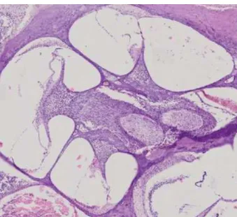
We performed H&E staining of rat cochlea to get a detailed view of the tissue (►Fig. 1).
The expression of calcineurin, after being evaluated with the immunohistochemistry method, showed an increased expression in Group 2 (►Fig. 2B), compared with other groups. The curcumin-treated groups showed lower density seen in the brown color, and less calcineurin-expressed fibroblasts than Group 2 (►Fig. 2C, D).
Data in ►Table 1 show significant differences for the expressions of calcineurin (p<0.05) in all groups. A dose of curcumin 100 mg per day showed statistically significant decreases in the expressions of calcineurin rather than a dose of curcumin at 50 mg per day.
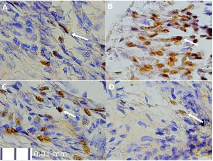
The expression of NFATc1 after being evaluated with the immunohistochemistry method showed an increased expression in Group 2 (►Fig. 3B) compared with other groups. The curcumin-treated groups showed less NFATc1-expressedfibroblasts than Group 2 (►Fig. 3C, D).
Fig. 1 The cochlear supporting tissues and lateral wall with H&E staining (20x zoom).
Data in►Table 2above showed significant differences for the expressions of NFATc1 (p<0.05) in all groups, except in Groups 1 and 4. A dose of curcumin of 100 mg per day showed statistically significant decreases in the expressions of NFATc1, rather than a dose of curcumin 50 mg per day.
The apoptotic index, after being evaluated with TUNEL assay showed an increased apoptotic index in group 2 (►Fig. 4 B) compared with other groups. The curcumin-treated groups showed less apoptotic cells-expressed fi -broblasts than group 2 (►Fig. 4C, D).
Data in ►Table 3 showed significant differences for the apoptotic index (p<0.05) in all groups. A dose of curcumin 100 mg per day showed statistically significant decreases in the apoptotic index rather than a dose of curcumin 50 mg per day.
Discussion
Acoustic overstimulation induces Ca2þ
overload and mediat-ed cell death pathways, involving activation of calcineurin.5,8 In this study, we found that the expression of calcineurin statistically increases in the cochlear fibroblast noise-exposed group (Group 2) when compared with the control group. Calcineurin belongs to the family of Ca2þ
/calmodulin-dependent protein phosphatases, protein phosphatase 2B. Calcineurin is activated by binding of Ca2þ
/calmodulin and the only protein phosphatase regulated by a second messen-ger Ca2þ
.9,10
It has been recently reported that calcineurin is activated in outer hair cells following noise exposure in mice exposed to broadband noise (2 - 20 kHz).8In another experimental study in guinea pigs, after intense noise exposure (4 - 10 kHz, 120 dB, for 5 hours), varying degrees of hair cells loss and calcineurin immunoreactivity were detected immunohisto-chemistrically in outer hair cells and concentrated at the cuticular plate.5
Fig. 3 The expression of NFATc1 in each group (1000x zoom): (A) Group 1/control; (B) Group 2; (C) Group 3; (D) Group 4. The white arrow indicates the expression of NFATc1 in cochlearfibroblasts marked by the brown color.
Table 1 ANOVA test results in terms of the expressions of calcineurin
Group Mean difference
Standard deviation
Pvalue
Group 1 Group 2 22.0001.392 0.000
Group 3 17.5001.392 0.000
Group 4 12.5001.392 0.000
Group 2 Group 3 4.5001.392 0.025
Group 4 9.5001.392 0.000
Group 3 Group 4 5.0001.392 0.011
The expression of NFATc1 was found to be statistically higher in the cochlearfibroblast noise-exposed group (Group 2) compared with the control group. Ca2þ
overload activates calcineurin-dephosphorylated NFATs, leading to their trans-location to the nucleus. In addition to thisfirst wave of NFAT activation, in a second step of NFATc1/αA generation, a short isoform of NFATc1 is strongly induced.11,12
The apoptotic cochlearfibroblasts were statistically higher in the cochlear fibroblast noise-exposed group (Group 2) compared with the control group. Calcineurin has a dual function and may exert its effects on apoptosis either by the
activation of specific transcriptional pathways or by direct dephosphorylation of proteins including Bad (B cell lympho-ma 2 antagonist of cell death) and caspase-9 involved in the apoptotic pathway.10,11
Calcineurin dephosphorylates Bad, resulting in the disrup-tion of the binding of Bax (B cell lymphoma 2 associated x protein) to Bcl-2 (B cell lymphoma 2) or Bcl-xL (B cell lymphoma 2-extra large) at the outer membrane of mito-chondria through intrinsic pathway. Free Bax translocate to mitochondria and activate the transport system to release cytochrome c.8,13,14
Fig. 4 The apoptotic index in each group (1000x zoom): (A) Group 1/control; (B) Group 2; (C) Group 3; (D) Group 4. The white arrow indicates the apoptotic cochlearfibroblasts marked by the brown color.
Table 2 ANOVA test results in terms of expressions of NFATc1
Group Mean difference
Standard deviation
Pvalue
Group 1 Group 2 10.8330.792 0.000
Group 3 5.6670.792 0.000
Group 4 1.8330.792 0.189
Group 2 Group 3 5.1670.792 0.000
Group 4 9.0000.792 0.000
Group 3 Group 4 3.8330.792 0.001
Denotes statistically signi ficant.
Table 3 ANOVA test results in terms of the apoptotic index
Group Mean difference
Standard deviation
Pvalue
Group 1 Group 2 12.3331.004 0.000
Group 3 6.8331.004 0.000
Group 4 3.6671.004 0.010
Group 2 Group 3 5.5001.004 0.000
Group 4 8.6671.004 0.000
Group 3 Group 4 3.1671.004 0.030
In the extrinsic pathway, outside the mitochondria, it has been previously described that expression of the membrane-bound death receptor ligand FasL is mediated by NFAT. When FasL binds to its receptor Fas, the intracellular machinery associated with the death receptor Fas is activated and eventually leads to apoptosis by caspase activation and subsequent DNA cleavage.2 Caspase-8 directly cleaves cas-pase-3 as well as cytosolic Bid (BH-3 interacting domain death agonist), its active fragment (tBid) translocates to mitochondria to release cytochrome c.15
Cytochrome c interacts with proteins such as Apaf-1 (apoptotic protease activating factor-1), dATP, and procas-pase-9 to produce apoptosome and then activates caspase-9. This complex degrades procaspase-3 to caspase-3.15In apoptotic cells, activated caspase-3 cleaves inhibitor of caspase activated DNAse (ICAD) to release CAD. CAD then degrades chromosomal DNA within the nuclei and causes chromatin condensation. Caspase-3 also induces cytoskel-etal reorganization and disintegration of the cell into apoptotic bodies.14
An experimental study in mice exposed to broadband noise (2 - 20 kHz) has discovered that noise exposure induces activation of mitochondria-mediated cell death pathways in outer hair cells of the cochlea through activation of Bad by calcineurin. The localization of Bad was analyzed by immunohistochemistry. Total Bad was observed both in the sensory cells and in the supporting cells and also in the nervefibers projecting to the sensory cells. The results of this study strongly support the Bad as a link between Ca2þ
influx after noise exposure and the death of outer hair cells.8In another study, after intense noise exposure (4 - 10 kHz, 120 dB, for 5 hours) in guinea pigs, some calcineurin-immunopositive hair cells demonstrated condensed and swollen nuclei, indicating that calcineurin is related to both apoptosis and necrosis.5
This study proved that curcumin was able to decrease the expression of calcineurin and NFATc1 in cochlearfibroblasts, where a dose of curcumin 100 mg per day showed statisti-cally significant decreases in the expression of calcineurin and NFATc1 compared with a dose of curcumin 50 mg per day. This is due to the speculation that curcumin inhibits the regulation and expression of calcineurin, and prevents the dephosphorylation of NFATc1 by calcineurin, thus, reducing its translocation to the nucleus. The expression of NFATc1 in Group 4 was statistically insignificant compared with the control group, indicating that curcumin administration at higher doses is able to prevent NFATc1 activation, thereby its expression was found to be nearly similar to the control group (without noise exposure).
Researchers have recently investigated the therapeutic efficacy of curcumin in attenuation of left ventricular hypertrophy and sought to delineate the associated signal-ing pathways in bluntsignal-ing the hypertrophic response in nephrectomized rats. Curcumin attenuates cardiac hyper-trophy and remodeling through deactivation of multiple hypertrophic signaling pathways. This study reported that cytosolic NFAT was significantly decreased in rats that underwent nephrectomy and was significantly attenuated
by curcumin. NFAT in the nucleus was decreased by curcumin with quantitative real-time reverse transcrip-tion-polymerase chain reaction (RT-PCR) analysis.16 Another study showed an action of curcumin as an NFAT inhibitor through the Ca2þ
signaling pathway blocking. This experiment also demonstrates that curcumin inhibits NFAT transcriptional activity by preventing its nuclear translo-cation from the cytoplasm into the nucleus upon phorbol myristate acetate (PMA)/ionomycin stimulation of Jurkat T-cells with laser scan microscopy (LSM) analysis.17
Curcumin significantly promoted nonischemic wound healing in a dose–response fashion compared with controls as judged by increased reepithelialization and granulation tissue formation. Improved wound healing was associated with significant decreases in pro-inflammatory cytokines interleukin (IL)-1 and IL-6 as well as the chemokine IL-8. Curcumin also significantly reduced hypertrophic scarring.18 Another study concluded that pre- and coreceiving curcumin can significantly protect the cochlear morphol-ogy and functions on paclitaxel-induced ototoxicity in rats using light microscopy and distortion product otoacoustic emissions (DPOAEs) to evaluate histopathological, immu-nohistochemical, and functional changes in hearing. Curcumin might be considered as a potential dietary supplement from a natural product given to patients undergoing paclitaxel chemotherapy.19
Curcumin can also be used as an efficient adjuvant to cisplatin cancer therapy. This treatment strategy in head and neck cancer could mediate cisplatin chemoresistance by modulating therapeutic targets (Signal transducer and activator of transcription 3 and NF-E2 p45-related factor 2) and, at the same time, reduce cisplatin-related ototoxic adverse effects.20
Preclinical studies demonstrated that systemic curcumin attenuates ototoxicity and provides molecular evidence for a role of hemeoxigenase (HO-1) as an additional mediator in attenuating cisplatin-induced hearing loss.21
A previous study found the effect of curcumin on perox-ynitrite (ONOO)-induced damage in rat spiral ganglion neu-rons. Pretreatment with curcumin abrogated cytochrome c release, blocked activation of caspase-3, and altered the expression of Bcl-2 family triggered by ONOO. Curcumin can attenuate ONOO-induced damage in spiral ganglion neurons by the anti-oxidative activity, as well as protect mitochondria from oxidative stress.22
Conclusion
This study indicates that curcumin is safe and effective as a therapeutic agent in the prevention and treatment of the damage offibroblasts within the supporting tissues and the cochlear lateral wall through the cell death pathway. More-over, the study provides more insight into the mechanism of curcumin against apoptosis and shows that curcumin inhibits multiple apoptosis signaling pathways, including calcineurin and NFATc1. The study may serve as a scientific basis in the traditional systems of medicine for the management of NIHL in the future.
References
1 Nandi SS, Dhatrak SV. Occupational noise-induced hearing loss in India. Indian J Occup Environ Med 2008;12(2):53–56
2 Harmadji S, Kabullah H. Noise induced hearing loss in steel factory workers. Folia Medica Indonesiana 2004;40(4):171–174 3 World Health Organization. WHO global estimates on prevalence
of hearing loss. Available at: http://www.who.int/pbd/deafness/ WHO_GE_HL.pdf. Accessed Dec 10, 2014
4 Kelly JJ, Forge A, Jagger DJ. Contractility in type III cochlearfibrocytes is dependent on non-muscle myosin II and intercellular gap junc-tional coupling. J Assoc Res Otolaryngol 2012;13(4):473–484 5 Minami SB, Yamashita D, Schacht J, Miller JM. Calcineurin
activa-tion contributes to noise-induced hearing loss. J Neurosci Res 2004;78(3):383–392
6 Trujillo J, Granados-Castro LF, Zazueta C, Andérica-Romero AC, Chirino YI, Pedraza-Chaverrí J. Mitochondria as a target in the therapeutic properties of curcumin. Arch Pharm (Weinheim) 2014;347(12):873–884
7 Prasad S, Gupta SC, Tyagi AK, Aggarwal BB. Curcumin, a component of golden spice: from bedside to bench and back. Biotechnol Adv 2014;32(6):1053–1064
8 Vicente-Torres MA, Schacht J. A BAD link to mitochondrial cell death in the cochlea of mice with noise-induced hearing loss. J Neurosci Res 2006;83(8):1564–1572
9 Morioka M, Hamada J, Ushio Y, Miyamoto E. Potential role of calcineurin for brain ischemia and traumatic injury. Prog Neuro-biol 1999;58(1):1–30
10 Zhu H, Gao W, Jiang H, Wu J, Shi YF, Zhang XJ. Calcineurin mediates acetylcholinesterase expression during calcium ionophore A23187-induced HeLa cell apoptosis. Biochim Biophys Acta 2007;1773(4):593–602
11 Alvarez S, Blanco A, Fresno M, Muñoz-Fernández MA. TNF-α contributes to caspase-3 independent apoptosis in neuroblastoma cells: role of NFAT. PLoS ONE 2011;6(1):e16100
12 Serfling E, Avots A, Klein-Hessling S, Rudolf R, Vaeth M, Berberich-Siebelt F. NFATc1/αA: The other Face of NFAT Factors in Lympho-cytes. Cell Commun Signal 2012;10(1):16
13 Precht TA, Phelps RA, Linseman DA, et al. The permeability transition pore triggers Bax translocation to mitochondria during neuronal apoptosis. Cell Death Differ 2005;12(3):255–265 14 Elmore S. Apoptosis: a review of programmed cell death. Toxicol
Pathol 2007;35(4):495–516
15 Maher S, Toomey D, Condron C, Bouchier-Hayes D. Activation-induced cell death: the controversial role of Fas and Fas ligand in immune privilege and tumour counterattack. Immunol Cell Biol 2002;80(2):131–137
16 Ghosh SS, Salloum FN, Abbate A, et al. Curcumin prevents cardiac remodeling secondary to chronic renal failure through deactiva-tion of hypertrophic signaling in rats. Am J Physiol Heart Circ Physiol 2010;299(4):H975–H984
17 Kliem C, Merling A, Giaisi M, Kohler R, Krammer PH, Li-Weber M. Curcumin suppresses T cell activation by blocking Ca2þ mobiliza-tion and nuclear factor of activated T cells (NFAT) activamobiliza-tion. J Biol Chem 2012;287(13):10200–10209
18 Jia S, Xie P, Hong SJ, et al. Intravenous curcumin efficacy on healing and scar formation in rabbit ear wounds under nonischemic, ischemic, and ischemia-reperfusion conditions. Wound Repair Regen 2015;22(6):730–739
19 Bucak A, Ozdemir C, Ulu S, et al. Investigation of protective role of curcumin against paclitaxel-induced inner ear damage in rats. Laryngoscope 2015;125(5):1175–1182
20 Fetoni AR, Paciello F, Mezzogori D, et al. Molecular targets for anticancer redox chemotherapy and cisplatin-induced ototoxicity: the role of curcumin on pSTAT3 and Nrf-2 signalling. Br J Cancer 2015;113(10):1434–1444
21 Fetoni AR, Eramo SLM, Paciello F, et al. Curcuma longa (curcumin) decreases in vivo cisplatin-induced ototoxicity through heme oxygenase-1 induction. Otol Neurotol 2014;35(5):e169–e177 22 Liu W, Fan Z, Han Y, et al. Curcumin attenuates