Pakistan Vet. J., 2006, 26(4): 176-178.
176
SEQUENCING AND SEQUENCE ANALYSIS OF MYOSTATIN GENE IN THE EXON 1 OF THE CAMEL (CAMELUS DROMEDARIUS)
M. G. SHAH, A. S. QURESHI1, M. REISSMANN2 AND H. J. SCHWARTZ3
Department of Anatomy and Histology, Sindh Agriculture University, TandoJam, Pakistan; 1
Department of Veterinary Anatomy, University of Agriculture, Faisalabad, Pakistan; 2
Institute of Animal Sciences, Humboldt-Universität zu Berlin, Germany; 3
Department of Livestock Ecology, Humboldt-Universität zu Berlin, Germany
ABSTRACT
Myostatin, also called growth differentiation factor-8 (GDF-8), is a member of the mammalian growth transforming family (TGF-beta superfamily), which is expressed specifically in developing an adult skeletal muscle. Muscular hypertrophy allele (mh allele) in the double muscle breeds involved mutation within the myostatin gene. Genomic DNA was isolated from the camel hair using NucleoSpin Tissue kit. Two animals of each of the six breeds namely, Marecha, Dhatti, Larri, Kohi, Sakrai and Cambelpuri were used for sequencing. For PCR amplification of the gene, a primer pair was designed from homolog regions of already published sequences of farm animals from GenBank. Results showed that camel myostatin possessed more than 90% homology with that of cattle, sheep and pig. Camel formed separate cluster from the pig in spite of having high homology (98%) and showed 94% homology with cattle and sheep as reported in literature. Sequence analysis of the PCR amplified part of exon 1 (256 bp) of the camel myostatin was identical among six camel breeds.
Key words: Myostatin gene, sequencing, camel.
INTRODUCTION
Myostatin, also called growth differentiation factor-8 (GDF-8), is a member of the mammalian growth transforming family (TGF-beta superfamily), which is expressed specifically in developing an adult skeletal muscle (Gonzalez-Cadavid and Bhasin, 2004). Mice which lack myostatin show a widespread increase in skeletal muscle mass, due to an increase in both myofiber size (hypertrophy) and myofiber number (hyperplasia; McPherron et al., 1997). Muscular hypertrophy (mh), also known as “double-muscling” in cattle, has been recognized as a heritable physiological character for decades (Arthur, 1995) and is present in Belgian Blue, Piedmontese and Asturiana de los Valles breeds of cattle (Smith et al., 1997). The commercial use of double-muscle beef breeds has been encouraged due to their high meat yield, and superior meat quality associated with a high proportion of white, glycolytic muscle fibers. Double-muscled cattle also deposit much less fat than other breeds (Potts et al., 2003). These animals have less bone, less fat, and 20% more muscle on an average (Shahin and Berg, 1985; Hanset, 1991). Muscular hypertrophy allele (mh allele) in the double muscle breeds involved mutation within the myostatin gene (Kambadur et al., 1997). Such a major effect of a single gene on processing yields opened a potential channel for improving processing yields of animals
using knockout technology (Arif et al., 2002). Therefore, sequencing of the myostatin locus from farm animals is important to produce genomic resources for development of knockout technology as well as for understanding the structure, function, and evolution of the gene. The sequence analysis of myostatin gene in exon 1 of one-humped camel (Camelus dromedarius) has been described in the present paper.
MATERIALS AND METHODS
Animals
Camels belonging to six different Pakistani breeds viz. Marecha, Dhatti, Larri, Kohi, Campbelpuri and Sakrai were included in this study. These camels belonged to two different ecological zones of Pakistan i.e., riverine and mountainous. Genomic DNA was isolated from camel hair using NucleoSpin Tissue kit (Macherey-Nagel, Germany) according to the manufacturer’s protocol. Two animals of each breed were used for sequencing.
PCR amplification and sequence analysis
177 Pakistan Vet. J., 2006, 26(4): 176-178.
dromedary sequence was used to design the specific primer pair MN-1B up (5'>CGC TCC GGG AAC TGA TTG A <3') and MN-1B low (5'>TGG GAA GGT TAC AGC AAG AT <3') which amplifed a 256 bp fragment of of dromedary myostatin gene exon 1.
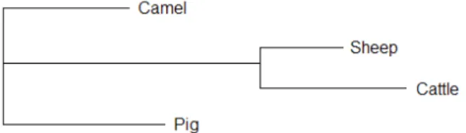
PCR reactions were carried out using UNO thermo cycler (Biometra, Germany) in a total volume of 25 µl containing 2.5 mM, MgCl2, 0.2 mM dNTP, 1U Taq DNA Polymerase (Genaxxon, Germany), 0.2 µM of forward and reverse primer and 100 ng genomic DNA. After an initial denaturation at 94°C for 2 min, 35 cycles were done, each consisting of 94°C for 1 min, 60°C (primer pairs MN-1B up and MN-1B low) for 30 sec and 72°C for 40 sec. The final step lasted for 10 min at 72°C. PCR amplified fragments were excide from 2% agarose gel and purified using Gene Clean II Kit (Q BIO gene, Canada). The fragment was sequenced in both directions using BigDye Terminator v1.1 Cycle Sequencing chemistry on an ABI Prism 310 Genetic Analyzer (Applied Biosystems, USA). All sequence alignments and distance calculations were made by Lasergene software (DNAStar, USA).To evaluate the evolutionary relationships of camel myostatin with the myostatin of other farm animals from the GenBank, a phylogenetic tree was constructed by using CLUSTAL W (1.8) computer software (Figure 1). All analyzed farm animals were divided into three subgroups at this gene. This work was carried out at the Molecular Genetics Laboratory, Institute of Animal Sciences, Humboldt-Universität zu Berlin, Germany.
Fig. 1: Phylogenetic tree showing the common ancestry at myostatin gene.
RESULTS
A part of the exon 1 of camel myostatin gene was PCR amplified using MN-1B up and MN-1B low primers. A fragment of 256 bp with primers position is shown in Figure 2.
The investigated sequence of the myostatin gene from six camel breeds showed no polymorphism. However, more than 90% homology of camel myostatin with that of the cattle, sheep and pig sequences published in the NCBI GenBank was observed. Camel formed separate cluster from the pig in spite of having high homology (98%), and showed 94% homology with
1 tgaatgagaa cagcgagcaa aaagaaaatg
tggaaaaaga ggggctgtgt aatgcatgta
61 tgtggagaca aaacactaaa tcttcaagac tagaagctat aaaaattcaa atcctcagta
121 aacttcgcct ggaaacagct cctaacatca gcaaagatgc tataagacaa cttttgccca 181 aagctcctcc gctccgggaa ctgattgatc
agtacgatgt ccagagagat gacagcagtg
241 acggctcctt ggaaga
Fig. 2: Sequence of camel myostatin gene and primers positions MN-1B up (desh) and MN-1B low (dots).
Table 1: Nucleotide identities in pair-wise compari-sons of farm animals’ sequences for myosta-tin gene (%).
Species Camel Cattle Sheep Pig Camel -- 94 94 98 Cattle -- 96 94 Sheep -- 94
Pig --
NCBI GenBank accession numbers for cattle, sheep and pig were AB076403, AF019622 and AY208121, respectively.
cattle and sheep (Table 1). Sheep and cattle shared the same cluster with 96% homology, indicating that these two species have had a small number of mutations in this gene since their break up evolution.
DISCUSSION
Sequence analysis of the PCR amplified part of exon 1 of the camel myostatin revealed similarity among six camel breeds. High homology of camel myostatin (more than 90%) with cattle, sheep and pig confirmed that vertebrate myostatin genes are highly conserved. The high level of sequence conservation among all known myostatin orthologs suggests the importance and conservation of its function in vertebrates. Myostatin was characterized as a negative regulator of skeletal muscle growth in mammals (McPherron et al., 1997).
Pakistan Vet. J., 2006, 26(4): 176-178. 178
2 (371 bp) and exon 3 (381 bp); intron 1 (363 bp) and intron 2 (811 bp) (Jeanplong et al., 2001; Ko et al., 2006; Liangyi et al., 2006). In Double Muscled cattle breeds, seven different mutations viz. nt821 (del 11), nt419 (del 7- ins 10), Q204X, E226X, C313Y, F94L and nt 414(C-T)) have been identified (Kambadur et al., 1997; McPherron and Lee, 1997; Grobet et al.,1997; Georges et al., 1998; Smith et al., 2000; Nishi et al., 2002).
Complete sequence of dromedary myostatin and sequence comparisons of different breeds are important to a better understanding of muscle growth and differentiation mechanisms in camels. Such knowledge will be helpful in further breeding selection strategy.
Acknowledgement
Appreciation is expressed to the Higher Education Commission, Islamabad and EU-Asia Link Project (UPHUMRAUF) for providing financial assistance in Pakistan and Germany, respectively.
REFERENCES
Arif, M. K., H. Kucuktas, R. A. Dunham and Z. Liu, 2002. Molecular characterization and differential expression of the myostatin gene in channel catfish (Ictalurus punctatus). Bioch. Biophys. Acta, 157(5): 99-107.
Arthur, P. F., 1995. Double muscling in cattle: a review. Aust. J. Agri. Res., 46: 1493-1515.
Gonzalez-Cadavid N. F. and S. Bhasin, 2004. Role of myostatin in metabolism. Curr. Opin. Clin. Nutr. Metab. Care, 7: 451-457.
Georges, M., L. Grobet., D. Poncelet., L. J. Royo, D. Pirottin and B. Brouwers, 1998. Positional candidate cloning of the bovine mh locus identifies an allelic series of mutations disrupting the myostatin function and causing double-muscling in cattle. Proc. 6th World Cong. Genetics Applied to Livestock Prod., 26: 195-204.
Grobet, L., L. J. R. Martin, D. Poncelet, D. Pirottin, B. Brouwers and J. Riquet, 1997. A deletion in the bovine myostatin gene causes the double-muscled phenotype in cattle. Nature Genetics, 17: 71-74. Hanset, R., 1991. The major gene of muscular
hypertrophy in the Belgian Blue cattle breed. In: “Breeding for Disease Resistance in Farm Animals” (ed. A. Owen). Commonwealth Agri. Bureaux International, London, UK, pp: 467-478. Jeanplong, F., M. Sharma, W. G. Somers, J. J. Bass and
R. Kambadur, 2001. Genomic organization and neonatal expression of the bovine myostatin gene. Mol. Cell Biochem., 220(1-2): 31-37.
Ji, S., R. L. Losinski, R. L. Cornelius, S. G. Frank, G. M. Willis, D. E. Gerrard, F. F. Depreux and M. E. Spurlock, 1998. Myostatin expression in porcine
tissues: tissue specificity and developmental and postnatal regulation. Amer. J. Physiol., 275: 1265-1273.
Kambadur, R., M. Sharma, T. P. L. Smith and J. J. Bass, 1997. Mutations in myostatin (GDF8) in Double-Muscled Belgian Blue and Piedmontese Cattle.Genome Res., 7: 910-915.
Ko, C. F, T. T. Chiou, T. T. Chen, J. L. Wu, J. C. Chen and J. K. Lu, 2006. Molecular cloning of myostatin gene and characterization of tissue-specific and developmental stage-specific expression of the gene in orange Spotted Grouper, Epinephelus coioides. Mar. Biotechnol., (NY). [article in press]. Liangyi, X., Q. Kaixian, Q. Hongqin, L. Lu, Y. Qiaoyi
and L. Mingyun, 2006. Molecular cloning and characterization of the myostatin gene in croceine croaker, Pseudosciaena crocea. Mol. Biol. Reprod., 33: 129-136.
McPherron, A. C., A. M. Lawler and S. J. Lee, 1997. Regulation of skeletal muscle mass in mice by a new TGF-beta superfamily member. Nature, 387: 83-90.
McPherron, A. C. and S. J. Lee, 1997. Double muscling in cattle due to mutations in the myostatin gene. J. Proc. Natl. Acad. Sci., 94(23):12457-12461.
Nishi, M., A. Yasue, S. Nishimatu, T. Nohno, T. Yamaoka, M. Itakura, K. Moriyama, H. Ohuchi and S. Noji, 2002. A missense mutant myostatin causes hyperplasia without hypertrophy in the mouse muscle. Biochem. Biophys. Res. Commun., 293(1): 247-251.
Potts, J. K., S. E. Echternkamp, T. P. Smith and J. M. Reecy, 2003. Characterization of gene expression in double-muscled and normal-muscled bovine embryos. Anim Genet., 34(6): 438-444.
Shahin, K. A. and R. T. Berg, 1985. Growth patterns of muscle, fat and bone, and carcass composition of double muscled and normal cattle. Can. J. Anim. Sci., 65: 279-293.
Sharma, M., R. Kambadur., K. G. Matthews., W. G. Somers., G. P. Devlin., J. V. Conaglen., P. J. Fowke and J. J. Bass, 1999. Myostatin, a transforming growth factor-beta superfamily member, is expressed in heart muscle and is upregulated in cardiomyocytes after infarction. J. Cell. Physiol., 180: 1-9.
Smith, T. P. L., N. L. Lopez-Corrales, S. M. Kappes and T. S. Sonstegard, 1997. Myostatin maps to the interval containing the bovine mh locus. Mammalian Genome, 8: 742-744.