Volume 2013, Article ID 635470,8pages http://dx.doi.org/10.1155/2013/635470
Research Article
Green Tea Extract Supplementation Induces
the Lipolytic Pathway, Attenuates Obesity, and Reduces
Low-Grade Inflammation in Mice Fed a High-Fat Diet
Cláudio A. Cunha,
1Fábio S. Lira,
2José C. Rosa Neto,
1Gustavo D. Pimentel,
3Gabriel I. H. Souza,
1Camila Morais Gonçalves da Silva,
4Cláudio T. de Souza,
2Eliane B. Ribeiro,
1Alexandra Christine Helena Frankland Sawaya,
5Cláudia M. Oller do Nascimento,
1Bruno Rodrigues,
6Patrícia de Oliveira Carvalho,
4and Lila M. Oyama
11Departamento de Fisiologia, Disciplina de Fisiologia da Nutric¸˜ao, Universidade Federal de S˜ao Paulo (UNIFESP),
04023-060 S˜ao Paulo, SP, Brazil
2Laboratory of Exercise Biochemistry and Physiology, Health Sciences Unit, University of Southern Santa Catarina,
88806-000 Crici´uma, SC, Brazil
3Department of Internal Medicine, State University of Campinas, 13083-887 Campinas, SP, Brazil
4Laboratory of Multidisciplinary Research, S˜ao Francisco University (USF), 12916-900 Braganc¸a Paulista, SP, Brazil
5Department of Plant Biology, Institute of Biology, State University of Campinas (UNICAMP), 13083-970 Campinas, SP, Brazil
6Human Moviment Laboratory, S˜ao Judas Tadeu University, 05503-001 S˜ao Paulo, SP, Brazil
Correspondence should be addressed to Lila M. Oyama; [email protected]
Received 23 August 2012; Accepted 23 November 2012
Academic Editor: Mireia Olivan Riera
Copyright © 2013 Cl´audio A. Cunha et al. his is an open access article distributed under the Creative Commons Attribution License, which permits unrestricted use, distribution, and reproduction in any medium, provided the original work is properly cited.
he aim of this study was to evaluate the efects of green teaCamellia sinensisextract on proinlammatory molecules and lipolytic
protein levels in adipose tissue of diet-induced obese mice. Animals were randomized into four groups: CW (chow diet and water); CG (chow diet and water + green tea extract); HW (high-fat diet and water); HG (high-fat diet and water + green tea extract). he
mice were fedad libitumwith chow or high-fat diet and concomitantly supplemented (oral gavage) with 400 mg/kg body weight/day
of green tea extract (CG and HG, resp.). he treatments were performed for eight weeks. UPLC showed that in 10 mg/mL green
tea extract, there were 15�g/mg epigallocatechin, 95�g/mg epigallocatechin gallate, 20.8�g/mg epicatechin gallate, and 4.9�g/mg
gallocatechin gallate. Green tea administered concomitantly with a high-fat diet increased HSL, ABHD5, and perilipin in mesenteric adipose tissue, and this was associated with reduced body weight and adipose tissue gain. Further, we observed that green tea
supplementation reduced inlammatory cytokine TNF�levels, as well as TLR4, MYD88, and TRAF6 proinlammatory signalling.
Our results show that green tea increases the lipolytic pathway and reduces adipose tissue, and this may explain the attenuation of low-grade inlammation in obese mice.
1. Introduction
Obesity is a serious health problem in developed countries, and the prevalence of obesity has increased dramatically for several decades. Both genetic and environmental factors are implicated in the development of obesity, in particular food overconsumption. Being severely overweight or obese
comprise main features relating to innate immunity [5]; this stage also is known to involve the toll-like receptors (TLRs) [6–9]. Stimulation of TLRs causes an immediate defensive response, including the production of an array of antimicrobial peptides and cytokines [10]; this response includes adaptor molecules, such as myeloid diferentiation primary response gene 88 (MyD88) and the tumour necrosis factor receptor-associated factor 6 (TRAF6) [11].
he adipose tissue is involved in metabolic, physiological, and immunological regulation including the cytokines. Adi-pose tissue fat stores are mainly dependent upon fatty acid (FA) supply, FA esteriication to triglycerides (TG), and TG breakdown, or lipolysis. Adipose triglyceride lipase (ATGL) and hormone-sensitive lipase (HSL) both have the capacity to degrade TG by cleaving the ester bond, thus governing the lipolysis pathway in adipose tissue [12]. Adipose tis-sue lipolysis has received much attention over the past 10 years because of its altered regulation in obesity. herefore, prevention and treatment of obesity should focus on anti-inlammatory efects, and various treatments have emerged, including phytoterapic therapy.
Green tea contains high levels of polyphenols, which may have a number of positive health efects in the prevention of lifestyle-related diseases [13]. Tea is one of the most popular beverages worldwide. Habitual consumption of green tea (Camellia sinensis), a popular beverage used in traditional Chinese medicine, has been associated with decreased risks for obesity [14], diabetes [15], hypertension [16], dyslipidemia [17], and CVD mortality [18] in several epidemiological studies. In selected clinical trials, green tea supplementation has been shown to signiicantly improve features of metabolic syndrome, such as decreased abdominal adiposity indicated by waist circumference in obese subjects [19].
Tea and tea components have been reported to pos-sess various biological and pharmacological efects, such as antibacterial actions [20] and lowering plasma lipids and glucose levels [21, 22]. Green tea catechins are eicacious in cell and animal models of obesity, and the proposed modes of action include: decreased adipocyte diferentiation and lipogenesis; increased beta-oxidation; and decreased lipid absorption [23]. However, relatively little is known about the underlying mechanism of action, in the regulation of body weight, lipolytic action and its relationship with inlammatory status. he aim of this study was to examine the efects of green tea extract on the body fat mass and lipolytic enzymes in adipose tissue of mice fed a high-fat diet and to observe whether reduction of fat mass is associated with diminished low-grade inlammation.
2. Experimental Methods
2.1. Animal, Diet, and Green Tea Supplementation. he Experimental Research Committee of the S˜ao Paulo Federal University approved (no. 1673/07) all procedures and the care of the animals used in this study. A total of 24 male Swiss mice ranging in age from 8 weeks were used. hey were housed four per cage, receiving a chow diet and waterad libitum, in an animal room under a 12 h light-dark cycle, at
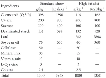
Table 1: Composition of standard chow and high-fat diet.
Ingredients Standard chow High-fat diet
g/kg−1 Kcal/kg−1 g/kg−1 Kcal/kg−1
Cornstarch (Q.S.P.) 398 1590 116 462
Casein 200 800 200 800
Sucrose 100 400 100 400
Dextrinated starch 132 528 132 528
Lard — — 312 2808
Soybean oil 70 630 40 360
Cellulose 50 — 50 —
Mineral mix 35 — 35 —
Vitamin mix 10 — 10 —
L-Cysteine 3 — 3 —
Choline 2.5 — 2.5 —
Total 1000 3948 1000 5358
22 ± 1∘C and60 ± 5% humidity. Ater the acclimatisation
period (1 week), the animals were randomly divided into four groups:(1)control mice (CW) fed on chow diet and placebo supplementation (0.1 mL water/day);(2)(CG) chow diet and green tea supplementation (0.1 mL water + 400 mg green tea extract per kg body weight/day); (3) (HW) a high-fat diet and placebo supplementation (0.1 mL water/day) for 2 months; (4) (HG) high-fat diet and green tea supplementation (0.1 mL water + 400 mg green tea commercial extract per kg body weight/day). he fatty acid composition of chow or high-fat diet diets is detailed in previous study from our group (Table 1) [24].
2.2. Composition of Green Tea by Ultra-Performance Liq-uid Chromatography (UPLC). We evaluated composition of green tea commercial extract by Ultra-performance Liquid Chromatography- Mass Spectrometry. An Acquity UPLC system (Waters, Milford, MA, USA) consisting of a binary solvent manager and a sample manager was coupled to an Acquity TQD Mass Spectrometer (Micromass Waters, Milford, MA, USA). Analyses were performed on a bridged ethylene hybrid (BEH) C18 analytical column (50 mm × 2.1 mm, 1.7�m, at a temperature of 25∘C, injecting 5�L of extract and standards. A gradient was applied at a low rate of 0.2 mL min−1 using two mobile phases—(A) puriied water with 0.1% formic acid; and (B) methanol—starting with 5% B, ramping to 100% B in 8 min, maintained until 8.50 min, returning to the initial conditions. Detection was carried out in the negative ion mode with an ESI source under the following conditions: capillary−3000 V, cone−30 volts, temperature 150∘C; ranging between m/z 100–1000. Data acquisition was carried out by MassLynx sotware. Our data showed that, in Green tea extract, there were 15�g/mg epi-gallocatechin, 95�g/mg epigallocatechin gallate, 20.8�g/mg epicatechin gallate, and 4.9�g/mg gallocatechin gallate.
samples were collected ater allowing the blood to clot on ice. Serum was stored frozen at−80∘C for analysis. Lab Test Kits were used to assess fasting total cholesterol, high-density lipoprotein (HDL-c), and triacylglycerol (TG) levels. he samples were analysed using an enzymatic method. LDL-c and VLDL-LDL-c were LDL-calLDL-culated aLDL-cLDL-cording to the Friedewald equation ((LDL-c = total cholesterol-(HDL-c)-(TG/5)) and (VLDL = TG/5)) [25]. he Zen-Bio Kit was used to assess free fatty acid.
2.4. TNF-�, Adiponectin, and IL-10 Protein Level Determina-tion by ELISA. Following decapitation, mesenteric adipose tissue was removed, dissected, homogenised, and centrifuged at 12,000 g for 40 min at 4∘C; the supernatant was saved, and the protein concentration was determined using the BCA assay (Bio-Rad, Hercules, California) with bovine serum albumin (BSA) as a reference. Quantitative assessment of adiponectin, TNF-�, and IL-10 proteins was carried out by ELISA (DuoSet ELISA, R and D Systems, Minneapolis, MN) following the recommendations of the manufacturer. All samples were run as duplicates, and the mean value is reported.
2.5. Protein Analysis by Western Blotting. Ater euthanasia, the epididymal, retroperitoneal, and mesenteric adipose tis-sue was dissected and weighed. Mesenteric adipose tistis-sue was homogenised in 1.0 mL solubilisation bufer at 4∘C (1% Triton X-100, 100 mM Tris-HCl (pH 7.4), 100 mM sodium pyrophosphate, 100 mM sodium luoride, 10 mM EDTA, 10 mM sodium orthovanadate, 2.0 mM phenylmethylsulpho-nyl luoride (PMSF), and 0.1 mg aprotinin/mL) with a Polytron (model 713T; Fisatom Equipamentos Cient´ıicos, S˜ao Paulo, SP, Brazil). Insoluble material was removed by centrifugation for 30 min at 9,000 g in a 70.Ti rotor (Beckman, Fullerton, CA, USA) at 4∘C. he protein concentration of the supernatants was measured by the BCA assay. Proteins were denatured by boiling (5 min) in Laemmli sample bufer [26] containing 100 mM DTT, run on 8, 10, or 12% SDS-PAGE gels in a Bio-Rad miniature slab gel apparatus. he electro-transfer of proteins from gels to nitrocellulose membranes was performed for∼1.30 h/4 gels at 15 V (constant) in a Bio-Rad semidry transfer apparatus. Nonspeciic protein binding to the nitrocellulose was reduced by preincubation for 2 h at 22∘C in blocking bufer (1% bovine serum albumine, 10 mM Tris, 150 mM NaCl, and 0.02% Tween 20). he nitrocellulose membranes were incubated overnight at 4∘C with antibodies against TLR4, myeloid diferentiation primary response gene (88) (MyD88), TNF receptor associated factor (TRAF6), hormone sensitive lipase (HSL), adipose triglyceride lipase (ATGL), comparative gene identiication-58 (CGI-58 or ABHD5), perilipin A, and alpha-tubulin obtained from Santa Cruz Biotechnology (Santa Cruz, CA, USA) diluted 1 : 1000 with blocking bufer supplemented with 1% BSA and then washed for 30 min in blocking bufer without BSA. he blots were subsequently incubated with peroxidase-conjugated secondary antibody for 1 h at 22∘C. For evaluation of protein loading, membranes were stripped and reblotted with an anti-alpha-tubulin antibody as appropriate. Speciic bands were
detected by chemiluminescence, and visualisation/capture was performed by exposure of the membranes to RX ilms. Band intensities were quantiied by optical densitometry of developed autoradiographs (Scion Image sotware-Scion Corporation, Frederick, MD, USA).
2.6. Statistical Analysis. he statistical analysis was per-formed using the GraphPad Prism statistics sotware package version 5.0 for Windows (GraphPad Sotware, San Diego, CA, USA). he data are expressed as the means ± SEM. Implementation of the Kolmogorov-Smirnov test revealed that the results of experiments were distributed normally. he data were analysed using ANOVA two ways for comparison between four groups. A value of� < 0.05 was considered statistically signiicant.
3. Results
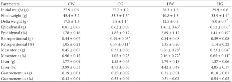
3.1. Body Mass and Tissue Weight. he relative weight (tissue weight/total body weight) of epididymal adipose tissue was increased in group HW compared to the GW group and decreased in the HG group compared to the HW group. he mesenteric adipose tissue showed an increase in group HW compared to the GW group and decrease in the HG group compared to the HW group. In retroperitoneal adipose tissue, only the CG group decreased compared with the GW group. Liver and gastrocnemius tissues showed no signiicant diference between groups (Table 2).
3.2. Lipid Proile and Serum Adiponectin. Serum triglycerides and total cholesterol did not difer between any of the groups. he serum concentration of LDL in the HW group was increased compared to the GW group. he concentration of serum HDL in the HG group showed an increase compared to the HW group. he serum concentration of FFA did not difer between any of the four groups. he serum adiponectin in CG group increased compared to the GW group, and the HG group increased compared to the HW group (Table 3).
3.3. Cytokines in the Adipose Tissue. he cytokine concentra-tion of adiponectin in the mesenteric adipose tissue in the CG group increased compared to the GW group, and the HG group increased compared to the HW group. he content of IL-10 showed a signiicant increase in group CG compared to the GW group. he TNF-�levels in the mesenteric adipose pad of the HW group showed a signiicant increase compared to the CW group. However, supplementation of green tea decreased this efect (HG versus HW groups;Table 4).
Table 2: Body weight and absolute and relative tissue weight.
Parameters CW CG HW HG
Initial weight (g) 27.9 ± 0.9 27.7 ± 1.2 28.3 ± 1.5 25.9 ± 0.6
Final weight (g) 45.4 ± 3.2 33.5 ± 1.5∗ 40.8 ± 1.3 33.9 ± 1.4#
Delta weight (g) 17.5 ± 1.3 5.8 ± 1.1∗ 12.5 ± 0.9 8.0 ± 0.7#
Epididymal (g) 0.81 ± 0.07 0.62 ± 0.09 1.45 ± 0.43$ 0.52 ± 0.08#
Epididymal (%) 1.78 ± 0.16 1.85 ± 0.17 2.89 ± 1.12 1.41 ± 0.19#
Retroperitoneal (g) 0.44 ± 0.07 0.19 ± 0.03∗ 0.54 ± 0.08 0.39 ± 0.08
Retroperitoneal (%) 1.05 ± 0.21 0.57 ± 0.11∗ 1.35 ± 0.20 1.14 ± 0.22
Mesenteric (g) 0.45 ± 0.07 0.33 ± 0.06 0.86 ± 0.28$ 0.23 ± 0.04#
Mesenteric (%) 0.96 ± 0.12 1.05 ± 0.23 2.16 ± 0.72$ 0.65 ± 0.11#
Liver (g) 1.77 ± 0.09 1.55 ± 0.05 1.79 ± 0.18 1.37 ± 0.06
Liver (%) 3.99 ± 0.33 4.73 ± 0.30 4.42 ± 0.49 4.05 ± 0.17
Gastrocnemius (g) 0.19 ± 0.01 0.17 ± 0.02 0.21 ± 0.01 0.18 ± 0.01
Gastrocnemius (%) 0.43 ± 0.04 0.53 ± 0.09 0.51±0.03 0.54 ± 0.05
∗� < 0.05chow diet and green tea (CG) group versus chow diet and water (CW) group (� = 12).$� < 0.05high-fat diet and water (HW) group versus CW
group (� = 12).#� < 0.05high-fat diet and green tea (HW) group versus HW group (� = 12). g: grams;%: percentage.
Table 3: Serum concentrations of triacylglycerol (TAG), total cholesterol (TC), high-density lipoprotein (HDL), low-density lipoprotein (LDL), free fatty acids (FFA), and adiponectin in diferent experimental groups.
Parameters CW CG HW HG
TAG (mmol/L) 1.58 ± 0.04 1.57 ± 0.03 1.60 ± 0.04 1.46 ± 0.05
CT (mmol/L) 3.12 ± 0.08 3.29 ± 0.13 3.58 ± 0.17 3.54 ± 0.06
HDL (mmol/L) 1.54 ± 0.10 1.85 ± 0.16 1.81 ± 0.10 2.31 ± 0.13#
LDL (mmol/L) 0.83 ± 0.04 0.71 ± 0.06 1.11 ± 0.09$ 0.87 ± 0.08
FFA (�M) 1.21 ± 0.08 1.26 ± 0.08 1.02 ± 0.11 1.16 ± 0.13
Adiponectin (ng/mL) 82.25 ± 1.76 106.49 ± 2.91∗ 85.82 ± 2.53 112.02 ± 7.64#
∗� < 0.05chow diet and green tea (CG) group versus chow diet and water (CW) group (� = 12).$� < 0.05high-fat diet and water (HW) group versus CW
group (� = 12).#� < 0.05high-fat diet and green tea (HW) group versus HW group (� = 12).
supplemented with green tea (Figure 1(b)). In the HW group, the ABHD5 (or CGI-58) protein levels were reduced when compared to the GW group. However, supplementation with green tea strikingly increased the ABHD5 protein levels in obese mice (HG group) when compared to the HW group. No signiicant diference was observed in the chow-diet groups (Figure 1(c)). he perilipin protein levels increased in mice fed with chow diet supplemented with green tea (CG group) compared to no supplementation (CW group). No diference was observed between the chow and high-fat diets without supplementation. However, green tea increased perilipin protein levels in the HG group compared to the HW group (Figure 1(d)).
3.5. Quantiication of Inlammatory Proteins. he TLR4 pro-tein levels in diet-induced obese mice (HW group) were signiicantly greater than chow-diet mice (CW group). Green tea treatment decreased this efect signiicantly (HG versus HW groups, � < 0.05; Figure 2(a)). he MyD88 protein levels in the HW group increased compared to the CW group. However, when obese mice were supplemented with green tea (HG group), this efect was attenuated (Figure 2(b)). he TRAF6 protein levels in diet-induced obese mice (HW group) were signiicantly greater than chow-diet mice (CW
group). Green tea treatment signiicantly decreased this efect (HG versus HW groups,� < 0.05;Figure 2(c)).
4. Discussion
Table 4: Content cytokines in mesenteric adipose tissue.
Adipokines CW CG HW HG
Adiponectin (pg/�g of protein) 0.24 ± 0.03 0.37 ± 0.02∗ 0.18 ± 0.01 0.40 ± 0.04#
IL-10 (pg/�g of protein) 1.91 ± 0.26 11.27 ± 1.33∗ 3.47 ± 0.50 5.34 ± 0.47
TNF-�(pg/�g of protein) 2.09 ± 0.79 1.92 ± 0.61 5.80 ± 0.47$ 2.62 ± 0.61#
∗� < 0.05chow diet and green tea (CG) group versus chow diet and water (CW) group (� = 12).$� < 0.05
high-fat diet and water (HW) group versus CW group (� = 12).#� < 0.05high-fat diet and green tea (HW) group versus HW group (� = 12).
HSL
CW CG HW HG
0 50 100 150 200
(a.u
.) #
∗
(a)
ATGL
CW CG HW HG
0 50 100 150 200
(a.u.)
∗
(b)
ABHD5
CW CG HW HG
0 50 100 150
(a.u
.)
#
$
(c)
Peri A
CW CG HW HG
0 50 100 150 200 250
(a.u
.) #
∗
(d)
HSL
ATGL
ABHD5
Peri A
𝛼-tubulin
(e)
Figure 1: Protein levels of HSL, ATGL, ABHD5, and perilipin A. Mesenteric adipose tissue extracts were immunoblotted with anti-HSL (a),
anti-ATGL (b), anti-ABHD5 (c), and Peri A (d). he results of scanning densitometry are expressed as arbitrary units. Bars represent means±
SEM of� = 6mice,∗� < 0.05chow and green tea (CG) group versus chow and water (CW) group,$� < 0.05high-fat and water (HW) group
versus CW group, and#
� < 0.05high-fat and green tea (HG) group versus HW group. In (e), the representative bands of the molecules are
shown. he membrane was stripped and immunoblotted with anti-�-tubulin antibody and used as loaded protein (lower panel in (e)).
In the study, we measured body weight of the animals at the beginning and end of the study. Our results demonstrate that a high-fat diet induced body-weight gain (as observed by delta weight) and epididymal and mesenteric adipose tissue pads. However, green tea promoted a reduced delta weight and adipose tissue pads. Further, green tea extract led to increased lipolytic pathway protein levels, adiponectin, and anti-inlammatory cytokine IL-10 and reduced proinlamma-tory cytokine TNF-�.
he therapeutic uses of tea are conined to alternative medicine. Although the anticarcinogenic, anti-inlammatory, and antimicrobial properties of tea have been known for many years, clinical medicine has not included its use in treatments, almost certainly due to the lack of knowledge about its exact mechanisms of action [21, 22]. In human experiments, acute ingestion of green tea extract, which is mainly composed of catechins, has been reported to increase the proportion of whole-body fat utilisation by augmenting oxidation and lipolysis [23,38,39]. Lee et al. [40]
demonstrated in an in vitro study that EGCG modulates the increase in lipolysis by directly increasing the gene expression of HSL, demonstrating its important role in lipid metabolism. Habitual consumption green tea extract has been reported to reduce body weight and body fat [32–36]; this may occur via increased lipolysis in adipose tissue, and our data support this.
he anti-inlammatory efect of green tea has been attributed to the polyphenol content [30, 31]. In Asian countries, green tea, which contains a class of polyphenols known as tea catechins, has been habitually consumed as one of the most popular beverages. Tea polyphenols have been shown to inhibit proteasome function, thereby terminating inlammation. Although tea polyphenols have been claimed to be the most potent constituents of tea, there is increasing evidence that these compounds are not the only constituents responsible for the beneicial efects on health from tea [41].
TLR4
CW CG HW HG
0 50 100 150 200
(a.u
.)
$
#
(a)
CW CG HW HG
0 50 100 150 200
(a.u
.)
$ MyD88
#
(b)
CW CG HW HG
TRAF6
0 50 100 150 200
(a.u
.)
$
#
(c)
TLR4
MyD88
TRAF6
𝛼-tubulin
(d)
Figure 2: Protein levels of TLR4, MyD88, and TRAF6. Mesenteric adipose tissue extracts were immunoblotted with TLR4 (a),
anti-MyD88 (b), and anti-TRAF6 (c). he results of scanning densitometry are expressed as arbitrary units. Bars represent means±SEM of� = 6
mice,#� < 0.05when compared to the high-fat diet and green tea (HG) group versus high-fat diet and water (HW) group. In (d), the
representative bands of the molecules are shown. he membrane was stripped and immunoblotted with anti-�-tubulin antibody and used as
loaded protein (lower panel in (d)).
lipolytic enzymes. hese conditions may favour reduced body weight and adipose tissues. In addition, we found that green tea reduced TLR4 expression, blocking proinlammatory efects. Youn et al. [42] showed that EGCG in cultured cells of the immune system had an anti-inlammatory efect, which was partly explained by the inhibition of the TLR. In summary, our results show that green tea extract intake increases expression of lipases, reduces adipose fat mass, and in parallel reduces inlammatory molecules and cytokines. Futures studies are needed to better understand the mecha-nism involved in the beneicial efects promoted by green tea extract intake, especially in mice fed a high-fat diet.
Conflict of Interests
All authors declare no conlict of interests.
Acknowledgments
his work was supported by Conselho Nacional de Desen-volvimento Cient´ıico e Tecnol´ogico (CNPq), Fundac¸˜ao de
Amparo `a Pesquisa do Estado de S˜ao Paulo (FAPESP), and Coordenac¸˜ao de Aperfeic¸oamento de Pessoal de N´ıvel Superior (CAPES). he authors would like to thank Paulo Mazzafera (BIOEN-FAPESP 08/58035-6) for the use of the UPLC-MS equipment.
References
[1] P. G. Kopelman, “Obesity as a medical problem,”Nature, vol.
404, no. 6778, pp. 635–643, 2000.
[2] A. Sch¨aler, J. Sch¨olmerich, and B. Salzberger, “Adipose tissue as an immunological organ: toll-like receptors, C1q/TNFs and
CTRPs,”Trends in Immunology, vol. 28, no. 9, pp. 393–399, 2007.
[3] K. E. Wellen and G. S. Hotamisligil, “Obesity-induced
inlam-matory changes in adipose tissue,” he Journal of Clinical
Investigation, vol. 112, no. 12, pp. 1785–1788, 2003.
[4] P. Trayhurn, “Endocrine and signalling role of adipose tissue:
new perspectives on fat,”Acta Physiologica Scandinavica, vol.
184, no. 4, pp. 285–293, 2005.
in adipocytes,”he Journal of Biological Chemistry, vol. 275, no. 32, pp. 24255–24263, 2000.
[6] D. Werling and T. W. Jungi, “TOLL-like receptors linking innate
and adaptive immune response,”Veterinary Immunology and
Immunopathology, vol. 91, no. 1, pp. 1–12, 2003.
[7] A. Shah, N. Mehta, and M. P. Reilly, “Adipose inlammation,
insulin resistance, and cardiovascular disease,”Journal of
Par-enteral and Enteral Nutrition, vol. 32, no. 6, pp. 638–644, 2008. [8] P. Cristofaro and S. M. Opal, “Role of toll-like receptors in
infection and immunity: clinical implications,”Drugs, vol. 66,
no. 1, pp. 15–29, 2006.
[9] S. Pandey and D. K. Agrawal, “Immunobiology of Toll-like
receptors: emerging trends,”Immunology and Cell Biology, vol.
84, no. 4, pp. 333–341, 2006.
[10] L. A. J. O’Neill, “Signal transduction pathways activated by the
IL-1 receptor/Toll-like receptor superfamily,”Current Topics in
Microbiology and Immunology, vol. 270, pp. 47–61, 2002.
[11] S. Akira, “Toll-like receptor signaling,”he Journal of Biological
Chemistry, vol. 278, no. 40, pp. 38105–38108, 2003.
[12] V. Bezaire, A. Mairal, C. Ribet et al., “Contribution of adipose triglyceride lipase and hormone-sensitive lipase to lipolysis in
hMADS adipocytes,”he Journal of Biological Chemistry, vol.
284, no. 27, pp. 18282–18291, 2009.
[13] S. Klaus, S. P¨ultz, C. h¨one-Reineke, and S. Wolfram, “Epi-gallocatechin gallate attenuates diet-induced obesity in mice by decreasing energy absorption and increasing fat oxidation,” International Journal of Obesity, vol. 29, no. 6, pp. 615–623, 2005. [14] C. H. Wu, F. H. Lu, C. S. Chang et al., “Relatioship among habitual tea consumption, percent body fat, and body fat
distribution,”Obesity Research, vol. 11, pp. 1088–1095, 2003.
[15] H. Iso, C. Date, K. Wakai et al., “he relationship between green tea and total cafeine intake and risk for self reported type 2
diabetes among Japanese adults,”Annals of Internal Medicine,
vol. 144, pp. 554–562, 2006.
[16] Y. C. Yang, F. H. Lu, J. S. Wu et al., “he protective efect of
habitual tea consumption on hypertension,”Annals of Internal
Medicine, vol. 164, pp. 1534–1540, 2004.
[17] S. Sasazuki, H. Kodama, K. Yoshimasu et al., “Relation between green tea consumption and the severity of coronary
atheroscle-rosis among Japanese men and women,”Annals of
Epidemiol-ogy, vol. 10, no. 6, pp. 401–408, 2000.
[18] H. D. Sesso, J. M. Gaziano, J. E. Buring, and C. H. Hennekens, “Cofee and tea intake and the risk of myocardial infarction,” American Journal of Epidemiology, vol. 149, no. 2, pp. 162–167, 1999.
[19] M. S. Westerterp-Plantenga, M. P. G. M. Lejeune, and E. M. R. Kovacs, “Body weight loss and weight maintenance in relation to habitual cafeine intake and green tea supplementation,” Obesity Research, vol. 13, no. 7, pp. 1195–1204, 2005.
[20] S. Shiota, M. Shimizu, T. Mizushima et al., “Marked reduction
in the minimum inhibitory concentration (MIC) of�- lactams
in methicillin-resistant Staphylococcus aureus produced by epi-catechin gallate, an ingredient of green tea (Camellia sinensis),” Biological and Pharmaceutical Bulletin, vol. 22, no. 12, pp. 1388– 1390, 1999.
[21] K. Muramatsu, M. Fukuyo, and Y. Hara, “Efect of green tea catechins on plasma cholesterol level in cholesterol-fed rats,” Journal of Nutritional Science and Vitaminology, vol. 32, no. 6, pp. 613–622, 1986.
[22] N. Matsumoto, F. Ishigaki, A. Ishigaki et al., “Reduction of blood
glucose levels by tea catechin,”Bioscience, Biotechnology, and
Biochemistry, vol. 57, pp. 525–527, 1993.
[23] S. Wolfram, Y. Wang, and F. hielecke, “Anti-obesity efects of
green tea: from bedside to bench,”Molecular Nutrition and Food
Research, vol. 50, no. 2, pp. 176–187, 2006.
[24] G. D. Pimentel, F. S. Lira, J. C. Rosa et al., “Yerba mate extract (Ilex paraguariensis) attenuates both central and peripheral
inlammatory efects of diet-induced obesity in rats,” he
Journal of Nutritional Biochemistry. In press.
[25] W. T. Friedewald, R. I. Levy, and D. S. Fredrickson, “Estimation of the concentration of low-density lipoprotein cholesterol in
plasma, without use of the preparative ultracentrifuge,”Clinical
Chemistry, vol. 18, no. 6, pp. 499–502, 1972.
[26] U. K. Laemmli, “Cleavage of structural proteins during the
assembly of the head of bacteriophage T4,”Nature, vol. 227, no.
5259, pp. 680–685, 1970.
[27] T. Rankinen, A. Zuberi, Y. C. Chagnon et al., “he human
obesity gene map: the 2005 update,”Obesity, vol. 14, no. 4, pp.
529–644, 2006.
[28] A. Festa, R. D’Agostino Jr., K. Williams et al., “he relation of body fat mass and distribution to markers of chronic
inlammation,” International Journal of Obesity, vol. 25, pp.
1407–1415, 2001.
[29] D. R. Cottam, S. G. Mattar, E. Barinas-Mitchell et al., “he chronic inlammatory hypothesis for the morbidity associated with morbid obesity: implications and efect of weight loss,” Obesity Surgery, vol. 14, no. 5, pp. 589–600, 2004.
[30] C. L. Shen, J. K. Yeh, C. Samathanam et al., “Green tea polyphenols attenuate deterioration of bone microarchitecture
in female rats with systemic chronic inlammation,”
Osteoporo-sis International, vol. 22, no. 1, pp. 327–337, 2011.
[31] C. L. Shen, J. K. Yeh, J. J. Cao, O. L. Tatum, R. Y. Dagda, and J. S. Wang, “Green tea polyphenols mitigate bone loss of female rats
in a chronic inlammation-induced bone loss model,”Journal of
Nutritional Biochemistry, vol. 21, no. 10, pp. 968–974, 2010. [32] P. Auvichayapat, M. Prapochanung, O. Tunkamnerdthai et al.,
“Efectiveness of green tea on weight reduction in obese hais: a
randomized, controlled trial,”Physiology and Behavior, vol. 93,
no. 3, pp. 486–491, 2008.
[33] C. H. Hsu, T. H. Tsai, Y. H. Kao, K. C. Hwang, T. Y. Tseng, and P. Chou, “Efect of green tea extract on obese women: a randomized, double-blind, placebo-controlled clinical trial,” Clinical Nutrition, vol. 27, no. 3, pp. 363–370, 2008.
[34] M. C. Lonac, J. C. Richards, M. M. Schweder, T. K. Johnson, and C. Bell, “Inluence of short-term consumption of the cafeine-free, epigallocatechin-3-gallate supplement, teavigo, on resting
metabolism and the thermic efect of feeding,”Obesity, vol. 19,
no. 2, pp. 298–304, 2011.
[35] T. Nagao, T. Hase, and I. Tokimitsu, “A green tea extract high in catechins reduces body fat and cardiovascular risks in humans,” Obesity, vol. 15, no. 6, pp. 1473–1483, 2007.
[36] O. J. Phung, W. L. Baker, L. J. Matthews, M. Lanosa, A. horne, and C. I. Coleman, “Efect of green tea catechins with or without cafeine on anthropometric measures: a systematic review and
meta-analysis,”American Journal of Clinical Nutrition, vol. 91,
no. 1, pp. 73–81, 2010.
[37] M. S. Westerterp-Plantenga, “Green tea catechins, cafeine and
body-weight regulation,”Physiology and Behavior, vol. 100, no.
1, pp. 42–46, 2010.
[38] A. Basu, K. Sanchez, M. J. Leyva et al., “Green tea supplementa-tion afects body weight, lipids, and lipid peroxidasupplementa-tion in obese
subjects with metabolic syndrome,”Journal of the American
[39] M. Boschmann and F. hielecke, “he efects of epigallocatechin-3-gallate on thermogenesis and fat oxidation
in obese men: a pilot study,”Journal of the American College of
Nutrition, vol. 26, no. 4, pp. 389S–395S, 2007.
[40] M. S. Lee, C. T. Kim, I. H. Kim, and Y. Kim, “Inhibitory efects of green tea catechin on the lipid accumulation in 3T3-l1
adipocytes,”Phytotherapy Research, vol. 23, no. 8, pp. 1088–1091,
2009.
[41] F. Pajonk, A. Riedisser, M. Henke, W. H. McBride, and B. Fiebich, “he efects of tea extracts on proinlammatory
signal-ing,”BMC Medicine, vol. 4, article 28, 2006.
[42] H. S. Youn, J. Y. Lee, S. I. Saitoh et al., “Suppression of MyD88-and TRIF-dependent signaling pathways of toll-like receptor by (-)-epigallocatechin-3-gallate, a polyphenol component of
green tea,”Biochemical Pharmacology, vol. 72, no. 7, pp. 850–
Submit your manuscripts at
http://www.hindawi.com
Stem Cells
International
Hindawi Publishing Corporation
http://www.hindawi.com Volume 2014
Hindawi Publishing Corporation
http://www.hindawi.com Volume 2014
INFLAMMATION
Hindawi Publishing Corporation
http://www.hindawi.com Volume 2014
Behavioural
Neurology
Endocrinology
International Journal ofHindawi Publishing Corporation
http://www.hindawi.com Volume 2014
Hindawi Publishing Corporation
http://www.hindawi.com Volume 2014
Disease Markers
Hindawi Publishing Corporation
http://www.hindawi.com Volume 2014
BioMed
Research International
Oncology
Journal ofHindawi Publishing Corporation
http://www.hindawi.com Volume 2014
Hindawi Publishing Corporation
http://www.hindawi.com Volume 2014
Oxidative Medicine and Cellular Longevity
Hindawi Publishing Corporation
http://www.hindawi.com Volume 2014
PPAR Research
The Scientiic
World Journal
Hindawi Publishing Corporationhttp://www.hindawi.com Volume 2014
Immunology Research Hindawi Publishing Corporation
http://www.hindawi.com Volume 2014
Journal of
Obesity
Journal ofHindawi Publishing Corporation
http://www.hindawi.com Volume 2014
Hindawi Publishing Corporation
http://www.hindawi.com Volume 2014 Computational and Mathematical Methods in Medicine
Ophthalmology
Journal of Hindawi Publishing Corporationhttp://www.hindawi.com Volume 2014
Diabetes Research
Journal ofHindawi Publishing Corporation
http://www.hindawi.com Volume 2014
Hindawi Publishing Corporation
http://www.hindawi.com Volume 2014
Research and Treatment
AIDS
Hindawi Publishing Corporation
http://www.hindawi.com Volume 2014
Gastroenterology Research and Practice
Hindawi Publishing Corporation
http://www.hindawi.com Volume 2014
Parkinson’s
Disease
Evidence-Based Complementary and Alternative Medicine
Volume 2014