Research Article
Preventive Effects of Chitosan Coacervate Whey Protein on Body
Composition and Immunometabolic Aspect in Obese Mice
Gabriel Inácio de Morais Honorato de Souza,
1,2Aline Boveto Santamarina,
1Aline Alves de Santana,
1Fábio Santos Lira,
3Rachel de Laquila,
4Mayara Franzoi Moreno,
1Eliane Beraldi Ribeiro,
1Claudia Maria da Penha Oller do Nascimento,
1Bruno Rodrigues,
2Elisa Esposito,
5and Lila Missae Oyama
11Departamento de Fisiologia, Disciplina de Fisiologia da Nutric¸˜ao, Universidade Federal de S˜ao Paulo, 04023-060 S˜ao Paulo, SP, Brazil 2Departamento de Ciˆencias Biol´ogicas, Laborat´orio de Movimento Humano da Universidade S˜ao Judas Tadeu,
03166-000 S˜ao Paulo, SP, Brazil
3Departamento de Educac¸˜ao F´ısica da Universidade Estadual Paulista, Faculdade de Ciˆencias e Tecnologia,
19060-900 Presidente Prudente, SP, Brazil
4Departamento de Nutric¸˜ao das Faculdades Integradas Corac¸˜ao de Jesus-FAINC, 09020-240 Santo Andr´e, SP, Brazil 5Instituto de Ciˆencias e Tecnologia da Universidade Federal de S˜ao Paulo, 04023-060 S˜ao Jos´e dos Campos, SP, Brazil
Correspondence should be addressed to Gabriel In´acio de Morais Honorato de Souza; [email protected] and Elisa Esposito; [email protected]
Received 23 May 2014; Revised 30 July 2014; Accepted 31 July 2014; Published 17 September 2014 Academic Editor: Jos´e Cesar Rosa Neto
Copyright © 2014 Gabriel In´acio de Morais Honorato de Souza et al. his is an open access article distributed under the Creative Commons Attribution License, which permits unrestricted use, distribution, and reproduction in any medium, provided the original work is properly cited.
Functional foods containing bioactive compounds of whey may play an important role in prevention and treatment of obesity. he aim of this study was to investigate the prospects of the biotechnological process of coacervation of whey proteins (CWP) in chitosan and test its antiobesogenic potential.Methods.CWP (100 mg⋅kg⋅day) was administered in mice with diet-induced obesity for 8 weeks. he animals were divided into four groups: control normocaloric diet gavage with water (C) or coacervate (C-CWP), and high fat diet gavage with water (HF) or coacervate (HF-CWP).Results.HF-CWP reduced weight gain and serum lipid fractions and displayed reduced adiposity and insulin. Adiponectin was signiicantly higher in HF-CWP group when compared to the HF. he level of LPS in HF-W group was signiicantly higher when compared to HF-CWP. he IL-10 showed an inverse correlation between the levels of insulin and glucose in the mesenteric adipose tissue in the HF-CWP group. CWP promoted an increase in both phosphorylation AMPK and the amount of ATGL in the mesenteric adipose tissue in HF-CWP group.Conclusion. CWP was able to modulate efects, possibly due to its high biological value of proteins. We observed a protective efect against obesity and improved the inlammatory milieu of white adipose tissue.
1. Introduction
Over the past decades, the incidence of obesity in the population has increased severely, and it has become a public health challenge. Its etiology is multifactorial, encompass-ing environmental, dietary, physical inactivity, and genetic factors. Obesity is a complex disease associated with a high-calorie diet, which contributes to the development
of several other chronic noncommunicable diseases [1].
Obesity is also associated with increased plasma endotoxin
(lipopolysaccharide-LPS), saturated fatty acids [2, 3], and
proinlammatory cytokines [3] all intricately involved in
the development of comorbidities such as diabetes mellitus, hypertension, dyslipidemia, and metabolic syndrome.
he fat tissue is not merely an energy storage organ, as it plays crucial endocrine and immune roles. White adipose tissue (WAT) is an endocrine organ secreting pro- and anti-inlammatory adipokines such as tumor necrosis factor
alpha (TNF-�) and interleukin-6 (IL-6), which are
impor-tant inlammatory markers that stimulate the production of Volume 2014, Article ID 281097, 13 pages
several proteins and proinlammatory cytokines in diferent
cell types, via nuclear factor�B activation (NF-�B) [4]. In
addition, endotoxin and saturated fatty acids act in the same
pathway of NF-�B activation. Many studies have shown that
LPS (endotoxin) can activate these proteins in adipocytes, thereby increasing the gene expression of proinlammatory
adipokines [5–7].
he main role of adipose lipolytic enzymes is to provide other tissues with FAs in case of energy demand. Triglyceride stored in the lipid droplet is irst hydrolyzed by the adipose triglyceride lipase enzyme (ATGL), also known as desnutrin, releasing a diacylglycerol moiety and FA, which requires an abhydrolase domain containing 5 (ABHD-5) promoter to be activated. Ater hydrolysis by ATGL, diacylglycerols are then hydrolyzed sequentially by hormone-sensitive lipase (HSL) and monoglyceride lipase (MGL), producing nonesteriied
fatty acids (NEFAs) and glycerol [8]. Diferent lipases gain
access to the lipid droplet when the proteins coating the vesicle (perilipins) are phosphorylated. Perilipin A normally prevents lipolysis of triglyceride by surrounding the lipid droplet, thus preventing the access of lipases.
Whey protein (WP) has been found to be an excellent prophylactic against obesity, because of the high biological value mediated by bioactive peptides. hese act as antimi-crobial agents, antihypertensive, and regulators of immune function, reducing body fat as well as a variety of related beneicial mechanisms for human health. hey also have additional functions; for example, they have appetite
sup-pressant efects [9], stimulate muscle protein synthesis, and
regulate of body energy homeostasis [10]. here is plenty
of evidence indicating the potential of the WP in
anti-inlammatory and antioxidant efects of exercise [9,11–13].
Chitosan complex coacervation with WP is composed of by-products from the processing of shrimp, crab (chitosan), and cheese, adding an environmental beneit to the product, as these by-products may be reused and not disposed of in
landill sites or released into rivers by producers [14]. In view
of the above, together with the development of fractionation technique and whey protein preservation, employing the method may contribute to the recovery of this valuable nutrient and increase the expression of the functional
prop-erties [15]. Complex coacervation is deined as a colloidal
separation forming two liquid phases. his process is essen-tially driven by attractive forces with opposite charges of a biopolymer. his phenomenon occurs by the formation of a system of balance between colloids and the diluted
supernatant [16,17]. hus, the purpose of the present study
was to investigate the prospect of a biotechnological process of complexation and separation of cheese whey proteins in chitosan and test their antiobesogenic potential through the modulation of inlammatory markers and lipolytic pathway present in obesity.
2. Methods
2.1. Coacervation and Characterization. In this study we
used sweet cheese whey (SW 1108 bag 25 kg) with 1.5% fat marketed by Company Alibra-PR. Dissolving 10 g of the whey
powder in 100 mL of distilled water. Chitosan was used for the coacervation medium molar mass with 75–85% degree of deacetylation and viscosity of 200–800 cps (Sigma-Aldrich 44887-7). A concentration of 0.75 mg/mL of Chitosan was used. Chitosan was dissolved in citric acid (208 mmol/L) and, ater this step, added to the cheese whey in a proportion of 1 : 1 under stirring at room temperature for 1 h. he pH of the solution of chitosan and WP was adjusted to 6 with NaOH
(250 mmol/L) and solubilized at room temperature (±25∘C)
with stirring. Solids coagulated with chitosan known as coacervate (CWP) were collected by centrifugation (1300 g).
In order to obtain on average 30% of the protein coac-ervate 3 L cheese whey was used. hus, samples of CWP were obtained for their chemical analysis of total lipids, total protein, and lactose. Finally, for measurements of samples of mineral micronutrients, Ca, K, Mg, and P, of cheese
whey, CWP, and Chitosan, 100�g was subjected to an optical
emission spectrometer for inductively coupled plasma (ICP OES, Perkin Elmer Optima 3000 DV, Norwalk, CT, USA). To determine the existing protein fractions in CWP the elec-trophoretic proile with reducing bufer containing 62.5 mM Tris-HCl, 20% glycerol, 2% sodium dodecyl sulfate(SDS)
(10%), 5%�-mercaptoethanol, and bromophenol blue at pH
6.8 was performed.
2.2. Animals and Treatment. his study was approved by the
Research Ethics Committee of the Universidade Federalde S˜ao Paulo, Escola Paulista de Medicina (UNIFESPEPM), as the search protocol number 0473/10. Experimental pro-cedures are in accordance with Principles of Laboratory Animal Care formulated by the National Institutes of Health (National Institutes of Health Publication number 96-23, revised 1996).
Forty-nine male Swiss mice twelve-week-old from CEDEME (Centro de Desenvolvimento de Modelos Experimentais da Universidade Federal de S˜ao Paulo) were housed ive in a cage in a standard experimental animal laboratory and kept under controlled conditions of light (12 h light-dark cycle with lights on at 6 am) and temperature
(24 ± 1∘C). All mice received water and food ad libitum.
he animals were divided as follows: control diet plus tap water (C-W); control diet plus coacervate (C-CWP); high fat diet plus coacervate (HF-CWP); and high fat diet plus tap water (HF-W). All diets were prepared according to the
recommendations of the American Institute of Nutrition [18]
(Table 1). Coacervate (CWP) containing 100 mg⋅kg⋅day was given by gavage. he body weight gain was recorded twice a week.
2.3. Composition and Aspect of the Coacervate. Figure 1(a)
shows the protein proile of the coacervate by the presence of the major proteins of larger fractions, namely,
alpha-lactalbumin (�-La-14 kDa), beta-lactoglobulin (�-Lg—
18 kDa monomer and 34 kDa dimer form), bovine serum albumin (BSA—66 kD), and lactoferrin (Lacf—86 kDa). he
CWP has a locculation aspect (Figure 1(b)) consisting of
proteins and micronutrients such as calcium, potassium,
Table 1: Composition of the control diet and diet enriched with saturated fatty acids according to AIN-93. Coacervate was resuspended in 300�L of water.
Components (%) Control diet (C) High fat diet (HF)
Corn starch 72.07 40.87
Casein 14.0 14.0
Soybean oil 4.0 4.0
Lard — 31.2
Cellulose 5.0 5.0
Vitamin mix 1.0 1.0
Mineral mix 3.5 3.5
L-cystine 0.18 0.18
Choline bitartrate 0.25 0.25
Butyl hydroquinone. g/kg 0.008 0.008
Energy. kcal/kg 3,802.8 5,362.8
Treatment by gavage
Coacervate (CWP) (C-CWP) 100 mg⋅kg⋅day (HF-CWP) 100 mg⋅kg⋅day
Water (W) (C-W) 300�L⋅day (HF-W) 300�L⋅day
Fatty acids (%)
Saturated (SFA) 17.12 34.13
Monounsaturated (MUFA) 25.63 39.14
Polyunsaturated (PUFA) 57.25 26.67
PUFA n3 4.32 6.37
PUFA n6 52.65 19.98
ST CWP
Lacfr BSA 75
50 37
25 20
15 �-La
�-Lg monomer
�-Lg dimer
(a) (b)
Figure 1: (a) Electrophoretic proile coacervate using 0.75 mg/ml of chitosan. ST: standard of diferent molecular weights,�-La: alpha-lactalbumin (14 kDa),�-Lg: beta-lactoglobulin monomer (18 kDa) and dimer (34 kDa), BSA: bovine albumin (66 kDa), and Lacfr: lactoferrin (86 kDa). (b) Aspect of CWP proteins in Chitosan, freeze-dried.
2.4. Fatty Acids Composition of Diets. For total lipid
extrac-tion, diet samples were homogenized in chloroform and methanol 2 : 1 (v/v), mixed, and incubated at room tem-perature for 5 min. hen, additional volumes of 1.25 mL
chloroform and 1.25 mL deionized H2O were added, and
inally, following being vigorously homogenized for 3 min, samples were centrifuged at 1000 rpm for 5 min at room
temperature. he chloroform layer was dried under N2, and
the total extract was converted into methyl esters and was analyzed in gas chromatography (GC), coupled with a lame ionizer detector (FID), (Varian GC 3900) and fatty acid proile was determined by calculating the retention time,
using a pattern of fatty acids with known retention time (Supelco, 37 Components). he addition was initiated at a
temperature of 170∘C maintained for 1 minute and then a
ramp of 2.5∘C/min to a inal temperature of 240∘C, which
was maintained for 5 minutes. he injector and detector
were maintained at 250∘C and 260∘C, respectively. We used a
column CP wax 52CB, with a thickness of 0.25 mm, internal
diameter of 0.25�m, and length of 30 m,with hydrogen as the
carrier gas at a linear velocity of 22 cm s−1.
2.5. Oral Glucose Tolerance Test (OGTT). Ater 12 hours
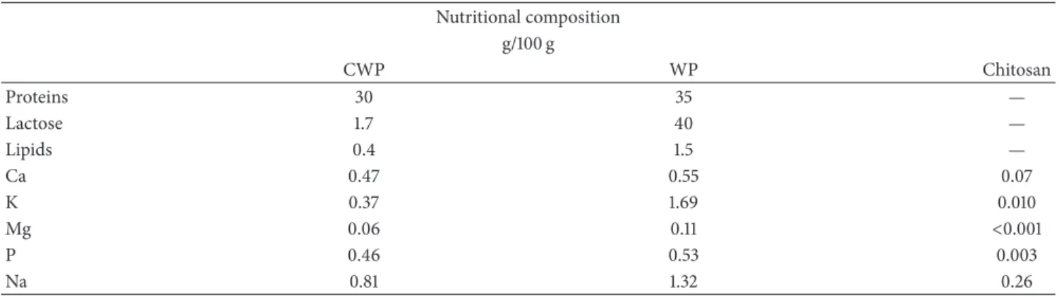
Table 2: Partial composition of micronutrients by ICP and chemical macronutrients of CWP, WP, and Chitosan. Nutritional composition
g/100 g
CWP WP Chitosan
Proteins 30 35 —
Lactose 1.7 40 —
Lipids 0.4 1.5 —
Ca 0.47 0.55 0.07
K 0.37 1.69 0.010
Mg 0.06 0.11 <0.001
P 0.46 0.53 0.003
Na 0.81 1.32 0.26
basal glucose concentration. hen, a glucose (Merck) solution (1.4 g/kg) was administrated by gavage. Blood samples were collected ater 15, 30, 45, 60, and 120 minutes to measure glucose concentration using a glucose analyzer (AccuCheck Roche).
2.6. Experimental Procedures. At the end of the experimental
period, animals were fasted for 12 h overnight prior to being sacriiced by decapitation. Trunk blood was collected
and immediately centrifuged (1125 g/15 min at 4∘C). Serum
was separated and stored at −80∘C for later biochemical
and hormonal determination. he adipose tissue depots, retroperitoneal (RET), mesenteric (MES), and epididymal (EPI), were dissected, weighed, immediately frozen in liquid
nitrogen, and stored at−80∘C.
2.7. Biochemical and Hormonal Serum Analyses. Serum
con-centrations of glucose, total cholesterol, triglycerides, and HDL-c were measured by an enzymatic colorimetric method using commercial kits (Labtest, Brazil). Concentrations of insulin and adiponectin were measured using speciic enzyme-linked immunosorbent assay (ELISA) kits (Milipore and R&D Systems). LPS was determined using a commercial kit (Lonza).
2.8. Mesenteric Adipose Tissue TNF-�, IL-6, and IL-10 Protein
Level Determined by ELISA. Following euthanasia,
mesen-teric adipose tissue was removed, homogenized into a speciic total protein extraction bufer [1% Triton X-100, 100 mm Tris-HCl (pH 7.4), 100 mm sodium pyrophosphate, 100 mm sodium luoride, 10 mm EDTA, 10 mm sodium orthovana-date, 2.0 mm phenylmethylsulfonyl luoride, and 0.1 mg
apro-tinin/mL], and centrifuged at 12,000 g for 30 min at 4∘C.
he supernatant was saved, and the protein concentration was determined using the BCA assay (Bio-Rad, Hercules, California) with bovine serum albumin (BSA) as a reference.
Quantitative assessment of TNF-�, IL-6, and IL-10 proteins
was carried out by ELISA (DuoSet ELISA, R&D Systems, Minneapolis, MN) following the recommendations of the manufacturer. All samples were run as duplicates and the mean value was reported.
2.9. Protein Analysis by Western Blotting. Ater euthanasia,
the mesenteric adipose tissue was dissected and homogenized
in 1.0 mL of solubilization bufer at 4∘C [1% Triton X-100,
100 mm Tris-HCl (pH 7.4), 100 mm sodium pyrophosphate, 100 mm sodium luoride, 10 mm EDTA, 10 mm sodium orthovanadate, 2.0 mm phenylmethylsulfonyl luoride, and 0.1 mg aprotinin/mL]. Insoluble material was removed by centrifugation for 30 min at 9000 g in a 70 Ti rotor (Beckman,
Fullerton, CA, USA) at 4∘C. he protein concentration of
the supernatants was determined using the BCA assay (Bio-Rad, Hercules, CA, USA). Proteins were denatured by boiling (5 min) in a Laemmli sample bufer containing 100 mM DTT and were run on 10% sodium dodecyl sulfate polyacrylamide gel electrophoresis in a Bio-Rad miniature slab gel apparatus. he proteins were electrotransferred from gels to
nitro-cellulose membranes for∼1.30 h/4 gels at 15 V (constant) in
a Bio-Rad semidry transfer apparatus. Nonspeciic protein binding to the nitrocellulose was reduced by preincubation
for 2 h at 22∘C in blocking bufer (1% BSA, 10 mM Tris,
150 mM NaCl, and 0.02% Tween 20). he nitrocellulose
membranes were incubated overnight at 4∘C with antibodies
against hormone-sensitive lipase (HSL), adipose triglyceride lipase (ATGL), abhydrolase domain containing protein 5
(ABHD-5), perilipin A, phospho 5�AMP-activated protein
kinase (p-AMPK� 1 e 2 - hr 172), and alpha-tubulin
obtained from Santa Cruz Biotechnology (Santa Cruz, CA, USA) diluted 1 : 1000 with blocking bufer supplemented with 1% BSA and then washed for 30 min in blocking bufer without BSA. he blots were subsequently incubated with
peroxidase-conjugated secondary antibody for 1 h at 22∘C.
To evaluate protein loading, the membranes were stripped and reblotted with an anti-alpha-tubulin antibody as appro-priate. Speciic bands were detected by chemiluminescence, and visualization/capture was performed by UVITEC gel-documentation system. Band intensities were quantiied by optical densitometry of developed autoradiographs (Scion Image sotware, Scion Corporation, Frederick, MD, USA).
2.10. Statistical Analyses. All results are presented as mean
±standard error of the mean (SEM). Statistical signiicances
were assessed using two-way analysis of variance (ANOVA)
40 42 44 46 48 50 52 54 56 58
1 3 6 8
g
Weeks C-W
C-CWP
HF-CWP HF-W
b, d b
Figure 2: Evolution of the average gain in body mass (g) of mice for eight weeks of treatment with high fat diet (HF) or control normocaloric (C) associated with gavage of coacervate (CWP) or water (W). Data submitted with an average±EPM. (b) C-W versus HF-W and (d) HF-CWP versus HF-W. (� < 0.05).
diferences among the groups.Pearson’scorrelation was used
to assess the associations between the analyzed variables.
Diferences were considered signiicant for (� ≤ 0.05) with
the StatsDirect sotware.
3. Results
3.1. Body and Tissue Weights. Ater six weeks of treatment,
the hyperlipidic diet promoted an increase in the body weight when compared to the control (C-W versus HF-W). On the other hand, HF-CWP showed a lower body weight
when compared to HF-W (Figure 2). he hyperlipid diet
increased the relative mass of epididymal and mesenteric
depot and adiposity (Σof epididymal, retroperitoneal, and
mesenteric relative weight) when compared to control group (C-W versus HF-W), while the association with coacervate reduced these parameters (HF-W versus HF-CWP). he retroperitoneal depot was increased in the HF-W group when
compared to C-W one (Table 3).
3.2. Levels of Serum Lipids, Insulin, Adiponectin,
Lipopolysac-charides, and OGTT. he hyperlipid diet increased the
triacylglycerol (TAG) and VLDL when compared to con-trol group (C-W versus HF-W), while the association with coacervate reduced these parameters (W versus HF-CWP). Insulin level and HOMA index were increased in the animals fed with hyperlipidic diet (C-W versus HF-W). When associated with coacervate, the hyperlipid diet promoted an increase in the adiponectin and a decrease in
LPS concentrations (HF-W versus HF-CWP) (Table 4).
he oral glucose tolerance test showed that the hyperli-pidic diet promoted an increase at 15 minutes when compared to control (HF-W versus C-W). he AUC (area under the curve) analysis increased HF-W compared with C-W (Figure 3).
3.3. Concentration of IL-6, IL-10, and TNF-Alpha in the
Mesenteric Adipose Tissue. here was a signiicant decrease
100
10000 20000 30000
0
120 140 160 180 200 220 240 260 280 300
B 15 30 60 90 120
(m
g/dL)
(Min)
AUC b
b
C-W C-CWP
HF-CWP HF-W
C-W
C-CWP
HF
-CWP HF
-W
Figure 3: OGTT and AUC (area under the curve) ater eight weeks of treatment with high fat diet (HF) or normocaloric control (C) associated with gavage of coacervate (CWP) or water (W). Glycemia in time zero (basal-B), 15, 30, 60, 90, and 120 minutes ater gavage of 0.2 g/100 g body weight of glucose. Data submitted with an average
±EPM. (b) C-W versus HF-W. (� < 0.05).
in IL-10 concentrations in the animal fed with high fat diet when compared to animals fed the control diet (HF-W versus C-W). he concentration of IL-6 in C-CWP group was lower
when compared to C-W group. he IL-10/TNF-� ratio in
mesenteric tissue showed no signiicant diferences (Table 5).
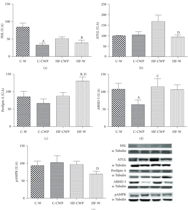
3.4. Expression of Proteins Involved in Lipolysis Pathway.
Figures4(a),4(b),4(c),4(d), and4(e)show the data of protein
expression of HSL, ATGL, Perilipin A, and ABHD-5 and AMPK activity, respectively, in mesenteric adipose tissue.
HSL protein expressions were reduced in C-CWP and
HF-W when compared to the C-W group (Figure 4(a)). here
was an increase in ATGL in HF-CWP group when compared
to HF-W group (Figure 4(b)). Perilipin A was signiicantly
higher in the HF-W group when compared to the C-W
and HF-CWP groups (Figure 4(c)). here was a signiicant
decrease in the protein expression of ABDH-5 in C-CWP
when compared to C-W and HF-CWP groups (Figure 4(d)).
he phosphorylation of AMPK (Figure 4(e)) was higher in
HF-CWP compared to HF-W group.
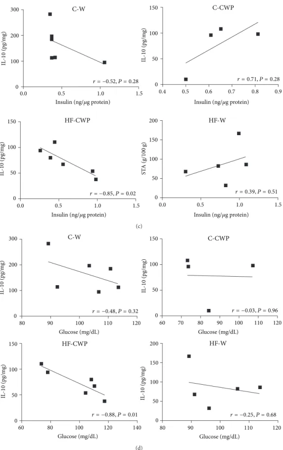
3.5. Correlations. A positive correlation between AUC and
TAG (� = 0.86 � < 0.05) was found in the HF-W
group (Figure 5(a)).Figure 5(b)shows a positive correlation
between insulin levels and STA in HF-W (� = 0.96 � = 0.006)
group. Figures 5(c) and 5(d) show an inverse correlation
between insulin (� = −0.85 � = 0.02) (Figure 5(c)) and
glucose (� = −0.88 � = 0.01) levels with IL-10 in the
mesenteric adipose tissue in HF-CWP group (Figure 5(d)).
4. Discussion
Numerous procedures for isolation and recovery of WP
C-W C-CWP HF-CWP HF-W 0
50 100 150
HS
L (U
.A)
A B
(a)
C-W C-CWP HF-CWP HF-W
D
0 50 100 150 200 250
A
T
GL (U
.A)
(b)
C-W C-CWP HF-CWP HF-W
0 50 100
150 B, D
P
er
ili
p
in A (U
.A)
(c)
C-W C-CWP HF-CWP HF-W
0 50 100 150
A
C
ABHD-5
(U
.A)
(d)
C-W C-CWP HF-CWP HF-W
0 50 100 150
D
pAMPK (U
.A)
HSL �-Tubulin
ATGL �-Tubulin
�-Tubulin
�-Tubulin
�-Tubulin Perilipin A
ABHD-5
pAMPK
(e)
Figure 4: Protein expression of HSL (a); ATGL (b); perilipin A (c); ABHD-5 (d); and pAMPK (e) in the mesenteric adipose tissue. he data are expressed in arbitrary units (A.U). Data submitted with an average±EPM. (A) C-W versus C-CWP; (B) C-W versus HF-W; (C) C-CWP versus HF-CWP; and (D) HF-CWP versus HF-W. (� < 0.05).
Table 3: Total mass (g), delta on the body mass gain (g/100 g body weight), and relative mass of tissue (RMT) (g/100 g body weight) of mice treated with high fat diet (HF) or control normocaloric (C) associated with gavage of coacervate (CWP) or water (W).
C-W (� = 10)
C-CWP (� = 10)
HF-CWP (� = 19)
HF-W (� = 19) RTM (g/100 g)
EPI 4.04 ± 0.17 3.52 ± 0.21 4.06 ± 0.28 4.93 ± 0.44b,d
MES 1.80 ± 0.24 1.29 ± 0.21 1.72 ± 0.1 2.6 ± 0.36b,d
RET 1.12 ± 0.10 0.82 ± 0.04 1.35 ± 0.12 1.48 ± 0.18b
Σadipose tissue 6.97 ± 0.38 5.64 ± 0.41 7.14 ± 0.46 9.02 ± 0.55b,d
16000 18000 20000 C-W
22000 24000
100 120 140 160
T
A
G (m
g/dL)
T
A
G (m
g/dL)
T
A
G (m
g/dL)
T
A
G (m
g/dL)
r = 0.15, P = 0.76
17000 18000 19000 20000 21000 22000 23000
r =−0.02, P = 0.97
95 100 105 110 115 120 125
C-CWP
AUC AUC
AUC AUC
16000 18000 20000 22000 24000 26000
90 100 110 120 130
r = 0.42, P = 0.40 HF-CWP
18000 20000 22000 24000 26000 28000 30000
r = 0.86, P = 0.05 HF-W
140 150 160 170 180
(a)
0.0 0.5 1.0 1.5
5 6 7 8 9
ST
A (g/
100
g)
4 5 6 7 8
ST
A (g/
100
g)
8 9 10 11 12
ST
A (g/
100
g)
r = 0.34, P = 0.49
r =−0.75, P = 0.05 r = 0.96, P = 0.006
C-W
Insulin (ng/𝜇g protein) Insulin (ng/𝜇g protein)
Insulin (ng/𝜇g protein) Insulin (ng/𝜇g protein)
0.0 0.5 1.0 1.5 0.0 0.5 1.0 1.5
0.4 0.5 0.6 0.7 0.8 0.9
4.0 4.5 5.0 5.5 6.0 6.5
ST
A (g/
100
g)
C-CWP
r =−0.33, P = 0.66
HF-CWP HF-W
(b)
0.0 0.5 1.0 1.5
C-W C-CWP
HF-CWP HF-W
0.0 0.5 1.0 1.5 0.0 0.5 1.0 1.5
r = −0.52, P = 0.28 r = 0.71, P = 0.28
r = 0.39, P = 0.51 0
100 200 300
IL
-10
(pg/m
g)
IL
-10
(pg/m
g)
0 50 100 150
IL
-10
(pg/m
g)
0 50 100 150
0 50 100 200
150
0.4 0.5 0.6 0.7 0.8 0.9
r = −0.85, P = 0.02
ST
A (g/
100
g)
Insulin (ng/𝜇g protein) Insulin (ng/𝜇g protein)
Insulin (ng/𝜇g protein) Insulin (ng/𝜇g protein)
(c)
0 100 200 300
IL
-10
(pg/m
g)
C-W
r = −0.48, P = 0.32
80 90 100 110 120
Glucose (mg/dL)
80 90 100 110 120
Glucose (mg/dL)
60 80 100 120 140
Glucose (mg/dL)
IL
-10
(pg/m
g)
0 50 100 150
IL
-10
(pg/m
g)
IL
-10
(pg/m
g)
0 50 100 150
C-CWP
80
60 70 90 100 110 120
Glucose (mg/dL)
r = −0.03, P = 0.96
r = −0.88, P = 0.01 r = −0.25, P = 0.68
HF-CWP HF-W
0 50 100 200
150
(d)
Table 4: Serum of triacylglycerol, total cholesterol, glucose, insulin, and adiponectin of mice treated with high fat diet (HF) or control normocaloric (C) associated with gavage of coacervate (CWP) or water (W).
Serum measurements C-W
(� = 10)
C-CWP (� = 11)
HF-CWP (� = 17)
HF-W (� = 17) Triacylglycerol (mg/dL) 133.37 ± 4.28 127.09 ± 4.85 129.30 ± 4.31 141.55 ± 3.94b,d
Total cholesterol (mg/dL) 128.75 ± 4.22 129.46 ± 4.76 128.99 ± 3.35 150.03 ± 6.00
LDL (mg/dL) 68.81 ± 1.75 73.18 ± 3.07 75.66 ± 1.64 80.3 ± 3.28
VLDL (mg/dL) 26.32 ± 0.86 25.42 ± 0.97 25.86 ± 0.86 28.92 ± 0.76b,d
HDL (mg/dL) 33.62 ± 1.75 36.71 ± 3.07 35.13 ± 1.64 40.83 ± 3.28
Glucose (mg/dL) 100.27 ± 2.69 89.84 ± 4.45 100.80 ± 3.58 105.28 ± 3.47
Insulin (ng/mL) 0.51 ± 0.03 0.64 ± 0.01 0.65 ± 0.01 0.75 ± 0.02b
HOMA-IR 0.89 ± 0.26 0.97 ± 0.15 1.12 ± 0.21 1.25 ± 0.19b
Adiponectin (ng/mL) 3.55 ± 0.47 3.98 ± 0.26 4.04 ± 0.10 3.34 ± 0.29d
Lipopolysaccharides (EU/mL) 2.58 ± 0.98 1.61 ± 0.66 1.49 ± 0.50 3.07 ± 0.97d
Data submitted with an average±EPM.bC-W versus HF-W anddHF-CWP versus HF-W. (� < 0.05).
Table 5: Concentrations of IL-6, TNF-�, IL-10, and IL-10/TNF-�(pg/mg of total protein content) in diferent experimental groups. C-W
(� = 6)
C-CWP (� = 7)
HF-CWP (� = 12)
HF-W (� = 12) IL-6 329.69 ± 43.64 132.08 ± 24.10a 215.80 ± 49.70 241.61 ± 29.27
TNF-� 111.93 ± 21.71 92.60 ± 22.86 54.48 ± 8.27 69.38 ± 17.35
IL-10 153.36 ± 27.01 119.30 ± 29.04 65.05 ± 9.96 100.74 ± 20.97b
IL-10/TNF-� 1.37 ± 0.027 1.28 ± 0.28 1.19 ± 0.10 1.45 ± 0.67
Data submitted with an average±EPM.aC-W versus C-CWP;bC-W versus HF-W. (� < 0.05).
physical, and chemical characteristics vary according to the procedures used to obtain these proteins. In our study, it was
possible to recover an average OF 30% in WP (Table 2). If
we compare these numbers with published data (which oten use conventional techniques such as ultrailtration (UF)), our
method seems to be a low eiciency process [19]. However,
cost-beneit considerations should be taken into account. For example, the UF method is expensive not only in terms of deployment but also in terms of operation. In addition to being cost-efective, the coacervation process promotes the separation of WP and obtains a low-calorie product.
he emergence of food compounds with health ben-eits may eventually become a good strategy to improve public health. In recent years, functional food has attracted the attention from scientiic community, consumers, and food manufacturers. he list of nutraceuticals compounds (vitamins, probiotics, bioactive peptides, and antioxidants among others) is extensive, and scientiic evidence seems to increasingly support the concept of health promotion
through food ingredients [20,21].
Functional foods are usually marketed as food containing ingredients technologically manipulated to perform a beneit
for health [22]. Our study lends support to previous studies
showing the efectiveness of CWP as a nutraceutical able to stabilize fat mass gain in animals fed with high fat diet. Our indings agree with the WP intake beneits extensively
reported in literature [23–27]. As demonstrated, there was
a decrease in body weight in the HF-CWP group when
compared to the HF-W group, accompanied by a reduction in the adiposity.
It is now well established that excessive consumption of saturated fat is related to the development of dyslipidemias
[28,29], and this study further corroborates it, as the animals
fed with high fat diet increased TAG and VLDL. Studies
have demonstrated the insulinotropic efect of WP [9, 11,
30]. In this study, we did not ind any signiicant diference
in blood glucose between the groups assessed. Regarding insulin and HOMA index, the hyperlipidic diet showed signiicantly higher values. Our results demonstrated the efectiveness of this experimental model of obesity. However, CWP treatment promoted improved glucose and insulin
tolerance. A study undertaken by Huang et al. [31] utilizing
diferent protein sources (cheese whey, soy, red meat, and milk) in obese mice (induced by high fat diet) has found increased adiponectin concentration and reduced insulin when the animals were fed with whey protein cheese. In the present study, we also detected the critical role of CWP in modulating adiponectin, as the values were higher for the HF-CWP group when compared to HF-W group. hese indings indicate that CWP may have functional efects. Yamauchi et
al. [32] have demonstrated the potential of adiponectin in
reducing insulin resistance by enhancing fatty acid oxidation, leading to a reduction in TAG content in obese diabetic rats. here is strong evidence of an immunomodulatory role
of WP [33]. Products containing WP may be beneicial in the
promotes improvement in the treatment of gastrointestinal symptoms of infant mice with rotavirus-induced diarrhea, a protective role in colorectal cancer in rats, reduced release of IL-6 in blood of rats undergoing transient ischemia/intestinal reperfusion and may provide protective efects against
exper-imentally induced breast cancer in animals [25,34,35]. Most
of the more recent studies in the literature, bothin vivoand
in vitro, have focused on the possible efects of these proteins
in macrophages and lymphocytes. Although most proteins are degraded during the gastric digestion, certain cheese
whey proteins, such as �-Lg, �-La, or GMP, are resistant
to digestion and remain intact. Such proteins can directly stimulate leukocyte ater their digestive absorption. hus, it is important to understand the mechanisms underlying the impact of these proteins on immunity, stimulating
anti-inlammatory processes in the body [33].
he IL-10 is a pleiotropic cytokine that controls inlammatory processes by eliminating the proinlammatory cytokines production such as IL-1, IL-6, and IL-8, and
TNF-� is produced mainly by monocytes, macrophages,
lymphocytes, mast cells, and mature adipocytes [14, 36].
he IL-10/TNF-� ratio has been considered an important
indicator of inlammatory status as low values are oten associated with increased morbidity and mortality risk. We
did not observe an increase of the IL-10/TNF-�ratio in MES
of group HF-CWP. We believe that because of the short period of the treatment the animals may not have developed their proinlammatory state. With increased length of treatment, we believe ind results more expressive. Another possibility is that the coacervate may have protected the mice from a proinlammatory state triggered by the treatment diet, leading to the counterbalance of IL-10 unnecessary in HF-CWP group. However, a study involving a longer treatment period may be required to discern this possible efect.
In this sense, there is an immunomodulatory mechanism underlying CWP, most likely the IL-10 cytokine, which has a homeostatic metabolic efect in the mesenteric adipose tissue. Our result suggests that IL-10 may be a positive regulator of insulin sensitivity and increased glucose uptake. his mecha-nism can protect the adipose tissue against insulin resistance. Although the precise origin of the unchecked inlammatory response in obesity is still unclear, it is well known that in obesity the overproduction of proinlammatory cytokines
afects metabolism. For example, TNF-� contributes to the
inability of cells to respond to insulin and to increased
levels of insulin [37], and IL-10 was associated with other
variables closely linked to insulin sensitivity, such as fasting and postload insulin concentrations, HDL cholesterol, and triglyceride levels. Besides the tissue-speciic efect of CWP, we showed a systemic efect in the decrease in the LPS serum level.
Regarding the composition of the CWP, we may highlight
the presence of�-La and �-Lg protein, the major protein
present in the coacervate, which has been proven efective in suppressing the release of proinlammatory cytokines
[27, 38]. he presence of these proteins is a great
indica-tion that the coacervate components are able to modulate the proinlammatory milieu promoted by hyperlipidic diet.
In another study by our group, mice, previously treated with high fat diet and fed with a supplementation of CWP (gavage, 36 mg protein/kg of body weight), showed a positive corre-lation between IL-10 and TNF-alpha in mesenteric adipose tissue, retroperitoneal adipose tissue, and liver tissue. We also observed a positive correlation between lipopolysaccharide and IL-10 in the liver tissue. herefore, pretreatment with high fat diet promoted metabolic alterations and
inlamma-tion, and CWP modulated the inlammatory milieu [14].
Evidence suggests that eating WP causes the decrease in calorie intake, increased basal energy expenditure, and modulates insulin sensitivity and glucose homeostasis, lead-ing to changes in lipid metabolism in adipose tissue, liver,
and muscle [39–42]. he AMPK and adiponectin are key
molecules to metabolic responses in diferent tissues [43,
44]; they are involved in the preventive response against
negative physiological processes caused by the consumption of a diet high in saturated fatty acids. he activation of AMPK by bioactive components of foods or medicines has been regarded as goal, since it may reverse the metabolic changes
associated with obesity and type 2 diabetes [45]. It is also
known that adiponectin can activate AMPK in white adipose
tissue [46]. he animals treated with coacervate showed an
increase in AMPK activation associated with the decrease in HSL and increase in ATGL protein expression in mesenteric adipose tissue. Similar results were reported in the review
undertaken by Bijland et al. [47].
A study conducted by Gaidhu et al. [43] showed that
AMPK activation stimulated by AICAR (5-aminoimidazol-4
-carboxamida ribonucleot´ıdeo)initially promoted inhibition of
lipolysis in adipocytes isolated asin vivo, relecting a decrease
in free fatty acid in serum. On the other hand, prolonged treatment with AICAR promoted an increase in lipolysis, which the authors attributed to an increase in the content
of ATGL and reduced activity of HSL [48]. However,
clear-cut conclusions are diicult to arrive at, due to a lack of tools for manipulating assays using speciic AMPK. Furthermore, the overall efect of AMPK activation of lipolysis is still controversial. he duration and mode of activation of AMPK may be of particular importance when it comes to a process aimed at reducing the proinlammatory state caused by
increased lipolysis [47]. In addition, adiponectin can suppress
the activation of HSL, without changing ATGL and ABHD-5 in adipocytes in order to modulate a homeostatic control of
lipolysis to avoid lipotoxicity [49].
Lipolysis does seem to play a crucial physiological role by recruiting a source of energy mobilized in times of stress and/or energy deprivation. Moreover, the very signiicant reduction in lipolysis is clearly harmful, as demonstrated in the clinical domain by the syndromes resulting from
deiciencies in the lipolytic apparatus [50]. Given that, it is
Another interesting inding was the signiicantly higher protein expression of perilipin A (52%) in HF-W group, which also refers to larger deposits of triglycerides, since this protein is primarily anchored around the droplets of neutral lipids in adipocytes. his is in line with studies showing that increased protein expression perilipin A leads to increased
storage of triglycerides by reducing its hydrolytic rate. [51].
Finally, there is plenty of evidence suggesting that the intake of WP may lower consumption of calories, increase baseline energy expenditure, and improve insulin sensitivity and glucose homeostasis, thus leading to changes in lipid
metabolism in adipose tissue, liver, and muscle [39–42].
5. Conclusion
CWP were able to promote nutritional and physiological improvements in HF-CWP group, such as reduction in body mass and decreased serum lipid levels followed by decreased serum insulin and LPS. In addition, intervention with CWP resulted in higher adiponectin contents and attenuated pro-cesses that would lead to glucose intolerance. herefore, CWP could play a beneicial role, in some way, in modulating lipolysis in animals treated with hyperlipidic diet.
Abbreviations
ABHD-5: Abhydrolase domain
containing 5
AUC: Area under the curve
CWP: Coacervate
C-CWP: Control diet plus coacervate
C-W: Control diet plus tap water
HF-CWP: High fat diet plus coacervate
HF-W: High fat diet plus tap water
HSL: Hormone-sensitive lipase
LPS: Lipopolysaccharide
MGL: Monoglyceride lipase
NEFAs: Nonesteriied fatty acid
p-AMPK�1 e 2 - hr 172: Phospho 5�AMP-activated
protein kinase
TAG: Triacylglycerol
OGTT: Oral glucose tolerance test
ATGL: Triglyceride lipase enzyme
WP: Whey protein.
Conflict of Interests
he authors declare that they have no competing interests.
Authors’ Contribution
All authors read and approved the inal paper.
Acknowledgments
he authors thank the staf of the LAMEROA Laboratory of USP for analytical assistance of diets. his work was
supported by FAPESP (Grants no. 2009/53801-5), CAPES, and CNPQ.
References
[1] M. F. Gregor and G. S. Hotamisligil, “Inlammatory mecha-nisms in obesity,”Annual Review of Immunology, vol. 29, pp. 415–445, 2011.
[2] M. L. Kueht, B. K. McFarlin, and R. E. Lee, “Severely obese have greater LPS-stimulated TNF-�production than normal weight African-American women,”Obesity, vol. 17, no. 3, pp. 447–451, 2009.
[3] P. H. C. Eilers, M. S. Westerterp-plantenga, T. Kooistra, H. Biology, and M. S. W. Maastricht, “Leptin and the proinlam-matory state associated with human obesity,”Journal of Clinical Endocrinology & Metabolism, vol. 89, no. 4, pp. 1773–1778, 2004. [4] M. H. Fonseca-Alaniz, J. Takada, M. I. C. Alonso-Vale, and F. B. Lima, “Adipose tissue as an endocrine organ: from theory to practice,”Jornal de Pediatria, vol. 83, no. 5, pp. S192–S203, 2007. [5] M. J. Song, K. H. Kim, J. M. Yoon, and J. B. Kim, “Activation of Toll-like receptor 4 is associated with insulin resistance in adipocytes,”Biochemical and Biophysical Research Communica-tions, vol. 346, no. 3, pp. 739–745, 2006.
[6] A. Chait and F. Kim, “Saturated fatty acids and inlammation: who pays the toll?”Arteriosclerosis, hrombosis, and Vascular Biology, vol. 30, no. 4, pp. 692–693, 2010.
[7] J. Park, S. C. Sung, A. H. Choi et al., “Increase in glucose-6-phosphate dehydrogenase in adipocytes stimulates oxidative stress and inlammatory signals,”Diabetes, vol. 55, no. 11, pp. 2939–2949, 2006.
[8] M. E. F. V´azquez-Vela, N. Torres, and A. R. Tovar, “White adipose tissue as endocrine organ and its role in obesity,” Archives of Medical Research, vol. 39, no. 8, pp. 715–728, 2008. [9] W. L. Hall, D. J. Millward, S. J. Long, and L. M. Morgan,
“Casein and whey exert diferent efects on plasma amino acid proiles, gastrointestinal hormone secretion and appetite,” British Journal of Nutrition, vol. 89, no. 2, pp. 239–248, 2003. [10] T. G. Anthony, B. J. McDaniel, P. Knoll, P. Bunpo, G. L.
Paul, and M. A. McNurlan, “Feeding meals containing soy or whey protein ater exercise stimulates protein synthesis and translation initiation in the skeletal muscle of male rats,”Journal of Nutrition, vol. 137, no. 2, pp. 357–362, 2007.
[11] J. Shi, E. Tauriainen, E. Martonen et al., “Whey protein isolate protects against diet-induced obesity and fatty liver formation,” International Dairy Journal, vol. 21, no. 8, pp. 513–522, 2011. [12] D. Rusu, R. Drouin, Y. Pouliot, S. Gauthier, and P. E. Poubelle,
“A bovine whey protein extract can enhance innate immunity by priming normal human blood neutrophils,” Journal of Nutrition, vol. 139, no. 2, pp. 386–393, 2009.
[13] B. Tranberg, L. I. Hellgren, J. Lykkesfeldt et al., “Whey protein reduces early life weight gain in mice fed a high-fat diet,”PLoS One, vol. 8, no. 8, Article ID e71439, 2013.
[14] M. F. Moreno, G. I. de Morais Honorato de Souza, A. C. L. Hachul et al., “Coacervate whey protein improves inlammatory milieu in mice fed with high-fat diet,”Nutrition & Metabolism, vol. 11, article 15, 2014.
[15] J. Hidalgo and P. M. T. Hansen,Selective Precipitation of Whey Proteins with Carboxymethylcellulose, Public Health, 1970. [16] V. B. Tolstoguzov, “Functional properties of food proteins and
[17] C. L. Cooper, P. L. Dubin, A. B. Kayitmazer, and S. Turksen, “Polyelectrolyte-protein complexes,”Current Opinion in Colloid and Interface Science, vol. 10, no. 1-2, pp. 52–78, 2005.
[18] P. G. Reeves, F. H. Nielsen, and G. C. Fahey Jr., “AIN-93 puriied diets for laboratory rodents: inal report of the American Insti-tute of Nutrition ad hoc writing committee on the reformulation of the AIN-76A rodent diet,”Journal of Nutrition, vol. 123, no. 11, pp. 1939–1951, 1993.
[19] J. N. de Wit,Lecturer’s Handbook on Whey and Whey Products, European Whey Products Association (Belgium), 2001. [20] L. Chen, G. E. Remondetto, and M. Subirade, “Food
protein-based materials as nutraceutical delivery systems,”Trends in Food Science and Technology, vol. 17, no. 5, pp. 272–283, 2006. [21] R. Caillard, R. Guillet-Nicolas, F. Kleitz, and M. Subirade,
“Tabletability of whey protein isolates,” International Dairy Journal, vol. 27, no. 1-2, pp. 92–98, 2012.
[22] M. Niva, ““All foods afect health”: understandings of func-tional foods and healthy eating among health-oriented Finns,” Appetite, vol. 48, no. 3, pp. 384–393, 2007.
[23] G. Bounous and P. Gold, “he biological activity of undenatured dietary whey proteins: role of glutathione,”Clinical and Inves-tigative Medicine, vol. 14, no. 4, pp. 296–309, 1991.
[24] M. M. Iskandar, N. Dauletbaev, S. Kubow, N. Mawji, and L. C. Lands, “Whey protein hydrolysates decrease IL-8 secretion in lipopolysaccharide (LPS)-stimulated respiratory epithelial cells by afecting LPS binding to Toll-like receptor 4,”British Journal of Nutrition, vol. 110, no. 1, pp. 58–68, 2013.
[25] R. Hakkak, S. Korourian, M. J. J. Ronis, J. M. Johnston, and T. M. Badger, “Dietary whey protein protects against azoxymethane-induced colon tumors in male rats,” Cancer Epidemiology Biomarkers and Prevention, vol. 10, no. 5, pp. 555–558, 2001. [26] C. F. Rosaneli, A. E. Bighetti, M. A. Antonio, J. E. Carvalho,
and V. C. Sgarbieri, “Eicacy of a whey protein concentrate on the inhibition of stomach ulcerative lesions caused by ethanol ingestion,”Journal of Medicinal Food, vol. 5, no. 4, pp. 221–228, 2002.
[27] S. Pal, V. Ellis, and S. Dhaliwal, “Efects of whey protein isolate on body composition, lipids, insulin and glucose in overweight and obese individuals,”British Journal of Nutrition, vol. 104, no. 5, pp. 716–723, 2010.
[28] S. Zhao, J. Wang, X. Song, X. Zhang, C. Ge, and S. Gao, “Impact of dietary protein on lipid metabolism-related gene expression in porcine adipose tissue,”Nutrition and Metabolism, vol. 7, article 6, 2010.
[29] D. E. C. Cintra, A. V. Costa, M. D. C. G. Peluzio, S. L. P. Matta, M. T. C. Silva, and N. M. B. Costa, “Lipid proile of rats fed high-fat diets based on laxseed, peanut, trout, or chicken skin,” Nutrition, vol. 22, no. 2, pp. 197–205, 2006.
[30] A. H. Frid, M. Nilsson, J. J. Holst, and I. M. E. Bj¨orck, “Efect of whey on blood glucose and insulin responses to composite breakfast and lunch meals in type 2 diabetic subjects,” he American Journal of Clinical Nutrition, vol. 82, no. 1, pp. 69–75, 2005.
[31] X.-F. Huang, Y. Liu, G. L. Rahardjo, P. L. Mclennan, L. C. Tapsell, and W. A. Buttemer, “Efects of diets high in whey, soy, red meat and milk protein on body weight maintenance in diet-induced obesity in mice,”Nutrition and Dietetics, vol. 65, no. 3, pp. S53– S59, 2008.
[32] T. Yamauchi, J. Kamon, H. Waki et al., “he fat-derived hor-mone adiponectin reverses insulin resistance associated with both lipoatrophy and obesity,”Nature Medicine, vol. 7, no. 8, pp. 941–946, 2001.
[33] D. Rusu, R. Drouin, Y. Pouliot, S. Gauthier, and P. E. Poubelle, “A bovine whey protein extract stimulates human neutrophils to generate bioactive IL-1Ra through a NF-�B- and MAPK-dependent mechanism,”Journal of Nutrition, vol. 140, no. 2, pp. 382–391, 2010.
[34] F. M. Wolber, A. M. Broomield, L. Fray, M. L. Cross, and D. Dey, “Supplemental dietary whey protein concentrate reduces rotavirus-induced disease symptoms in suckling mice,”Journal of Nutrition, vol. 135, no. 6, pp. 1470–1474, 2005.
[35] M. Yamaguchi and M. Uchida, “�-lactalbumin suppresses interleukin-6 release ater intestinal ischemia/reperfusion via nitric oxide in rats,”Inlammopharmacology, vol. 15, no. 1, pp. 43–47, 2007.
[36] C. E. Juge-Aubry, E. Somm, A. Pernin et al., “Adipose tissue is a regulated source of interleukin-10,”Cytokine, vol. 29, no. 6, pp. 270–274, 2005.
[37] J. M. Han, S. J. Patterson, M. Speck, J. A. Ehses, and M. K. Levings, “Insulin inhibits IL-10-mediated regulatory T cell function: implications for obesity,”he Journal of Immunology, vol. 192, no. 2, pp. 623–629, 2014.
[38] M. Yamaguchi and M. Uchida, “�-Lactalbumin suppresses interleukin-6 release ater intestinal ischemia/reperfusion via nitric oxide in rats,”Inlammopharmacology, vol. 15, no. 1, pp. 43–47, 2007.
[39] M. B. Zemel, “Role of calcium and dairy products in energy partitioning and weight management,”he American Journal of Clinical Nutrition, vol. 79, no. 5, pp. 907S–912S, 2004.
[40] B. L¨onnerdal, “Nutritional and physiologic signiicance of human milk proteins,”he American Journal of Clinical Nutri-tion, vol. 77, no. 6, pp. 1537S–1543S, 2003.
[41] J.-C. J. Bouthegourd, S. M. Roseau, L. Makarios-Lahham, P. M. Leruyet, D. G. Tom´e, and P. C. Even, “A preexercise � -lactalbumin-enriched whey protein meal preserves lipid oxi-dation and decreases adiposity in rats,” American Journal of Physiology: Endocrinology and Metabolism, vol. 283, no. 3, pp. E565–E572, 2002.
[42] L. McAllan, P. D. Cotter, H. M. Roche, R. Korpela, and K. N. Nilaweera, “Bioactivity in whey proteins inluencing energy balance,”Journal of Metabolic Syndrome, vol. 1, article 107, 2012. [43] M. P. Gaidhu, N. M. Anthony, P. Patel, T. J. Hawke, and R. B. Ceddia, “Dysregulation of lipolysis and lipid metabolism in visceral and subcutaneous adipocytes by high-fat diet: Role of ATGL, HSL, and AMPK,”he American Journal of Physiology— Cell Physiology, vol. 298, no. 4, pp. C961–C971, 2010.
[44] T. Kadowaki and T. Yamauchi, “Adiponectin and adiponectin receptors,”Endocrine Reviews, vol. 26, no. 3, pp. 439–451, 2005. [45] B. B. Zhang, G. Zhou, and C. Li, “AMPK: an emerging drug tar-get for diabetes and the metabolic syndrome,”Cell Metabolism, vol. 9, no. 5, pp. 407–416, 2009.
[46] P. Huypens, E. Quartier, D. Pipeleers, and M. van de Casteele, “Metformin reduces adiponectin protein expression and release in 3T3-L1 adipocytes involving activation of AMP activated protein kinase,”European Journal of Pharmacology, vol. 518, no. 2-3, pp. 90–95, 2005.
[47] S. Bijland, S. J. Mancini, and I. P. Salt, “Role of AMP-activated protein kinase in adipose tissue metabolism and inlammation,” Clinical Science, vol. 124, no. 8, pp. 491–507, 2013.
[49] L. Qiao, B. Kinney, J. Schaack, and J. Shao, “Adiponectin inhibits lipolysis in mouse adipocytes,”Diabetes, vol. 60, no. 5, pp. 1519– 1527, 2011.
[50] S. Santamarina-Fojo, “he familial chylomicronemia syn-drome,”Endocrinology and Metabolism Clinics of North Amer-ica, vol. 27, no. 3, pp. 551–567, 1998.
Submit your manuscripts at
http://www.hindawi.com
Stem Cells
International
Hindawi Publishing Corporationhttp://www.hindawi.com Volume 2014
Hindawi Publishing Corporation
http://www.hindawi.com Volume 2014
INFLAMMATION
Hindawi Publishing Corporation
http://www.hindawi.com Volume 2014
Behavioural
Neurology
Endocrinology
International Journal of Hindawi Publishing Corporationhttp://www.hindawi.com Volume 2014
Hindawi Publishing Corporation
http://www.hindawi.com Volume 2014
Disease Markers
Hindawi Publishing Corporation
http://www.hindawi.com Volume 2014
BioMed
Research International
Oncology
Journal ofHindawi Publishing Corporation
http://www.hindawi.com Volume 2014
Hindawi Publishing Corporation
http://www.hindawi.com Volume 2014
Oxidative Medicine and Cellular Longevity
Hindawi Publishing Corporation
http://www.hindawi.com Volume 2014
PPAR Research
The Scientiic
World Journal
Hindawi Publishing Corporationhttp://www.hindawi.com Volume 2014
Immunology Research
Hindawi Publishing Corporation
http://www.hindawi.com Volume 2014 Journal of
Obesity
Journal ofHindawi Publishing Corporation
http://www.hindawi.com Volume 2014
Hindawi Publishing Corporation
http://www.hindawi.com Volume 2014
Computational and Mathematical Methods in Medicine
Ophthalmology
Journal ofHindawi Publishing Corporation
http://www.hindawi.com Volume 2014
Diabetes Research
Journal ofHindawi Publishing Corporation
http://www.hindawi.com Volume 2014
Hindawi Publishing Corporation
http://www.hindawi.com Volume 2014
Research and Treatment
AIDS
Hindawi Publishing Corporation
http://www.hindawi.com Volume 2014
Gastroenterology Research and Practice
Hindawi Publishing Corporation
http://www.hindawi.com Volume 2014
Parkinson’s
Disease
Evidence-Based Complementary and Alternative Medicine
Volume 2014