Research Article
TRPV1 Antagonism by Capsazepine Modulates Innate Immune
Response in Mice Infected with
Plasmodium berghei
ANKA
Elizabeth S. Fernandes,
1,2Carolina X. L. Brito,
1Simone A. Teixeira,
3Renato Barboza,
4Aramys S. dos Reis,
3Ana Paula S. Azevedo-Santos,
5Marcelo Muscará,
3Soraia K. P. Costa,
3Claudio R. F. Marinho,
3Susan D. Brain,
2and Marcos A. G. Grisotto
1,61Universidade CEUMA, 65075-120 S˜ao Lu´ıs, MA, Brazil
2Cardiovascular Division, King’s College London, London, UK
3Universidade de S˜ao Paulo, S˜ao Paulo, Brazil
4Universidade Federal de S˜ao Paulo, Diadema, Brazil
5Universidade Federal do Maranh˜ao, S˜ao Lu´ıs, Brazil
6Instituto Florence de Ensino Superior, S˜ao Lu´ıs, Brazil
Correspondence should be addressed to Elizabeth S. Fernandes; elizabeth.soares@ceuma.br
Received 3 June 2014; Accepted 8 July 2014; Published 24 August 2014
Academic Editor: Mauricio Martins Rodrigues
Copyright © 2014 Elizabeth S. Fernandes et al. his is an open access article distributed under the Creative Commons Attribution License, which permits unrestricted use, distribution, and reproduction in any medium, provided the original work is properly cited.
housands of people sufer from severe malaria every year. he innate immune response plays a determinant role in host’s defence to malaria. Transient receptor potential vanilloid 1 (TRPV1) modulates macrophage-mediated responses in sepsis, but its role in other pathogenic diseases has never been addressed. We investigated the efects of capsazepine, a TRPV1 antagonist, in malaria.
C57BL/6 mice received 105red blood cells infected withPlasmodium bergheiANKA intraperitoneally. Noninfected mice were used
as controls. Capsazepine or vehicle was given intraperitoneally for 6 days. Mice were culled on day 7 ater infection and blood and
spleen cell phenotype and activation were evaluated. Capsazepine decreased circulating but not spleen F4/80+Ly6G+cell numbers
as well as activation of both F4/80+and F4/80+Ly6G+cells in infected animals. In addition, capsazepine increased circulating but
not spleen GR1+and natural killer (NK) population, without interfering with natural killer T (NKT) cell numbers and blood NK
and NKT activation. However, capsazepine diminished CD69 expression in spleen NKT but not NK cells. Infection increased lipid
peroxidation and the release of TNF�and IFN�, although capsazepine-treated group exhibited lower levels of lipid peroxidation
and TNF�. Capsazepine treatment did not afect parasitaemia. Overall, TRPV1 antagonism modulates the innate immune response
to malaria.
1. Introduction
Malaria is an infectious disease caused by intracellular protozoans of the genusPlasmodiumand transmitted from person to person through bites of infected mosquitoes. It afects millions of people annually and is a leading cause of child mortality in underdevelopment countries [1]. Severe malaria such as cerebral malaria is frequently fatal and outcome of infection depends on host’s immune response, with innate immunity playing a determinant role in it [2,
3]. Available antimalarial therapy targets the Plasmodium. However, elimination of the parasite does not halt the clinical
consequences of disease as surviving patients from severe malaria can develop a range of neurological deicits [4, 5]. In this context, an efective immune response is essential for patient’s recovery. Innate response is the major immunity component of patients who have been infected with Plasmod-iumfor the irst time, being essential to the development of an efective acquired immune response [6].
Recently, a protective role for transient receptor vanilloid 1 (TRPV1), a nonselective cation channel found on both neuronal and nonneuronal cells, was suggested in bacteria-induced sepsis [7–10]. Indeed, in the absence of TRPV1 activation, macrophage functions such as their ability to
phagocytose and to release inlammatory mediators (nitric oxide (NO), reactive oxygen species (ROS), and cytokines) are impaired [9]. Also, TRPV1 has been linked to macrophage survival [9]. Evidence suggests a feedback between TRPV1 activation and ROS production may exist; in addition to modulating oxidative stress by downregulating ROS gener-ation, this receptor can be directly activated by hydrogen peroxide (H2O2) [11] and regulated by superoxide anion (O2−)release [12–14]. Oxidative stress generation has a direct impact on macrophage-erythrocyte-endothelium interac-tions and imbalances of this pathway may trigger excessive damage and impaired host’s immune response to malaria [15,16].
Herein, the role of TRPV1 in malaria was investigated for the irst time. We used the TRPV1 antagonist, capsazepine, to assess whether TRPV1 is able to modulate the innate immune response to malaria in animals infected with Plasmodium
bergheiANKA.
2. Materials and Methods
2.1. Animals. Inbred male C57BL/6 mice (8 weeks old) were
used. Mice were obtained from the animal’s facility of the Department of Parasitology, Institute of Biomedical Sciences, University of S˜ao Paulo. Mice were kept in a climatically controlled environment and given food and waterad libitum. All procedures were approved by the Ethics Committee of the University of S˜ao Paulo and carried out in accordance with the Brazilian society for animal welfare (SBCAL).
2.2. Malaria Induction. Malaria was induced by a single
intraperitoneal (i.p.) injection of 105 red blood cells (RBCs) infected with Plasmodium berghei ANKA (clone 1.49L) as described by Elias et al. [17]. Parasitaemia was assessed daily in a blood smear stained by Giemsa, by microscopy, from day 5 to day 7 following infection and was expressed as % of infected RBCs. Mice were terminally anaesthetised with a mixture of ketamine (75 mg/kg; Dopalen, Ceva, Brazil) and xylazine (1 mg/kg; i.p.; Sigma-Aldrich, Brazil), and exsanguinated by cardiac puncture on day 7 ater infection (premortality end point; [18,19]). heir blood and spleen were collected for further analysis. he plasma was separated and stored at−70∘C for further quantitation of plasma aldehydes and cytokines. Cell population phenotype and activation were evaluated by low cytometry. Noninfected mice were used as controls.
2.3. Pharmacological Treatment. In order to assess the role of
TRPV1 in malaria, animals received the TRPV1 antagonist, capsazepine (Sigma-Aldrich, Brazil;� = 5, uninfected (con-trol) group and� = 8, infected group), intraperitoneally from 24 h following infection, for 6 days (2x day, 50�g/animal; [9]). Vehicle (10% DMSO in saline, 120�L/animal;� = 5, uninfected (control) group and� = 8, infected group) was used as controls.
2.4. Flow Cytometry Analysis. Blood and spleen samples
from infected and uninfected mice and single-cell suspen-sions were prepared. Peripheral blood cells were isolated by Percoll gradient (Sigma-Aldrich, Brazil). Spleens were homogenized and passed through a nylon mesh of 70�m to create a single-cell suspension. Cells were stained with Trypan blue (Sigma-Aldrich, Brazil) and assessed for viability in a haemocytometer. Cells (106) were washed, resuspended in low cytometry bufer (2% foetal calf serum (Invitrogen, Brazil) in phosphate bufered saline-PBS (Sigma-Aldrich, Brazil)), and stained with directly conjugated monoclonal antibodies (BD Biosciences or eBiosciences, Brazil): anti-F4/80 FITC, anti-IAb PE, anti-Ly6G PerCP, anti-GR1 Pe-Cy7, CD3 PE and APC, NK1.1 FITC, and anti-CD69 PECy5. Events were acquired on a BD FACSCanto (BD Biosciences-Immunocytometry Systems) and analyzed using FlowJo sotware (Tree Star Inc.). In order to ana-lyze monocyte/macrophage and neutrophil populations, cells expressing CD3+, CD4+, CD8+, and CD19+ were gated out and the phenotypic F4/80, GR1, and Ly6G lineage markers were evaluated. Results are expressed as representative two-colour dot-plots as well as number of cells (×106), except for IAb, expressed as mean luorescence.
2.5. Plasma Cytokine Levels. he plasma levels of TNF�,
IFN�, IL-4, IL-6, IL-2, IL-10, and IL-17 were evaluated by using a cytometric bead array (CBA) mouse h1/h2/h17 cytokine kit (BD Biosciences, Brazil) according to man-ufacturer’s instructions. Analysis was performed on a Facscalibur cytometer low cytometer (BD Biosciences-Immunocytometry Systems). Results were calculated in CBA FCAP Array sotware (BD Biosciences, Brazil) as pg/mL and are expressed as fold-increase relative to uninfected controls.
2.6. Plasma Aldehydes. Total plasma aldehyde (mainly
mal-ondialdehyde) concentrations were quantiied as an index of lipid peroxidation and oxidative stress, as previously described [20,21]. Briely, 100�L of sample were incubated with 100�L of PBS and 400�L of thiobarbituric acid (0.67%; Sigma-Aldrich, Brazil), at 90∘C for 45 min. Samples were then centrifuged at 1,000 xg for 10 min. hree hundred�L of the supernatant was incubated with 300�L of butanol (Sigma-Aldrich, Brazil) and 30�L of a saturated solution of sodium chloride (Sigma-Aldrich, Brazil). Samples were mixed in a vortex, centrifuged at 1,000 xg for 2 min and then added to a 96-well plate (200�L/well). Absorbance was read at 535 and 572 nm and the diference between the absorbance was used to calculate the aldehyde concentrations, using the molar extinction coeicient of the chromophore (1.56× 10⁵M−1/cm−1). Results are expressed as fold-increase relative to uninfected controls.
2.7. Data Analysis. he results are presented as the mean±
by Bonferroni and unpaired�-test when appropriate. he� values<0.05 were considered signiicant.
3. Results
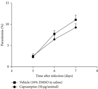
3.1. Capsazepine Does Not Afect Parasitaemia. Figure1shows
parasitaemia levels up to day 7 ater infection, in mice treated with either vehicle or capsazepine. Parasitaemia progres-sively increased in both groups. Repeated treatment with capsazepine had no efect on parasitaemia. Experiments were performed with a premortality end-point. hus, no signs of cerebral malaria such as reduced responsiveness to stimula-tion, ataxia, respiratory distress or prostrastimula-tion, paralysis, and convulsions were observed in either capsazepine- or vehicle-infected mice. However, both groups of animals displayed piloerection and/or abnormal posture, as a result of infection.
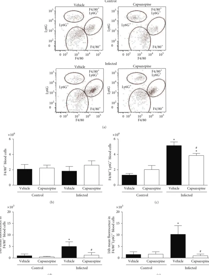
3.2. Capsazepine Alters Circulating Monocyte but Not Spleen
Macrophage Population Number and Activation. As
repre-sented in Figures 2(a)–2(e), three distinct populations of peripheral blood leukocytes were detected in all groups of uninfected and infected animals: F4/80+, F4/80+Ly6G+ and Ly6G+ cells. Malaria induction had no efect on Ly6G+ cell population as no statistical signiicance was found between any of the evaluated groups. Mean±SD values for Ly6G+ populations are as follows: vehicle-uninfected group 2.0 ± 0.6, capsazepine uninfected group 1.3 ± 0.4, vehicle-infected group 0.9 ± 1.1, and capsazepine infected group 1.3 ± 0.8. On the other hand, P. berghei ANKA infection increased F4/80+Ly6G+ cell numbers in both vehicle- (3.9-fold increase) and capsazepine- (1.9-(3.9-fold increase) treated groups when compared to uninfected controls, whilst no efects were observed on F4/80+cell population (Figures2(b)
and 2(c)). In addition, malaria caused increased activation of both F4/80+ (5.1-fold increase) and F4/80+Ly6G+ (6.6-fold increase) circulating cells when compared to uninfected animals, as denoted by expression of IAb on these cells (Fig-ures2(d)and2(e)). Repeated administration of capsazepine in infected animals caused reduction of F4/80+Ly6G+ pop-ulation expansion by25.0 ± 5.2% but did not afect F4/80+ cell numbers (Figures2(b)and2(c)). In addition,P. berghei
ANKA-induced activation of F4/80+and F4/80+Ly6G+cells was halted by capsazepine. As depicted in Figures 2(d)
and 2(e), capsazepine treatment reduced by 75 ± 22.7% and 90.3 ± 7.1%, the expression of IAb on F4/80+ and F4/80+Ly6G+ cells, respectively, when compared to vehicle-treated infected controls. As infection raised the number of circulating F4/80+Ly6G+ cells, a decline was noticed in the GR1+ cell population (34.1 ± 15.1%). However, infected mice treated with capsazepine exhibited a higher number of these cells when compared with both their uninfected-(1.8-fold increase) and infected- (2.4-fold increase) control animals. Mean ± SD values for GR1+ populations are as follows: vehicle-uninfected group 2.9 ± 0.6, capsazepine uninfected group 2.5 ± 0.4, vehicle-infected group 1.9 ± 0.4, and capsazepine infected group4.6 ± 0.5 (� < 0.05). Capsazepine treatment in uninfected animals had no efects in regard to expansion or activation of both F4/80+ and
4 5 6 7 8
15
10
5
0
Vehicle (10%DMSO in saline) Capsazepine (50 �g/animal)
Time ater infection (days)
P
arasi
temia (%)
Figure 1: Efect of capsazepine on parasitaemia levels. Parasitaemia was measured daily, from day 5 to day 7 ater infection in blood
smear samples obtained from C57BL/6 mice infected with
Plasmod-ium bergheiANKA (105 infected RBCs/animal; i.p.) treated with
either capsazepine (50�g/animal, 2x day, for 6 days) or vehicle (10%
DMSO in saline) from 24 h ater infection (� = 8per group).
F4/80+Ly6G+ cells (Figures2(a)–2(e)) and neither on GR1+ cells.
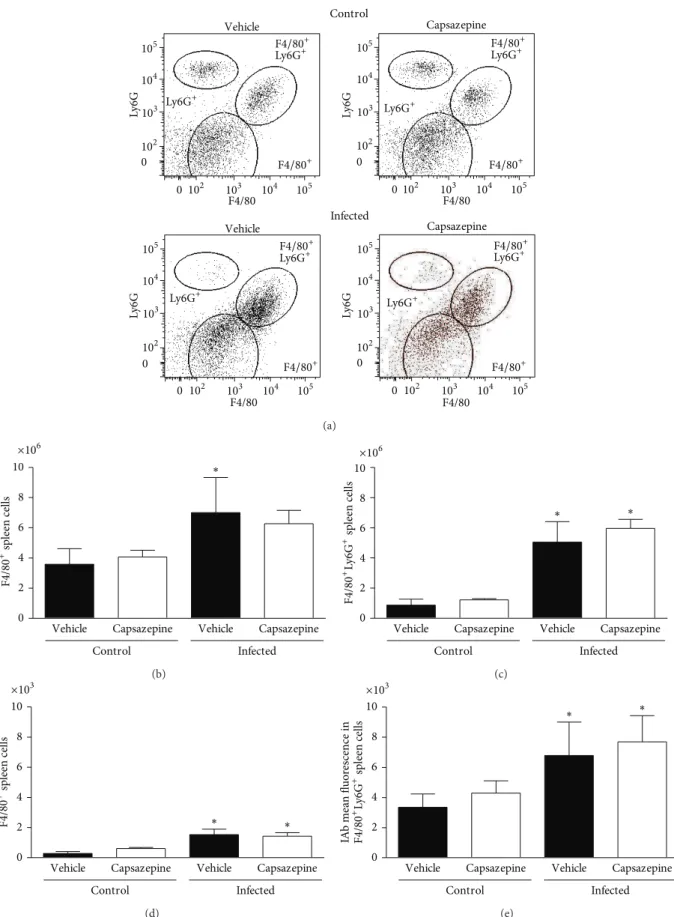
Similarly, F4/80+, F4/80+Ly6G+ and Ly6G+ cells were detected in spleen samples obtained from both infected and uninfected mice whether or not they were treated with capsazepine (Figures3(a)–3(e)). As observed for circulating Ly6G+ cells, malaria induction had no efects on spleen Ly6G+cells. Mean±SD values for spleen Ly6G+populations are as follows: vehicle-uninfected group0.7±0.2, capsazepine uninfected group 1.1 ± 0.4, vehicle-infected group 1.0 ± 0.6, and capsazepine infected group 0.9 ± 0.3. However,
P. berghei ANKA injection raised the numbers of F4/80+
and F4/80+Ly6G+ cells in both vehicle- (2.0- and 5.8-fold increase, resp.) and capsazepine- (1.5- and 4.9-fold increase, resp.) treated groups when compared to their respective uninfected controls (Figures3(b)and3(c)). Also,P. berghei
ANKA infection augmented F4/80+ and F4/80+Ly6G+cells in both vehicle- (5.5- and 2.0-fold increase, resp.) and capsazepine- (2.3- and 1.8-fold increase, resp.) treated groups when compared to their respective uninfected controls (Fig-ures 3(d) and 3(e)). Capsazepine had no efects on the number or activation of spleen F4/80+and F4/80+Ly6G+cells (Figures 3(a)–3(e)). Spleen GR1+ cell population remained unaltered irrespective of treatments and mean±SD values are as follows: vehicle-uninfected group0.9±0.2, capsazepine uninfected group 1.3 ± 0.6, vehicle-infected group 0.8 ± 0.3, and capsazepine infected group 1.3 ± 0.4. Moreover, capsazepine had no efects on F4/80+and F4/80+Ly6G+cells (Figures3(a)–3(e)) when administered to uninfected mice.
3.3. Capsazepine Modulates Blood and Spleen NK and
F4/80
F4/80 F4/80
F4/80
L
y6G
L
y6G Ly6G
L
y6G
Control
Infected Vehicle
Vehicle Capsazepine
Capsazepine
105
105 104
104 103
103 102
102 0
0 105
105 104
104 103
103 102
102 0
0
105
105 104
104 103
103 102
102 0
0
105
105 104
104 103
103 102
102 0
0
F4/80+
F4/80+
Ly6G+
Ly6G+ F4/80+
F4/80+
Ly6G+
Ly6G+
F4/80+
F4/80+
Ly6G+
Ly6G+
F4/80+
F4/80+
Ly6G+
Ly6G+
(a)
0 2 4 6
Control Infected
Vehicle Capsazepine Vehicle Capsazepine
F4/80
+ b
lo
o
d
cells
×106
(b)
#
∗ ∗
0 2 4 6
Control Infected
Vehicle Capsazepine Vehicle Capsazepine
×106
F4/80
+L
y6G
+b
lo
o
d cells
(c)
#
∗
Control Infected
Vehicle Capsazepine Vehicle Capsazepine
0 5 10 15 20
IA
b me
an
fl
u
o
res
cence in
F4/80
+ b
lo
o
d cells
×103
(d)
#
∗
Control Infected
Vehicle Capsazepine Vehicle Capsazepine
0 5 10 15 20
IA
b me
an
fl
u
o
res
cence in
F4/80
+ L
y6G
+ b
lo
o
d cells
×103
(e)
Figure 2: Efect of capsazepine on peripheral blood F4/80+, F4/80+Ly6G+, and Ly6G+ cells. (a) Representative two-colour dot-plots for
peripheral blood F4/80+, F4/80+Ly6G+, and Ly6G+ cell populations from uninfected andPlasmodium bergheiANKA-infected mice (105
infected RBCs/animal; i.p.). Circulating (b) F4/80+and (c) F4/80+Ly6G+cell numbers in uninfected andPlasmodium bergheiANKA-infected
mice (105infected RBCs/animal; i.p.). Expression of IAb (mean luorescence) on circulating (d) F4/80+and (e) F4/80+Ly6G+cell populations.
Capsazepine (50�g/animal, 2x day, for 6 days) was administered from 24 h infection. Vehicle- (10% DMSO in saline) treated animals were
used as controls. Data are expressed as mean±SD,� = 5per group. ∗� < 0.05compared with respective uninfected (control) groups;
F4/80
F4/80 F4/80
F4/80
L
y6G
L
y6G Ly6G
L
y6G
Control
Infected Vehicle
Vehicle Capsazepine
Capsazepine
105
105 104
104 103
103 102
102 0
0 105
105 104
104 103
103 102
102 0
0
105
105 104
104 103
103 102
102 0
0
105
105 104
104 103
103 102
102 0
0
F4/80+
F4/80+
Ly6G+
Ly6G+ F4/80+
F4/80+
Ly6G+
Ly6G+
F4/80+
F4/80+
Ly6G+
Ly6G+
F4/80+
F4/80+
Ly6G+
Ly6G+
(a)
Control Infected
Vehicle Capsazepine Vehicle Capsazepine
F4/80
+sp
leen cells
∗ ×106
10
8
6
4
2
0
(b)
∗ ∗
Control Infected
Vehicle Capsazepine Vehicle Capsazepine
×106
F4/80
+ L
y6G
+ sp
leen cells
10
8
6
4
2
0
(c)
∗ ∗
Control Infected
Vehicle Capsazepine Vehicle Capsazepine
IA
b me
an
fl
u
o
res
cence in
F4/80
+sp
leen cells
10
8
6
4
2
0 ×103
(d)
∗ ∗
Control Infected
Vehicle Capsazepine Vehicle Capsazepine
IA
b me
an
fl
u
o
res
cence in
F4/80
+ L
y6G
+ sp
leen cells
10
8
6
4
2
0 ×103
(e)
Figure 3: Efect of capsazepine on spleen F4/80+, F4/80+Ly6G+, and Ly6G+cells. (a) Representative two-colour dot-plots for spleen F4/80+,
F4/80+Ly6G+, and Ly6G+cell populations from uninfected andPlasmodium bergheiANKA-infected mice (105infected RBCs/animal; i.p.).
Spleen (b) F4/80+and (c) F4/80+Ly6G+cell numbers in uninfected andPlasmodium bergheiANKA-infected mice (105infected RBCs/animal;
i.p.). Expression of IAb (mean luorescence) on spleen (d) F4/80+and (e) F4/80+Ly6G+cell populations. Capsazepine (50�g/animal, 2x day,
for 6 days) was administered from 24 h ater infection. Vehicle- (10% DMSO in saline) treated animals were used as controls. Data are expressed
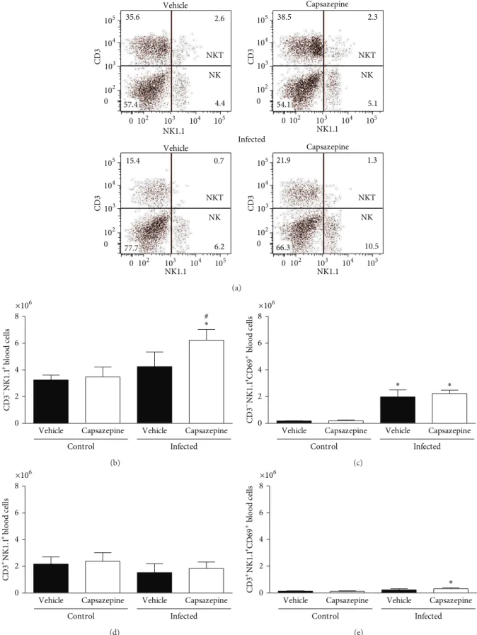
the efects of capsazepine on circulating and spleen NK (CD3−NK1.1+) and NKT (CD3+NK1.1+) cells. Peripheral blood NK and NKT cells were detected in all groups of uninfected and infected animals (Figures4(a)–4(e)). Malaria induction had no efects on either NK or NKT cell numbers when compared to uninfected control mice (Figures4(b)and
4(d)). On the other hand, NK, but not NKT activation via CD69 expression, was increased in both vehicle- (11.1-fold increase) and capsazepine- (11.5-fold increase) treated groups when compared to their respective uninfected controls (Figure4(c)). However, capsazepine treatment signiicantly increased NK cell population (Figure4(b)) when compared to either its uninfected control group (1.8-fold increase) or vehicle-treated infected animals (1.5-fold increase). Also, capsazepine was able to enhance activation of NKT-infected cells (2.7-fold increase; Figure4(e)). Importantly, NK cells account for the majority of circulating and activated NK1.1+ cells in infected animals (Figure4).
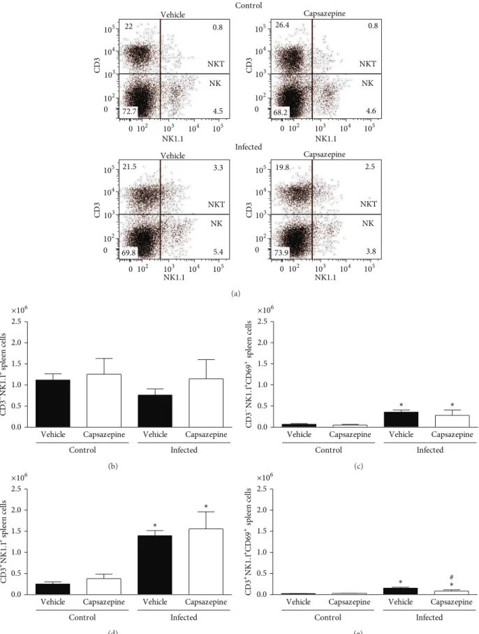
Similarly, spleen NK and NKT cells were detected in all groups of uninfected and infected animals (Figures 5(a)–
5(e)). Malaria induction caused spleen NKT but not NK cell population expansion when compared to uninfected control mice (Figures 5(b) and 5(d)). his was observed for both vehicle- (5.6-fold increase) and capsazepine- (4.1-fold increase) treated mice (Figure 5(d)). Figures5(c) and
5(e)demonstrate that CD69 expression was augmented on both spleen NK and NKT cells obtained from infected mice treated with either vehicle (5.3- and 6.9-fold increase, resp.) or capsazepine (5.6- and 3.1-fold increase, resp.). However, NKT activation was45.7±17.5% lower in capsazepine-treated mice when compared to vehicle-infected controls (Figure 5(e)). Capsazepine treatment in infected mice had no efects on spleen NK and NKT cell numbers or NK activation (Figure5). Similarly, capsazepine did not alter spleen NK and NKT proile (cell number and activation) in samples obtained from uninfected mice (Figure5).
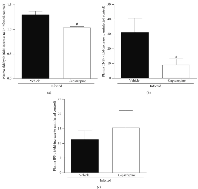
3.4. Capsazepine Reduces Lipid Peroxidation and Plasma
TNF� Levels in P. berghei ANKA-Infected Mice. P. berghei
ANKA infection increased lipid peroxidation in both vehicle-and capsazepine-treated mice, as demonstrated by the levels of plasma aldehydes (Figure6(a)). However, this increase was less pronounced in the capsazepine-treated group (20.5±6.4% reduction). Malaria also triggered the release of TNF� and IFN�in both infected groups (Figures6(b)and6(c)). TNF� production was markedly reduced by capsazepine treatment (70.8 ± 14.5%; Figure6(b)). On the other hand, capsazepine treatment did not afect IFN� release triggered by malaria (Figure 6(c)). Vehicle- and capsazepine-treated uninfected mice exhibited similar levels of plasma aldehydes, TNF�, and IFN�(data not shown). Production of IL-4, IL-6, IL-2, IL-10, and IL-17 was not detected in any of the evaluated groups.
4. Discussion
Since its discovery, evidence has accumulated that TRPV1 has the potential to play a key role in a variety of patholo-gies, especially those associated with imbalances of the
immune and inlammatory response, such as asthma [22–
24] and rheumatoid arthritis [25–27]. Recent reports demon-strated that TRPV1 is expressed on immune cells such as macrophages and peripheral-blood mononuclear cells in inlammatory conditions [9,28–30]. More recently, TRPV1 was suggested to modulate a range of macrophage-mediated responses to bacterial infection [9]. Indeed, TRPV1 deletion or antagonism has been associated with poorer outcome of experimental sepsis as TRPV1 blockade increases pathogen load and also facilitates the transition from a local to a systemic inlammatory response to bacteria [7,9,10,31–34]. In addition, TRPV1 knockout (TRPV1 KO) mice challenged with intestinal bacteria or LPS present with a dysregulated production of inlammatory mediators, including NO, ROS, and cytokines such as TNF�, IL-10, and IL-6 [7–9]. So far, there are no reports of TRPV1 playing any roles in the immune response to other pathogens.
Here, we show for the irst time that TRPV1 antagonism by capsazepine, a nonselective antagonist, modulates the innate immune response to malaria. Reports have shown that capsazepine presents species- and modality-speciic activity on TRPV1 and also inhibits acetylcholine receptors, voltage-gated calcium channels, and hyperpolarization-activated cyclic nucleotide-gated channels, in addition to TRPV1 [35–
38]. However, it is important to highlight that capsazepine was administered in this study, as described by Fernandes and collaborators [9] who showed that repeated treatment with this drug in vivo produces a similar proile to that of TRPV1 KO mice in response to bacterial infection [9]. Herein, we induced malaria by injectingP. bergheiANKA in a susceptible strain of mice known to develop cerebral malaria-like symptoms and to present with 60–100% mortality within the second week following injection (for review see [19]). Our studies were carried out at a premortality end-point, that is, 7 days following infection. At this time point, parasitaemia had reached 11%, in agreement with previous studies [17,22].
Evidence suggests that the innate immune response plays an important role during the early phase of infection, with activated monocytes and neutrophils releasing nonspeciic inlammatory mediators such as ROS and cytokines [2,39] and exerting their roles as phagocytes and antigen-presenting cells when in contact with circulating infected RBCs [40]. Indeed, phagocytosis of infected RBCs by peripheral blood and tissue phagocytes is suggested to be the major mechanism
ofPlasmodiumremoval [41]. As a result of this interaction,
phagocytes may damage the endothelium, thus contributing to the collapse of the circulation and in the case of cerebral malaria, damaging the brain microvasculature [4,42]. During infection, monocytes diferentiate into macrophages in the spleen and also in the brain, becoming available in the brain microvasculature [4]. Activated macrophages contribute to pathogen clearance and activation of lymphocytes, in an attempt to stimulate the generation of an acquired immune response capable of improving parasite removal and ighting a secondary infection [2,43].
NK1.1 NK1.1
NK1.1 NK1.1
CD3
CD3 CD3
CD3
NKT
NK
35.6 2.6
57.4 4.4
NKT
NK
38.5 2.3
54.1 5.1
NKT
NK
15.4 0.7
77.7 6.2
NKT
NK
21.9 1.3
66.3 10.5
Control
Infected Vehicle
Vehicle Capsazepine
Capsazepine
105
105 104
104 103
103 102
102 0
0 105
105 104
104 103
103 102
102 0
0
105
105 104
104 103
103 102
102 0
0
105
105 104
104 103
103 102
102 0
0
(a)
#
∗
Control Infected
Vehicle Capsazepine Vehicle Capsazepine
6
4
2
0 8 ×10⁶
CD3
− NK1.1
+ b
lo
o
d
cells
(b)
∗ ∗
Control Infected
Vehicle Capsazepine Vehicle Capsazepine
6
4
2
0 8 ×10⁶
CD3
−NK1.1 +CD69 +b
lo
o
d cells
(c)
Control Infected
Vehicle Capsazepine Vehicle Capsazepine
6
4
2
0 8 ×10⁶
CD3
+NK1.1 +b
lo
o
d cells
(d)
∗
Control Infected
Vehicle Capsazepine Vehicle Capsazepine
6
4
2
0 8 ×10⁶
CD3
+NK1.1 +CD69 +b
lo
o
d cells
(e)
Figure 4: Efect of capsazepine on peripheral blood CD3−NK1.1+ and CD3+NK1.1+ cells. (a) Representative two-colour dot-plots for
peripheral blood CD3−NK1.1+(NK) and CD3+NK1.1+(NKT) cell populations from uninfected andPlasmodium bergheiANKA-infected mice
(105infected RBCs/animal; i.p.). Circulating (b) CD3−NK1.1+and (c) CD3−NK1.1+cell numbers in uninfected andPlasmodium berghei
ANKA-infected mice (105infected RBCs/animal; i.p.). Expression of CD69 on circulating (d) CD3−NK1.1+and (e) CD3−NK1.1+cells. Capsazepine
(50�g/animal, 2x day, for 6 days) was administered from 24 h ater infection. Vehicle- (10% DMSO in saline) treated animals were used as
NKT
NK
22 0.8
72.7 4.5
NKT
NK
26.4 0.8
68.2 4.6
NKT
NK
21.5 3.3
69.8 5.4
NKT
NK
19.8 2.5
73.9 3.8
NK1.1 NK1.1
NK1.1 NK1.1
CD3
CD3 CD3
CD3
Control
Infected Vehicle
Vehicle Capsazepine
Capsazepine
105
105 104
104 103
103 102
102 0
0 105
105 104
104 103
103 102
102 0
0
105
105 104
104 103
103 102
102 0
0
105
105 104
104 103
103 102
102 0
0
(a)
2.5
2.0
1.5
1.0
0.5
0.0
Control Infected
Vehicle Capsazepine Vehicle Capsazepine
×106
CD3
− NK1.1 + sp
leen cells
(b)
∗ ∗
2.5
2.0
1.5
1.0
0.5
0.0
Control Infected
Vehicle Capsazepine Vehicle Capsazepine
×106
CD3
−NK1.1 +CD69 +sp
leen cells
(c)
∗
∗ 2.5
2.0
1.5
1.0
0.5
0.0
Control Infected
Vehicle Capsazepine Vehicle Capsazepine
×106
CD3
+NK1.1 +sp
leen cells
(d)
#
∗ ∗
2.5
2.0
1.5
1.0
0.5
0.0
Control Infected
Vehicle Capsazepine Vehicle Capsazepine
×106
CD3
+NK1.1 +CD69 +sp
leen cells
(e)
Figure 5: Efect of capsazepine on spleen CD3−NK1.1+and CD3+NK1.1+cells. (a) Representative two-colour dot-plots for spleen CD3−NK1.1+
(NK) and CD3+NK1.1+(NKT) cell populations from uninfected andPlasmodium bergheiANKA-infected mice (105infected RBCs/animal;
i.p.). Spleen (b) CD3−NK1.1+and (c) CD3−NK1.1+cell numbers in uninfected andPlasmodium bergheiANKA-infected mice (105infected
RBCs/animal; i.p.). Expression of CD69 on circulating (d) CD3−NK1.1+and (e) CD3−NK1.1+cells. Capsazepine (50�g/animal, 2x day, for 6
days) was administered from 24 h ater infection. Vehicle- (10% DMSO in saline) treated animals were used as controls. Data are expressed
Infected
Vehicle Capsazepine
# 1.5
1.0
0.5
0.0
Plasma aldeh
yde (f
o
ld-incr
ea
se t
o uninf
ec
te
d co
n
tr
o
l)
(a)
Infected
Vehicle Capsazepine
# 50
40
30
20
10
0
Plasma TNF
�
(f
o
ld-incr
ea
se
t
o
uninf
ec
ted co
n
tr
o
l)
(b)
Infected
Vehicle Capsazepine
25
20
15
10
5
0
Plasma IFN
𝛾
(f
o
ld-incr
ea
se
t
o
uninf
ec
te
d co
n
tr
o
l)
(c)
Figure 6: Efect of capsazepine on systemic lipid peroxidation and cytokine release. (a) Aldehyde; (b) TNF�and (c) IFN�levels in plasma
samples obtained from uninfected andPlasmodium bergheiANKA-infected mice (105infected RBCs/animal; i.p.). Capsazepine (50�g/animal,
2x day, for 6 days) was administered from 24 h ater infection. Vehicle- (10% DMSO in saline) treated animals were used as controls. Data are
expressed as mean±SD,� = 5–8 per group.#� < 0.05compared with vehicle-treated infected group.
is activation of both monocytes and spleen macrophages (F4/80+). Interestingly, we detected a group of circulating F4/80+Ly6G+ cells which became markedly expanded and activated (as denoted by IAb expression) following infection. his is the irst report to our knowledge of their contribution to malaria. However, it was recently suggested in a model of infection caused by vaccinia virus inoculation in C57BL/6 mice that F4/80+Ly6G+ cells are indeed monocytes with a great capacity of producing ROS and IFN� whilst Ly6G− monocytes produce NO and TNF� [44]. In addition, the same study suggested that F4/80+Ly6G+cells replace Ly6G− monocytes as infection progresses. Herein, we show the existence of F4/80+Ly6G+ cells also in the spleen and that they exhibit a similar proile (in terms of number and
activation pattern) to that of Ly6G−cells in this tissue. As the population of F4/80+Ly6G+cells rises, GR1+ cells (myeloid-derived suppressor cells) decline with infection. his is expected as an increase of mature monocytes and neutrophils normally occurs as a result of infection [45,46]. However, at 7 days ater infection, these changes in the balance and/or activation of F4/80+, F4/80+Ly6G+, and GR1+cells could not be noticed at spleen level. However, it is possible that these alterations may occur in this organ at a diferent time point not addressed by this study.
markedly increased in the same group of animals, suggesting that, as capsazepine shuts down F4/80+- and F4/80+Ly6G+ -mediated responses, either by deactivating them or decreas-ing their availability in the circulation, these cells are pro-gressively replaced by GR1+ cells in order to restore the immune responses that may be dependent on F4/80+- and F4/80+Ly6G+monocytes. Indeed, GR1+cells are suggested to be recruited in order to compensate “the loss” of monocytes that undergo polarization followingPlasmodiumor bacterial infection [47, 48]. hese cells are also called regulatory monocytes/macrophages and have a potent ability to suppress T cell proliferation and h1 responses [49]. On the other hand, the deinite role of these cells in critical illness is still of debate (for review see [47]).
To evaluate the impact this shit on monocyte proile had on inlammatory mediator release, we measured the levels of plasma aldehydes (index of lipid peroxidation secondary to oxidative stress) and cytokines such as TNF�and IFN�. Indeed, high systemic levels of inlammatory mediators such as TNF�, IFN�, and aldehydes are correlated with severe malaria in humans [50]. In fact, lipid peroxidation is produced during malaria as a result of the interactions between monocytes/macrophages and infected erythrocytes, and also the endothelium [51,52]. In addition, cytokines are also released in response to malaria, triggering suppression of erythropoiesis and activation of a variety of circulating and spleen cells [2,53,54]. We found that whilst diminished levels of lipid peroxidation-derived aldehydes and TNF� were detected in animals with malaria that had been treated with capsazepine, IFN�production remained similar to that observed for samples obtained from vehicle-infected mice. A reduction of oxidative stress was expected as capsazepine was previously suggested to inhibit oxidative stress in cultured RAW264 monocytes/macrophages in a TRPV1 independent manner, although this data was obtained from cells that had not been challenged with any pathogen product [55]. Later, TRPV1 deletion was shown to decrease ROS production by macrophages in sepsis [9] and to modulate the release of these mediators in other inlammatory conditions [14,
56]. However, loss of TRPV1 function has been associated with increased production of TNF�upon bacterial infection [7, 9, 32]. It is possible that TRPV1 diferently modulates monocytes and macrophages with respect to their ability to produce TNF�. Also, TRPV1 efects on TNF� production may vary at diferent stages of malaria.
NK and NKT cells are important efectors of the innate immune response to malaria, directly recognizing Plasmod-ium-infected RBCs and malarial antigens, in addition to producing IFN�in order to contain parasitaemia [2,18,40]. Indeed, these cells rise early during malaria and have been suggested to mediate the diferentiation of h1/h2 responses and to be essential for the traicking of leukocytes to the brain in cerebral malaria (for review see [2]). In addition, similarly to macrophages, they can accumulate into the brain during cerebral malaria, contributing to a worse outcome [18]. NK cells have also been linked to dendritic cell maturation and T cell activation in the spleen by releasing of cytokines [53,57]. Also, evidence has shown that during malaria, NK cells may undergo an intense turnover or even migrate out of the spleen
[18]. his was investigated at a similar time-point to that used in our study (1 week following infection). Here, we assessed the dynamics between circulating and spleen NK and NKT cells. We found that P. berghei ANKA-induced infection caused expansion of spleen NKT but not NK cell population. his was not accompanied by any change on circulating NK and NKT cell numbers. It is possible that at this time-point, NK and NKT cells have already migrated to the brain. Indeed, Hansen and collaborators showed that NK cells accumulate into the brain of C57BL/6 mice as early as 4 days following infection withP. bergheiANKA, triggering the migration of T cells to the brain microvasculature [18]. On the other hand, we show that both NK and NKT cells became activated in response to infection, with NK cells representing the majority of activated cells. Disappointingly, expansion of spleen NKT cells did not translate in their activation as only few of these cells expressed CD69. Capsazepine treatment in infected mice led to a further expansion of the circulating NK population, but its activation was similar to that of vehicle-infected group. In addition, capsazepine treatment reduced by half the activation of spleen NKT cells when compared to its infected-control. We would expect that the reduction of activation of spleen NKT cells and F4/80+Ly6G+monocytes by capsazepine would impair IFN� release. However, we show capsazepine inhibitory efects on these populations do not afect IFN�production in malaria, indicating that either TRPV1 may not play a role on IFN�production or NK cells become the sole source of IFN�once TRPV1 is blocked. In addition, the lack of efect of capsazepine on parasitaemia may be related to the similar levels of IFN�detected in both groups of animals.
Our study provides the irst evidence that TRPV1 modu-lates malaria by mediating innate immune response, specii-cally by interfering with the expansion and activation of efec-tor cells, especially monocytes. We also show that TRPV1 reg-ulates the immunological balance between diferent mono-cyte populations in addition to modulating mediator release by them. It is possible that blocking TRPV1 may be either beneicial, as a reduction of oxidative stress may relect on reduced vascular dysfunction, or deleterious, as impairment of innate response may lead to an ineicient removal of the parasite in addition to an ineicient acquired immune response to malaria. However, the impact TRPV1 antagonism may have on severe malaria outcome is of importance and remains to be investigated.
Conflict of Interests
he authors declare no conlict of interests.
Acknowledgments
Brazil), and Fundac¸˜ao de Amparo `a Pesquisa do Estado de S˜ao Paulo (FAPESP; Brazil).
References
[1] World Health Organization,World Malaria Report: 2013, 2013.
[2] L. Schoield, “Intravascular iniltrates and organ-speciic
inlammation in malaria pathogenesis,”Immunology and Cell
Biology, vol. 85, no. 2, pp. 130–137, 2007.
[3] V. Pradhan and K. Ghosh, “Immunological disturbances
asso-ciated with malarial infection,”Journal of Parasitic Diseases, vol.
37, no. 1, pp. 11–15, 2013.
[4] H. J. Shikani, B. D. Freeman, M. P. Lisanti, L. M. Weiss, H. B. Tanowitz, and M. S. Desruisseaux, “Cerebral malaria: we have
come a long way,”he American Journal of Pathology, vol. 181,
no. 5, pp. 1484–1492, 2012.
[5] A. Moulick, S. Maitra, B. S. Sarkar, A. Jana, and S. Sarkar, “Vivax
malaria presenting with myelitis: a rare complication,”Journal of
Clinical and Diagnostic Research, vol. 7, no. 5, pp. 914–916, 2013.
[6] M. M. Stevenson and E. M. Riley, “Innate immunity to malaria,”
Nature Reviews Immunology, vol. 4, no. 3, pp. 169–180, 2004.
[7] I. A. Clark, “How TNF was recognized as a key mechanism of
disease,”Cytokine and Growth Factor Reviews, vol. 18, no. 3-4,
pp. 335–343, 2007.
[8] E. S. Fernandes, M. A. Fernandes, and J. E. Keeble, “he functions of TRPA1 and TRPV1: moving away from sensory
nerves,”British Journal of Pharmacology, vol. 166, no. 2, pp. 510–
521, 2012.
[9] E. S. Fernandes, L. Liang, S. Smillie et al., “TRPV1 deletion enhances local inlammation and accelerates the onset of
sys-temic inlammatory response syndrome,”Journal of
Immunol-ogy, vol. 188, no. 11, pp. 5741–5751, 2012.
[10] V. Guptill, X. Cui, A. Khaibullina et al., “Disruption of the tran-sient receptor potential vanilloid 1 can afect survival, bacterial clearance, and cytokine gene expression during murine sepsis,”
Anesthesiology, vol. 114, no. 5, pp. 1190–1199, 2011.
[11] K. Susankova, K. Tousova, L. Vyklicky, J. Teisinger, and V. Vla-chova, “Reducing and oxidizing agents sensitize heat-activated
vanilloid receptor (TRPV1) current,”Molecular Pharmacology,
vol. 70, no. 1, pp. 383–394, 2006.
[12] E. S. Fernandes, C. T. Vong, S. Quek et al., “Superoxide generation and leukocyte accumulation: key elements in the mediation of leukotriene B4-induced itch by transient receptor potential ankyrin 1 and transient receptor potential vanilloid 1,”
he FASEB Journal, vol. 27, no. 4, pp. 1664–1673, 2013.
[13] P. Puntambekar, D. Mukherjea, S. Jajoo, and V. Ramkumar, “Essential role of Rac1/NADPH oxidase in nerve growth factor
induction of TRPV1 expression,”Journal of Neurochemistry, vol.
95, no. 6, pp. 1689–1703, 2005.
[14] A. Starr, R. Graepel, J. Keeble et al., “A reactive oxygen
species-mediated component in neurogenic vasodilatation,”
Cardiovas-cular Research, vol. 78, no. 1, pp. 139–147, 2008.
[15] C. K. B. Ferrari, P. C. S. Souto, E. L. Franc¸a, and A. C. Honorio-Franc¸a, “Oxidative and nitrosative stress on phagocytes’ func-tion: from efective defense to immunity evasion mechanisms,”
Archivum Immunologiae et herapiae Experimentalis, vol. 59,
no. 6, pp. 441–448, 2011.
[16] S. Perc´ario, D. R. Moreira, B. A. Q. Gomes et al., “Oxidative
stress in Malaria,”International Journal of Molecular Sciences,
vol. 13, no. 12, pp. 16346–16372, 2012.
[17] R. M. Elias, M. Correa-Costa, C. R. Barreto et al., “Oxidative stress and modiication of renal vascular permeability are associated with acute kidney injury during P. berghei ANKA
infection,”PLoS ONE, vol. 7, no. 8, Article ID e44004, 2012.
[18] D. S. Hansen, N. J. Bernard, C. Q. Nie, and L. Scholeld, “NK
cells stimulate recruitment of CXCR3+ T cells to the brain
duringPlasmodium berghei-mediated cerebral malaria,”Journal
of Immunology, vol. 178, no. 9, pp. 5779–5788, 2007.
[19] L. R´enia, S. W. Howland, C. Claser et al., “Cerebral malaria
Mysteries at the blood-brain barrier,”Virulence, vol. 3, no. 2, pp.
193–201, 2012.
[20] R. P. Bird and H. H. Draper, “Comparative studies on diferent
methods of malonaldehyde determination,”Methods in
Enzy-mology, vol. 105, pp. 299–305, 1984.
[21] L. Sharma, J. Kaur, and G. Shukla, “Role of oxidative stress and apoptosis in the placental pathology of plasmodium berghei
infected mice,”PLoS ONE, vol. 7, no. 3, Article ID e32694, 2012.
[22] R. Nassini, P. Pedretti, N. Moretto et al., “Transient receptor potential ankyrin 1 channel localized to non-neuronal airway
cells promotes non-neurogenic inlammation,”PLoS ONE, vol.
7, no. 8, Article ID e42454, 2012.
[23] F. Tsuji and H. Aono, “Role of transient receptor potential
vanilloid 1 in inlammation and autoimmune diseases,”
Phar-maceuticals, vol. 5, no. 8, pp. 837–852, 2012.
[24] J. Yang, H. M. Yu, X. D. Zhou, V. P. Kolosov, and J. M. Perelman, “Study on TRPV1-mediated mechanism for the hypersecretion
of mucus in respiratory inlammation,”Molecular Immunology,
vol. 53, no. 1-2, pp. 161–171, 2013.
[25] E. S. Fernandes, F. A. Russell, D. Spina et al., “A distinct role for transient receptor potential ankyrin 1, in addition to transient receptor potential vanilloid 1, in tumor necrosis factor
�-induced inlammatory hyperalgesia and Freund’s complete
adjuvant-induced monarthritis,”Arthritis and Rheumatism, vol.
63, no. 3, pp. 819–829, 2011.
[26] J. Keeble, F. Russell, B. Curtis, A. Starr, E. Pinter, and S. D. Brain, “Involvement of transient receptor potential vanilloid 1 in the vascular and hyperalgesic components of joint inlammation,”
Arthritis and Rheumatism, vol. 52, no. 10, pp. 3248–3256, 2005.
[27] F. A. Russell, E. S. Fernandes, J. Courade, J. E. Keeble, and S. D.
Brain, “Tumour necrosis factor�mediates transient receptor
potential vanilloid 1-dependent bilateral thermal hyperalgesia
with distinct peripheral roles of interleukin-1�, protein kinase C
and cyclooxygenase-2 signalling,”Pain, vol. 142, no. 3, pp. 264–
274, 2009.
[28] T. K. Finney-Hayward, M. O. Popa, P. Bahra et al., “Expression of transient receptor potential C6 channels in human lung
macrophages,” he American Journal of Respiratory Cell and
Molecular Biology, vol. 43, no. 3, pp. 296–304, 2010.
[29] C. I. Saunders, R. G. Fassett, and D. P. Geraghty, “Up-regulation of TRPV1 in mononuclear cells of end-stage kidney disease
patients increases susceptibility toN-arachidonoyl-dopamine
(NADA)-induced cell death,”Biochimica et Biophysica Acta:
Molecular Basis of Disease, vol. 1792, no. 10, pp. 1019–1026, 2009.
[30] J. Zhao, L. Ching, Y. R. Kou et al., “Activation of TRPV1
pre-vents OxLDL-induced lipid accumulation and TNF-�-Induced
inlammation in macrophages: role of liver X receptor �,”
Mediators of Inlammation, vol. 2013, Article ID 925171, 14 pages,
2013.
[31] P. Bryant, M. Shumate, G. Yumet, C. H. Lang, T. C. Vary, and R. N. Cooney, “Capsaicin-sensitive nerves regulate the metabolic
response to abdominal sepsis,”he Journal of Surgical Research,
[32] N. Clark, J. Keeble, E. S. Fernandes et al., “he transient receptor potential vanilloid 1 (TRPV1) receptor protects against the onset
of sepsis ater endotoxin,”he FASEB Journal, vol. 21, no. 13, pp.
3747–3755, 2007.
[33] T. Iida, I. Shimizu, M. L. Nealen, A. Campbell, and M. Caterina, “Attenuated fever response in mice lacking TRPV1,”
Neuroscience Letters, vol. 378, no. 1, pp. 28–33, 2005.
[34] Y. Wang, M. Novotn´y, V. Quaiserov´a-Mocko, G. M. Swain, and D. H. Wang, “TRPV1-mediated protection against
endotoxin-induced hypotension and mortality in rats,”American Journal of
Physiology: Regulatory Integrative and Comparative Physiology,
vol. 294, no. 5, pp. R1517–R1523, 2008.
[35] R. J. Docherty, J. C. Yeats, and A. S. Piper, “Capsazepine block of voltage-activated calcium channels in adult rat dorsal root
ganglion neurones in culture,”British Journal of Pharmacology,
vol. 121, no. 7, pp. 1461–1467, 1997.
[36] L. Liu and S. A. Simon, “Capsazepine, a vanilloid receptor antagonist, inhibits nicotinic acetylcholine receptors in rat
trigeminal ganglia,”Neuroscience Letters, vol. 228, no. 1, pp. 29–
32, 1997.
[37] C. H. Gill, P. J. Strijbos, S. A. Bates, W. Cairns, D. Owen, and C. H. Davies, “Capsazepine inhibits a recombinant human
HCN1-mediated current,”British Journal of Pharmacology, vol. 135, no.
5, p. 251, 2001.
[38] P. McIntyre, L. M. McLatchie, A. Chambers et al., “Phar-macological diferences between the human and rat vanilloid
receptor 1 (VR1),”British Journal of Pharmacology, vol. 132, no.
5, pp. 1084–1094, 2001.
[39] U. Frevert and A. Nacer, “Immunobiology of Plasmodium in
liver and brain,”Parasite Immunology, vol. 35, no. 9-10, pp. 267–
282, 2013.
[40] C. Coban, K. J. Ishii, T. Horii, and S. Akira, “Manipulation of
host innate immune responses by the malaria parasite,”Trends
in Microbiology, vol. 15, no. 6, pp. 271–278, 2007.
[41] V. Gallo, O. A. Skorokhod, E. Schwarzer, and P. Arese,
“Simul-taneous determination of phagocytosis of Plasmodium
falci-parum-parasitized and non-parasitized red blood cells by low
cytometry,”Malaria Journal, vol. 11, article 428, 2012.
[42] F. M. S. de Leoratti, S. C. Trevelin, F. Q. Cunha et al., “Neutrophil
paralysis inPlasmodium vivaxmalaria,”PLoS Neglected Tropical
Diseases, vol. 6, no. 6, Article ID e1710, p. e1710, 2012.
[43] C. L. L. Chua, G. Brown, J. A. Hamilton, S. Rogerson, and P. Boeuf, “Monocytes and macrophages in malaria: protection or
pathology?”Trends in Parasitology, vol. 29, no. 1, pp. 26–34,
2013.
[44] M. A. Fischer, M. L. Davies, I. E. Reider et al., “CD11b+, Ly6G+ cells produce type I interferon and exhibit tissue
pro-tective properties following peripheral virus infection,”PLoS
Pathogens, vol. 7, no. 11, Article ID e1002374, 2011.
[45] J. A. Villadangos, “Hold On, the Monocytes Are Coming!,”
Immunity, vol. 26, no. 4, pp. 390–392, 2007.
[46] Q. Yang, P. Ghose, and N. Ismaila, “Neutrophils mediate immunopathology and negatively regulate protective immune responses during fatal bacterial infection-induced Toxic Shock,”
Infection and Immunity, vol. 81, no. 5, pp. 1751–1763, 2013.
[47] J. N. Fullerton, A. J. O’Brien, and D. W. Gilroy, “Pathways mediating resolution of inlammation: when enough is too
much,”he Journal of Pathology, vol. 231, no. 1, pp. 8–20, 2013.
[48] J. A. van Ginderachter, A. Beschin, P. D. Baetselier, and G. Raes, “Myeloid-derived suppressor cells in parasitic infections,”
European Journal of Immunology, vol. 40, no. 11, pp. 2976–2985,
2010.
[49] D. I. Gabrilovich and S. Nagaraj, “Myeloid-derived suppressor
cells as regulators of the immune system,” Nature Reviews
Immunology, vol. 9, no. 3, pp. 162–174, 2009.
[50] K. E. Lyke, R. Burges, Y. Cissoko et al., “Serum levels of
the proinlammatory cytokines interleukin-1 beta (IL-1�),
IL-6, IL-8, IL-10, tumor necrosis factor alpha, and IL-12(p70) in
Malian children with severePlasmodium falciparummalaria
and matched uncomplicated malaria or healthy controls,”
Infec-tion and Immunity, vol. 72, no. 10, pp. 5630–5637, 2004.
[51] S. L. Hazen, “Oxidized phospholipids as endogenous pattern
recognition ligands in innate immunity,”Journal of Biological
Chemistry, vol. 283, no. 23, pp. 15527–15531, 2008.
[52] E. Schwarzer, H. K¨uhn, E. Valente, and P. Arese, “Malaria-parasitized erythrocytes and hemozoin nonenzymatically gen-erate large amounts of hydroxy fatty acids that inhibit monocyte
functions,”Blood, vol. 101, no. 2, pp. 722–728, 2003.
[53] M. Vitale, M. Della Chiesa, S. Carlomagno et al.,
“NK-dependent DC maturation is mediated by TNF� and IFN�
released upon engagement of the NKp30 triggering receptor,”
Blood, vol. 106, no. 2, pp. 566–571, 2005.
[54] F. Gimenez, S. B. De Lagerie, C. Fernandez, P. Pino, and D.
Mazier, “Tumor necrosis factor�in the pathogenesis of cerebral
malaria,”Cellular and Molecular Life Sciences, vol. 60, no. 8, pp.
1623–1635, 2003.
[55] M. J. Garle, A. Knight, A. T. Downing, K. L. Jassi, R. H. Clothier, and J. R. Fry, “Stimulation of dichloroluorescin oxidation by capsaicin and analogues in RAW 264 monocyte/macrophages:
lack of involvement of the vanilloid receptor,” Biochemical
Pharmacology, vol. 59, no. 5, pp. 563–572, 2000.
[56] T. Schilling and C. Eder, “Importance of the non-selective cation channel TRPV1 for microglial reactive oxygen species
generation,”Journal of Neuroimmunology, vol. 216, no. 1-2, pp.
118–121, 2009.
[57] F. Gerosa, B. Baldani-Guerra, C. Nisii, V. Marchesini, G. Carra, and G. Trinchieri, “Reciprocal activating interaction
between natural killer cells and dendritic cells,”he Journal of
Submit your manuscripts at
http://www.hindawi.com
Stem Cells
International
Hindawi Publishing Corporationhttp://www.hindawi.com Volume 2014
Hindawi Publishing Corporation
http://www.hindawi.com Volume 2014 INFLAMMATION
Hindawi Publishing Corporation
http://www.hindawi.com Volume 2014
Behavioural
Neurology
Endocrinology
International Journal ofHindawi Publishing Corporation
http://www.hindawi.com Volume 2014
Hindawi Publishing Corporation
http://www.hindawi.com Volume 2014
Disease Markers
Hindawi Publishing Corporation
http://www.hindawi.com Volume 2014
BioMed
Research International
Oncology
Journal ofHindawi Publishing Corporation
http://www.hindawi.com Volume 2014
Hindawi Publishing Corporation
http://www.hindawi.com Volume 2014
Oxidative Medicine and Cellular Longevity Hindawi Publishing Corporation
http://www.hindawi.com Volume 2014
PPAR Research
The Scientiic
World Journal
Hindawi Publishing Corporation
http://www.hindawi.com Volume 2014
Immunology Research
Hindawi Publishing Corporation
http://www.hindawi.com Volume 2014
Journal of
Obesity
Journal ofHindawi Publishing Corporation
http://www.hindawi.com Volume 2014
Hindawi Publishing Corporation
http://www.hindawi.com Volume 2014
Computational and Mathematical Methods in Medicine
Ophthalmology
Journal ofHindawi Publishing Corporation
http://www.hindawi.com Volume 2014
Diabetes Research
Journal ofHindawi Publishing Corporation
http://www.hindawi.com Volume 2014
Hindawi Publishing Corporation
http://www.hindawi.com Volume 2014 Research and Treatment
AIDS
Hindawi Publishing Corporation
http://www.hindawi.com Volume 2014
Gastroenterology Research and Practice
Hindawi Publishing Corporation
http://www.hindawi.com Volume 2014
Parkinson’s
Disease
Evidence-Based Complementary and Alternative Medicine
Volume 2014