Evaluation of the venous reflux of the great saphenous vein by
Evaluation of the venous reflux of the great saphenous vein by
Evaluation of the venous reflux of the great saphenous vein by
Evaluation of the venous reflux of the great saphenous vein by
Evaluation of the venous reflux of the great saphenous vein by
duplex scan after surgical treatment of the saphenofemoral
duplex scan after surgical treatment of the saphenofemoral
duplex scan after surgical treatment of the saphenofemoral
duplex scan after surgical treatment of the saphenofemoral
duplex scan after surgical treatment of the saphenofemoral
junction insufficiency
junction insufficiency
junction insufficiency
junction insufficiency
junction insufficiency
Avaliação da ocorrência do refluxo venoso da safena magna pela
Avaliação da ocorrência do refluxo venoso da safena magna pela
Avaliação da ocorrência do refluxo venoso da safena magna pela
Avaliação da ocorrência do refluxo venoso da safena magna pela
Avaliação da ocorrência do refluxo venoso da safena magna pela
ultrassonografia com doppler colorido após tratamento cirúrgico da
ultrassonografia com doppler colorido após tratamento cirúrgico da
ultrassonografia com doppler colorido após tratamento cirúrgico da
ultrassonografia com doppler colorido após tratamento cirúrgico da
ultrassonografia com doppler colorido após tratamento cirúrgico da
insuficiência da junção safeno-femoral
insuficiência da junção safeno-femoral
insuficiência da junção safeno-femoral
insuficiência da junção safeno-femoral
insuficiência da junção safeno-femoral
GUTENBERGDO AMARAL GURGEL1; ALDEMAR ARAÚJO CASTRO2; MARCELO ARAÚJO,ACBC-BA3; JORGE EDUARDO AMORIM4; GUILHERME BENJAMIN BRANDÃO PITTA5; FAUSTO MIRANDA JÚNIOR,TCBC-SP6
A B S T R A C T A B S T R A C T A B S T R A C T A B S T R A C T A B S T R A C T
Objective Objective Objective Objective
Objective: To evaluate the occurrence of reflux from the great saphenous vein by color Doppler ultrasonography in subjects undergoing treatment of insufficiency of the saphenofemoral junction by simple ligation or ligation with section of the saphenous arch. MethodsMethodsMethodsMethodsMethods: We performed 60 operations (in 45 subjects) of varicose insufficiency of the saphenofemoral junction (SFJ), belonging to the CEAP clinical classification of 2-5, who were randomly divided into two groups. A group called C, with ligature and section of the saphenous arch, and a group called L, with simple ligation of the saphenous vein and no sectioning of its arch. We then investigated the occurrence of reflux from the great saphenous vein in groups C and L through postoperative color Doppler ultrasonography at intervals of six months to one year. ResultsResultsResultsResults: Of the 60 limbs submitted to the approach of the saphenous arch,Results 57 were evaluated by postoperative doppler ultrasound, since two subjects (three limbs) did not return and were excluded from the study. The mean age was 54 years, with 93% females and predominance of CEAP classification 2 in 60.5%. Of the 57 operations for the treatment of reflux of the saphenous arch, 43.9% had reflux postoperatively,14.1% in group C and 29.8% in group L (p < 0,05). The relative risk of reflux of the saphenous arch in group L was 2.03 times higher compared with group C. ConclusionConclusionConclusionConclusionConclusion: the section of the arch of the great saphenous vein causes less postoperative reflux than simple ligation in treatment of insufficiency of the great saphenous vein.
Key words: Key words: Key words: Key words:
Key words: Varicose veins. Venous insufficiency. Surgical procedures, operative. Saphenous vein. Ultrasonography, Doppler, color.
1. Professor, Cardio-Renal-Vascular Physiology, Potiguar University, Natal (RN), Brazil; 2. Assistant Professor, Department of Social Medicine, Health Sciences State University of of Alagoas, Maceió (AL), Brazil; 3. Assistant Professor, Angiology, Department of Health, State University of Santa Cruz, Ilheus (Bahia), Brazil; 4. Associate Professor, Vascular Surgery, Department of Surgery, Federal University of São Paulo, São Paulo (SP), Brazil; 5. Associate Professor, Surgical Clinics, Health Sciences State University of of Alagoas, Maceió (AL), Brazil; 6. Professor, Vascular Surgery, Department of Surgery, Federal University of São Paulo, São Paulo (SP), Brazil.
INTRODUCTION
INTRODUCTION
INTRODUCTION
INTRODUCTION
INTRODUCTION
V
enous disease affects millions of people worldwide, and varicose veins are the most obvious and known feature of its spectrum. It is considered the most common vascular diseases, with high prevalence, especially in females1,2.The treatment for the correction of trunk varicose veins and prevention of its complications is surgical. Most surgical techniques involve the approach of the saphenous vein, as reflux is usually the main pathophysiological substrate. Options range from preservation to truncal ablation to radical operation, with saphenous stripping and ligation of insufficient perforating veins and resection of varices3.
With the development and improvement of noninvasive diagnostic methods in the preoperative evaluation, it was possible to improve the indications of surgical treatment of primary varicose veins by different techniques3-8. These diagnostic methods were able to
identify the points of reflux of the superficial and deep systems and classify the degree of venous reflux to the saphenous veins4,9-12.
Color Doppler Ultrasonography, considered the gold standard, allows the accurate study of venous reflux of the lower limbs10,13. Due to this exam, various techniques
of treatment of insufficiency of the saphenofemoral junction (SFJ) were improved over time5,14-18 aiming at the efficiency
Ligation of saphenous vein and resection of varicose veins14 and ligation and section of the JSF with
treatment of varicose veins16 are two of the most used
approaches that preserve the saphenous vein. However, the overall recurrence rate of varicose veins still varies between 20% and 30%19. This may be related to errors
when ligating the saphenous arch and to the absence of treatment of other reflux sites20-22.
The aim of this study was to evaluate the occurrence of reflux of the great saphenous vein by color Doppler ultrasonography in subjects undergoing treatment of insufficiency of the saphenofemoral junction by simple ligation or ligation and section of the arch.
METHODS
METHODS
METHODS
METHODS
METHODS
This study was approved by the Ethics in Research Committee of UNCISAL (Health Sciences State University of Alagoas) under authorization no 53/2004. Patients were informed of the purpose of the research, its expected results, risks and benefits of procedures, in accordance with Resolution 196/1996 of the National Research Council. After agreeing with the research and providing a written consent, all were treated according to the ethical concepts of the Declaration of Helsinki.
The sample was calculated by estimating the difference between two proportions, with a statistical power of 80%, for an alpha of 5% and a beta of 20%, 30 limbs having been calculated for each group. Forty- five consecutive subjects (60 limbs) underwent approach to saphenofemoral junction insufficiency and for the treatment of varicose veins. We included subjects with CEAP clinical classification23 (based on clinical, etiology, anatomy and
pathophysiology) C2-5, patients with great saphenous arch insufficiency confirmed on ultrasound with color Doppler. We excluded subjects under age 18 and JSF with diameter greater than 12mm, pregnant women, indigenous people, people with post-thrombotic syndrome, deep vein thrombosis, arterial and lymphatic insufficiency and patients difficult to follow.
Two groups were formed through the draw with opaque envelopes opened sequentially (containing 30 letters “C” and 30 letters “L”) intraoperatively, indicating the surgical technique to be used for each limb. Group C was submitted to ligation and section of the arch of the great saphenous vein, and Group L, simple ligation without sectioning the arch. All participants were subjected to the treatment of the primary varicose veins and ligation of insufficient perforating and communicating veins.
The primary variable was the presence of reflux in the great saphenous vein after surgical procedure of the arch by one of the techniques, considering defined as the reverse flow in high speed, with high peak wave e” 30cm/ s and time longer than 0.5 second4. To this end, we
performed clinical evaluation and indication of surgical
treatment, patients being sent to the sonographer, who performed all tests before and after the operation, without knowing which group they belonged to. We used the color Doppler ultrasound machine with a linear transducer of 4.0 to 7.0 MHz
With the patient in supine position, we evaluated the patency of the superficial and deep system with compression maneuvers in the femoropopliteal segment, JSF, junction of the short saphenous with the popliteal vein, tributaries of the saphenous arch and of the femoral vein. We recorded the flow of the following veins: common, su-perficial and deep femoral; popliteal; anterior and posteri-or tibial; and fibular. We also verified the patency and flow of communicating and perforating veins found in the examined limbs.
With the patient standing, we studied the patency, with venous compressibility and measurement of the diameter of the saphenous vein from the JSF to the ankle. In the arch of the great saphenous, we verified the presence of tributaries (medial and lateral accessory saphenous veins, superficial circumflex iliac vein, superfici-al epigastric vein and superficisuperfici-al externsuperfici-al pudendsuperfici-al vein), and the presence or absence of reflux (Figure 1). We adopted the rating for the insufficiency of the great saphenous vein proposed by Engelhorn et al.4.
To approach the insufficient arch of the great saphenous, we performed an incision in the groin, with 3cm in length, dissection of all tributaries of the saphenous arch, followed by their ligation with 3-0 cotton suture, one ligature with cotton at the 2-0 SFJ and two other ligatures, separated from each other 1.0cm distally to the tributaries.
In Group L (simple ligation) we only performed the ligatures, then followed by wound synthesis with 3-0 polyglycolic suture and the skin with 4-3-0 nylon. In Group C (section of the arch), we sectioned the saphenous vein between the ligatures distal to its tributaries (Figure 2).
Statistical analysis was performed using the chi-square test, which allowed to establish whether the proportion of postoperative SFJ reflux was higher in group L than in group C, with p < 0.05. We also evaluated the relative risk of postoperative reflux between groups.
RESULTS
RESULTS
RESULTS
RESULTS
RESULTS
Of the 43 subjects followed, 93.0% (40/43) were female. The age ranged from 22 to 82 years, with an average of 54 years. After patients clinical assessment, we found the following distribution: C2: 60.5%; C3: 30.2%, C4: 7.0%, and C5: 2.3%. Preoperatively we found: Type I: 1.8%; Type II: 68.4%, Type III: 0%; Type IV: 0%; and Type V: 29.8%. Postoperatively we found: limbs which were Type II and became type IV with 51.3%, twice as more in
group C – 22.8% (13/57) – than in group L – 13.3% (7/ 57); limbs who were Type V – 29.8% (17/57) – preoperatively and became Type IV – 41% (7/17), and limbs type V which remained in the same – 47% (8/17) classification, there being no statistical difference between the two techniques applied.
Of the total operated limbs, reflux was present postoperatively in JSF by 29.8% in group L and 14.1% in group C (p <.05). Of the limbs from group C, 29% had postoperative reflux as opposed to 59.0% in group L, a statistically significant difference (p < 0.05) (Table 1). The relative risk of group L of displaying reflux after surgery was 2.03 times greater than the C group, and in group C, the relative risk of not presenting with reflux was 1.73 ti-mes than in group L.
DISCUSSION
DISCUSSION
DISCUSSION
DISCUSSION
DISCUSSION
This study was designed to evaluate the presence of reflux in the great saphenous arch by color Doppler ultrasonography, comparing two surgical techniques: ligation and section of the arch and simple ligation without sectioning the arch of the great saphenous vein, substantiated by evidence that the persistence of reflux of the great saphenous arch after varicose vein surgery is due to the persistence of reflux from its sites and inadequate SFJ ligation19,24. However, once implemented the
standardization of surgical technique for both approaches and color Doppler ultrasonography performed in the same service, we reduced the likelihood of technical error, both in the surgical point of view and in the postoperative evaluation, thus increasing the accuracy of the acquired data.
The calculation of sample size for the two surgical groups was determined according to published studies, one by Pitta in which he held resection of the saphenous arch and found 37% reflux postoperatively25, and another by
Dwurryhouse, who applied simple ligation of the arch, detecting a SFJ reflux of 71%26.
The research subjects were homogeneous in their preoperative clinical evaluation, with 89.4% of them rated the CEAP 2 and 3, the average age being 54 years (22-82
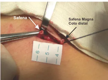
Figure 2 -Figure 2 -Figure 2 Figure 2
-Figure 2 - Section of the great saphenous vein between ligatures distal to the tributaries.
Figure 1 -Figure 1 -Figure 1 Figure 1
-Figure 1 - Doppler ultrasound image of the insufficient saphenofemoral junction.
Table 1 -Table 1 -Table 1 Table 1
-Table 1 - Reflux of the arch of saphenous vein. Conditional proportions (n = 57).
Type of Operation Type of OperationType of Operation Type of Operation
Type of Operation R e f l u xR e f l u xR e f l u xR e f l u xR e f l u x T o t a lT o t a lT o t a lT o t a lT o t a l P o s i t i v e
P o s i t i v eP o s i t i v e P o s i t i v e
P o s i t i v e N e g a t i v eN e g a t i v eN e g a t i v eN e g a t i v eN e g a t i v e
Ligation with section 0.29 0.71 1.00
Simple ligation 0.59 0.41 1.00
Reflux - reflux of the great saphenous vein at the arch Type of operation used in the arch of the saphenous
years), predominantly female (93%), probably due to the given inherent aesthetic social representation related to primary varicose veins, which is often the major motivation for treatment, a fact commonly seen in our country.
Among the limbs of the group C, 71% showed no SFJ reflux postoperatively, but in 29% (8/28) we observed a lesser rate of reflux than that found in the literature, of 37%25. Sheppard, in 1978, reported that there is suggestive
evidence that reflux of the saphenous arch could occur even after an effective treatment of saphenofemoral junction24.
This can be explained by neovascularization, cited by many authors as a predictor of high recurrence of reflux26,27. In
our study, neovascularization was observed in 87.5% (7/8) of patients in group C who had reflux. In one patient in group C (1/8), we found that one of the tributaries of the saphenous arch remained with reflux.
In relation to the group L, we observed almost the opposite of what happened with group C. In this sample, 59.6% (17/29) of patients had postoperative reflux on color Doppler ultrasonography, and in 41.4% (12/29) there was no reflux. In the former, there was 100% recanalization visualized by Doppler ultrasonography, suggesting that the flux somehow flowed by the ligated arch. With approximately 60% of segmental reflux in cases of ligation, our results do not differ from published works, which show high levels of reflux of the great saphenous arch after simple ligation10,26,28. The relative risk in the absence of reflux was
1.73 times higher in the group with associated section of the arch. Consequently, because of the difficulty encountered in surgical rapprochement of SFJ reflux, this evidence leads us to avoid techniques that predispose to this condition.
Among the criticisms about this study, we can point out the use of cotton sutures in the ligation of the saphenous vein and its tributaries. However, it should be noted that we used the same suture in the two surgical techniques, so the risk of failure of ligation would be the same. What differs between the two techniques is the permanence of the path of the great saphenous vein in group L, thereby possibly promoting recanalization and the subsequent permanence of SFJ reflux.
Regarding the reflux of the greater saphenous vein, classified by Engelhorn et al.4, we compared the
pre-with the postoperative periods, and observed the predominance of type II, with 68.4% (39/57), preoperatively. Postoperatively, we found patients who were
type II and became type IV, with 51.3% (20/39), being more frequent in the group with section of the arch 22.8% (13/57), higher than seen in the ligature 12.3% (7 /57). We noted that the saphenous vein reflux, when not maintained by the arch, which appeared insufficient postoperatively, was due the saphenous’s tributaries and perforating veins.
In all seven segments of the great saphenous, accompanied by color Doppler ultrasonography, we observed reduction in caliber, even among those who showed reflux postoperatively, showing a probable decrease of pressure in the saphenous venous flow through stopping reflux from the deep to the superficial venous systems (p <0.05).
The goal set was to compare the efficiency between the simple ligation and crossectomy. In this sense, a study by Hammarsten showed equivalence between the evidence of variceal recurrent between simple ligation and resection of the arch16.
In our research, we found postoperative reflux in 59% of patients with simple ligation, and 29% in those with ligation and section, an evidence suggestive that the preservation of the saphenous vein is feasible, especially when considered the crossectomy, which in our study was the technique that showed the best results.
In the context of research, when we compare these two techniques that preserve the saphenous vein, we find that the most conservative approach, simple ligation, got an unfavorable outcome of 59%, while the technique with associated section of the saphenous arch showed 71% success (p <0.05) in the treatment of reflux from the arch; most cases of reflux, 87.5% (7/8), had Doppler ultrasonography with signs of neovascularization, considered by Dwerryhouse and Jones as an important factor for saphenous reflux26,27.
Such evidence converge to the line therapy in which the approach to the reflux of the great saphenous arch is made with more efficient techniques, such as with its section, as well as applying a “barrier” between the stump of the saphenofemoral junction and the remnant saphenous vein, which will contribute to an increase in the rate of surgical success and reduce the recurrence of postoperative reflux.
R E S U M O R E S U M O R E S U M O R E S U M O R E S U M O
Objetivo: Objetivo: Objetivo: Objetivo:
Objetivo: avaliar a ocorrência do refluxo da safena magna através da ultrassonografia com Doppler colorido em sujeitos submeti-dos ao tratamento da insuficiência da junção safeno-femoral por ligadura simples ou por ligadura com secção da crossa. Métosubmeti-dosMétodosMétodosMétodosMétodos: foram realizadas 60 operações (45 sujeitos) de varizes com insuficiência da junção safeno-femoral (JSF), pertencentes à classificação clínica do CEAP 2 a 5, que foram distribuídos aleatoriamente em dois grupos. Um grupo denominado C, com ligadura e secção da crossa, e um grupo denominado L, com ligadura simples sem secção da crossa da veia safena magna. Foi então pesquisada a ocorrência do refluxo da safena magna nos grupos C e L através da ultrassonografia doppler colorida após o tratamento cirúrgico com intervalos de seis meses a um ano. Resultados:Resultados:Resultados:Resultados: dos 60 membros submetidos à abordagem da crossa da safena magna, 57Resultados: foram avaliados pela ultrassonografia doppler pós-operatório, pois dois sujeitos (três membros) não retornaram e foram excluídos do estudo. A média de idade foi 54 anos, 93% do sexo feminino e predominância da classificação (CEAP) C2 de 60,5%. Das 57 operações para o tratamento do refluxo da crossa da safena, 43,9% apresentaram refluxo no pós-operatório, sendo 14,1% do grupo C e 29,8% no grupo L (p<0,05). O risco relativo de apresentar refluxo da crossa da safena no grupo L foi 2,03 vezes maior em comparação com o grupo C. Conclusão:Conclusão:Conclusão:Conclusão: a secção da crossa da safena magna apresenta menos refluxo pós-operatório do que aConclusão: ligadura simples no tratamento da insuficiência da crossa da veia safena magna.
Descritores: Descritores: Descritores: Descritores:
Descritores: Varizes. Insuficiência venosa. Procedimentos cirúrgicos operatórios. Veia safena. Ultrassonografia Doppler em cores.
REFERENCES
REFERENCES
REFERENCES
REFERENCES
REFERENCES
1. Maffei FHA, Silveira PAM. Varizes dos membros inferiores: epidemiologia, patologia, etiopatogenia e fisiopatologia. In: Maffei FHA, Lastória S, Yoshida WB, Rollo HA, Giannini M, Moura R, editores. Doenças vasculares periféricas. Rio de Janeiro: Guanabara Koogan; 2008. p. 1713-28.
2. Cabral ALS. Insuficiência venosa crônica de membros inferiores; prevalência, sintomas e marcadores preditivos [tese]. São Paulo: Universidade Federal de São Paulo, Escola Paulista de Medicina; 2000.
3. Medeiros JJ, Mansilha A. Estratégia terapêutica na doença veno-sa crônica. Angiol Cir Vasc. 2012;8(3):110-26.
4. Engelhorn CA, Engelhorn AL, Cassou MF, Zanoni CC, Gosalan CJ, Ribas E. Classificação anátomo-funcional da insuficiência das veias safenas baseado no esoDoppler colorido, dirigida para o planeja-mento da cirurgia de varizes. J Vasc Br. 2004;3(1):13-9. 5. Rollo HA, Lastória S, Yoshida WB, Moura R, Maffei FHA. Cirurgia
de varizes com preservação da veia safena magna: avaliação pelo mapeamento duplex, resultados preliminares. Cir Vasc Angiol. 1996;12(4 supl):63-8.
6. Sarquis AL. Avaliação pré e pós-operatória no tratamento cirúrgi-co cirúrgi-conservador de varizes tronculares cirúrgi-com o duplex scan a cirúrgi-cores. Cir Vasc Angiol. 1996;12(4 supl):9-11.
7. Lastória S, Rollo HA. Tratamento de varizes de membros inferio-res. In: Maffei FHA, Lastória S, Yoshida WB, Rollo HA, Giannini M, Moura R, editores. Doenças vasculares periféricas. Rio de Janeiro: Guanabara Koogan; 2008. p.1739-50.
8. Pitta GBB, Teixeira LR. Ultra-som na recidiva de varizes In: Nectoux JLF, Cunha SS, Paglioli SA, Souza GG, Pereira AH, editores. Ultra-sonografia vascular. Rio de Janeiro: Revinter; 2000. p.201-7. 9. Thibault PK, Lewis WA. Recurrent varicose veins. Part 1: Evaluation
utilizing duplex venous imaging. J Dermatol Surg Oncol. 1992;18(7):618-24.
10. Maesener MG. Strategies to minimize the effect of neovascularization at the saphenofemoral junction after great saphenous vein surgery: an overview. Phlebolymphology. 2006;13(4):207-13.
11. Labropoulos N, Touloupakis E, Giannoukas AD, Leon M, Katsamouris A, Nicolaides AN. Recurrent varicose veins: investigation of the pattern and extent of reflux with color flow duplex scanning. Surgery. 1996;119(4):406-9.
12. Turton EP, Scott DJ, Richards SP, Weston MJ, Berridge DC, Kent PJ, et al. Duplex-derived evidence of reflux after varicose vein surgery: neoreflux or neovascularisation? Eur J Vasc Endovasc Surg. 1999;17(3):230-3.
13. Bradbury AW, Stonebridge PA, Ruckley CV, Beggs I. Recurrent varicose veins: correlation between preoperative clinical and hand-held Doppler ultrasonographic examination, and anatomical findings at surgery. Br J Surg. 1993;80(7):849-51.
14. Munn SR, Morton JB, Macbeth WA, Mcleish AR. To strip or not to strip the long saphenous vein? A varicose veins trial. Br J Surg. 1981;68(6):426-8.
15. Large J. Surgical treatment of saphenous varices, with preservation of the main great saphenous trunk. J Vasc Surg. 1985;2(6):886-91.
16. Hammarsten J, Pederson P, Cederlund CG, Campanello M. Long saphenous vein saving surgery for varicose veins. A long-term follow-up. Eur J Vasc Surg. 1990;4(4);361-4.
17. Fonseca FP, Sarquis AL, Evangelista SSM; Union Internationale de Phlebologie. Surgery for primary troncular varicose without stripping the saphenous vein – pre- and post-operative evaluation by duplex scan and photoplethysmography. In: Phlebology ’95: Proceedings of the XII Congress Union Internationale de Phlebologie. September 3-8, 1995. London; 1995. p.419-21.
18. Corcos L, De Anna D, Zamboni P, Gasbarro V, Bresadola V, Procacci T, et al. Reparative surgery of valves in the treatment of superfi-cial venous insufficiency. External banding valvuloplasty versus high ligation or disconnection. A prospective multicentric trial. J Mal Vasc. 1997;22(2):128-36.
19. Gibbs PJ, Foy DM, Darke SG. Reoperation for recurrent saphenofemoral incompetente: a prospective randomise trial using a reflected flan of pectineus fascia. Eur J Vasc Endovasc Surg. 1999;18(6):494-8.
20. Campbell WB, Vijay Kumar A, Collin TW, Allington KL, Michaels JA; Randomised and Economic Analysis of Conservative and Therapeutic Interventions for Varicose veins Study. The outcome of varicose vein surgery at 10 years: clinical findings, symptoms and patient satisfaction. Ann R Coll Surg Engl. 2003;85(1):52-7. 21. Stonebridge PA, Chalmers N, Beggs I, Bradbury AW, Ruckley CV.
Recurrent varicose veins: a varicographic analysis leading to a new practical classification. Br J Surg. 1995;82(1):60-2.
22. Canonico S, Campitiello F, Lauletta V, Pacifico F, Sciaudone G. Diagnostic and surgical approaches to recurrent varicose veins of lower limbs. Panminerva Med. 1997;39(4):287-90.
23. Eklöf B, Rutherford RB, Bergan JJ, Carpentier PH, Gloviczki P, Kistner RL, et al. Revision of the CEAP classification for chronic venous disorders: consensus statement. J Vasc Surg. 2004;40(6):1248-52.
25. Pitta GBB. Preservação da veia safena magna na cirurgia das varizes tronculares primárias [tese]. São Paulo: Universidade Fe-deral de São Paulo, Escola Paulista de Medicina; 1998.
26. Dwerryhouse S, Davies B, Harradine K, Earnshaw JJ. Stripping the long saphenous vein reduces the rate of reoperation for recurrent varicose veins: five-years results of a randomized trial. J Vasc Surg. 1999;29(4):589-92.
27. Jones L, Braithwaite BD, Selwyn D, Cooke S, Earnshaw JJ. Neovascularisation is the principal cause of varicose vein recurrence: results of a randomised trial of stripping the long saphenous vein. Eur J Vasc Endovasc Surg. 1996;12(4):442-5.
28. Sarin S, Scurr JH, Coleridge Smith PD. Stripping of the long saphenous vein in the treatment of primary varicose veins. Br J Surg. 1994;81(10):1455-8.
Received on 20/08/2012
Accepted for publication 23/10/2012 Conflict of interest: none
Source of funding: none
How to cite this article: How to cite this article: How to cite this article: How to cite this article: How to cite this article:
Gurgel GA, Castro AA, Araújo M, Amorim JE, Pitta GBB, Miranda Júnior F. Avaliação da ocorrência do refluxo venoso da safena magna pela ultrassonografia com Doppler colorido após cirurgia da insuficiên-cia da junção safeno-femoral. Rev Col Bras Cir. [periódico na Internet] 2013;40(5). Disponível em URL: http://www.scielo.br/rcbc