arteries in the second and third trimesters for
the prediction of gestational outcome
Doplervelocimetria da artéria uterina no segundo e terceiro
trimestres para predição dos resultados gestacionais
haMid reza haghighatkhah2
MohaMMad fakhri3
nina MaSooM4
Abstract
PURPOSE: The aim of this longitudinal study was to investigate the value of uterine artery Doppler sonography during the second and third trimesters in the prediction of adverse pregnancy outcome in low-risk women. METHODS: From July 2011 to August 2012, a total of 205 singleton pregnant women presenting at our antenatal clinic were enrolled in this prospective study and were assessed for baseline demographic and obstetric data. They underwent ultrasound evaluation at the time of second and third trimesters, both included Doppler assessment of bilateral uterine arteries to determine the values of the pulsatility index (PI) and resistance index (RI) and presence of early diastolic notch. The endpoint of this study was assessing the sensitivity, speciicity, positive predictive value (PPV) and negative predictive value (NPV) of Doppler ultrasonography of the uterine artery, for the prediction of adverse pregnancy outcomes including preeclampsia, stillbirth, placental abruption and preterm labor. RESULTS: The mean age of cases was 26.4±5.11. The uterine artery PI and RI values for both second (PI: 1.1±0.42 versus 1.53±0.59, p=0.002; RI: 0.55±0.09 versus 0.72±0.13, p=0.000 respectively) and third-trimester (PI: 0.77±0.31 versus 1.09±0.46, p=0.000; RI: 0.46±0.10 versus 0.60±0.14, p=0.010 respectively) evaluations were signiicantly higher in patients with adverse pregnancy outcome than in normal women. Combination of PI and RI >95th percentile and presence of
bilateral notch in second trimester get sensitivity and speciicity of 36.1 and 97% respectively, while these measures were 57.5 and 98.2% in third trimester. CONCLUSIONS: According to our study, it seems that uterine artery Doppler may be a valuable tool for the prediction of a variety of adverse outcomes in second and third trimesters.
Resumo
OBJETIVO: O objetivo do presente estudo longitudinal foi avaliar o valor da ultrassonograia Doppler das artérias uterinas no segundo e terceiro trimestres de gestação para a predição de desfecho adverso da gravidez em mulheres de baixo risco. MÉTODOS: De julho de 2011 até agosto de 2012, 205 gestantes de feto único atendidas em nossa clínica de pré-natal foram incluídas no presente estudo prospectivo e avaliadas em termos de dados demográicos e obstétricos. As pacientes foram submetidas à avaliação de ultrassom durante o segundo e terceiro trimestres, incluindo avaliação Doppler das artérias uterinas bilaterais, visando determinar os valores do índice de pulsatilidade (IP) e do índice de resistência (IR), bem como a presença de incisura diastólica precoce. O desfecho do presente estudo foi a avaliação da sensibilidade, especiicidade, valor preditivo positivo (VPP) e valor negativo preditivo (VNP) da ultrassonograia Doppler das artérias uterinas para a predição de desfechos adversos da gravidez, incluindo pré-eclâmpsia, natimortalidade, descolamento prematuro da placenta e trabalho de parto prematuro. RESULTADOS: A média de idade das gestantes foi de 26,4±5,11 anos. Os valores de IP e IR das artérias uterinas para o primeiro (IP: 1,1±0,42 versus 1,53±0,59, p=0,002; IR: 0,55±0,09 versus 0.72±0.13, p=0,000, respectivamente) e para o terceiro trimestre (IP: 0,77±0,31
versus 1,09±0,46, p=0,000; IR: 0,46±0,10 versus 0,60±0,14, p=0,010, respectivamente) foram signiicativamente maiores em pacientes com desfecho adverso da gravidez em relação às mulheres com desfecho normal. A combinação de IP e IR > percentil 95 e a presença de incisura bilateral apresentou sensibilidade e especiicidade de 36,1 e 97%, respectivamente, no segundo trimestre e de 57,5 e 98,2% no terceiro trimestre. CONCLUSÕES: Com base no presente estudo, o Doppler das artérias uterinas parece ser ferramenta valiosa para a predição de uma variedade de desfechos adversos no segundo e terceiro trimestres de gestação.
Study carried out at Shahid Beheshti University of Medical Sciences – Theran, Iran.
1Department of Obstetrics and Gynecology; Tajrish Hospital; Shahid Beheshti University of Medical Sciences – Tehran, Iran. 2Department of Radiology; Tajrish Hospital; Shahid Beheshti University of Medical Sciences – Tehran, Iran.
3School of Medicine; Shahid Beheshti University of Medical Sciences – Tehran, Iran. 4School of Medicine; Tehran University of Medical Sciences – Tehran, Iran.
Conlict of interests: none.
Keywords
Ultrasonography, Doppler Laser-doppler lowmetry Uterine artery/ultrasonography Pregnancy trimester, second Pregnancy trimester, third Pregnancy outcome
Palavras-chave
Ultrassonograia Doppler Fluxometria por laser-doppler Artéria uterina/ultrassonograia
Segundo trimestre da gravidez Terceiro trimestre da gravidez Resultado da gravidez
Correspondence Aida Moeini Department of Obstetrics and Gynecology, Tajrish Hospital Tajrish Square, Tehran, Iran
Received 11/11/2013
Introduction
Preeclampsia, Intrauterine Growth Restriction (IUGR), abruption and stillbirth and other related complications are the consequence of defect in normal placentation during pregnancy1. In fact impaired placentation results in in-creased impedance to low in uteroplacental circulation leading to hypoxemia and necrosis2. These complications are major causes of maternal and perinatal mortality3. The epidemiologic study of adverse pregnancy outcomes shows a higher rate of these complications in developing countries than in developed ones4. Predicting the risk of these outcomes helps obstetricians to consider appropriate antenatal surveillance and therapeutic intervention5.
In normal pregnancy the resistance in the uterine artery low decreases with advancing gestational age. Failure to get a low resistant circulation is associated with a subsequent risk of pregnancy adverse outcome1. Uteroplacental perfusion adequacy can be examined in-directly by uterine artery Doppler examination6. The fact that uterine artery Doppler velocimetry is a non-invasive technique which can be easily added to the current routine ultrasound examination without signiicant extra costs, plus its ability to determine a group of at-risk patients who would beneit from increased care, makes it an eligible candidate for a potential screening tool in predicting adverse pregnancy outcomes7-9. Previous studies have shown that uterine artery Doppler could improve screening eficacy for the prediction of adverse pregnancy outcomes in the irst10,11 and second12,13 trimester of pregnancy with dif-ferent sensitivity and speciicity. Recently, some studies have continued uterine artery indices measurement in the third trimester of pregnancy1,14. However, many studies were retrospective or cross-sectional in design and could not truly show the relationship between uterine artery indices and the risk of adverse outcome2,15,16. It seems reasonable that serial evaluation of uterine artery Doppler velocimetry in second and third trimesters could help to determine normal development of fetal growth and deserve as a good predictor of pregnancy complications. The aim of this prospective longitudinal study was to investigate the sensitivity, speciicity, positive and negative predictive value of uterine artery Doppler ul-trasonography indices including Resistance Index (RI) and Pulsatility Index (PI) in second and third trimesters for prediction of pregnancy complications.
Methods
Study population
Between July 2011 and August 2012, to a total of 250 singleton pregnant women attending our perinatal clinic for routine antenatal care were offered to participate in
the project. Inclusion criteria were singleton pregnancies with normal fetuses, not taking aspirin, heparin, metfor-min or antihypertensive drugs. Cases with concomitant maternal diseases (e.g. morbid obesity, chronic hyperten-sion, renovascular or connective tissue diseases) and those pregnancies that resulted in fetuses with structural or chromosomal abnormalities or Rh immunization were excluded from the study. Of 250 recruited participants who fulilled the entry criteria, 205 women were able to complete the study and their data were included in the inal analyses. Forty-ive patients were excluded for different reasons, including lack of satisfaction for incorporation in the study and lack of complete records. The entire study protocol has been reviewed and approved by the Ethics Committee of the Shahid Beheshti University of Medical Sciences. Informed written consent was obtained from all study participants.
All recruited women were assessed at the 1st trimester screening for baseline demographic and obstetric data including age, parity, Body Mass Index (BMI) and past medical events. Smoking, alcohol and drug use were also determined.
Study design
This was a single center observational prospective study conducted in the gynecology department of Tajrish Hospital in Tehran, Iran. Sample size was determined after consideration of type 1 statistical error <5% and type 2 statistical error <20%.
Sonographic assessment
occasions that each uterine artery was sampled. The mean PI and RI of the two uterine arteries were calculated manually by arithmetic mean between the PI and RI values of the left and right arteries respectively.
Study outcomes
All pregnancy outcomes were obtained from the deliv-ery suite database. Fetal and neonatal status and morbidity including baby Apgar scores, fetal distress or fetal death and admission to the neonatal intensive care unit were de-termined. All the examinations and data recording were performed by two senior resident physicians. The primary endpoint of this study is assessing the sensitivity and speciic-ity of Doppler ultrasonography of the uterine artery, for the prediction of adverse pregnancy outcomes. Adverse outcomes included developments of preeclampsia (deined according to the guidelines of the International Society for the Study of Hypertension in Pregnancy17; recording of two diastolic blood pressure greater than 90 mmHg at least 4 hours apart in previously normotensive women, and proteinuria with minimum of 300 mg in 24 hours, or 2 readings of at least 2+ protein on dipstick analysis of urine), and birth weight less than the tenth percentile for gestation requiring delivery before 34 weeks. Other adverse outcomes were stillbirth, placental abruption (deined as presence of retro placental clot at delivery or vaginal bleeding leading to emergency delivery), preterm labor (labor 20–37 weeks of gestation), Apgar scores at one and ive minutes less than 7, and ges-tational hypertension (blood pressure of 140/90 mmHg persistent after delivery).
Statistical analysis
The sensitivity, speciicity, Positive Predictive Value (PPV) and Negative Predictive Values (NPV) of uterine artery RI and PI in the prediction of adverse pregnancy outcomes were calculated by using the IBM SPSS version 18® software package (SPSS, Chicago, IL, USA). Continuous variables were presented as mean ± standard deviation (SD); categorical data were expressed as numbers and percent-ages. Student t-test, Mann-Whitney U-test, and χ2 test or Fisher’s exact test were used as appropriate.
Results
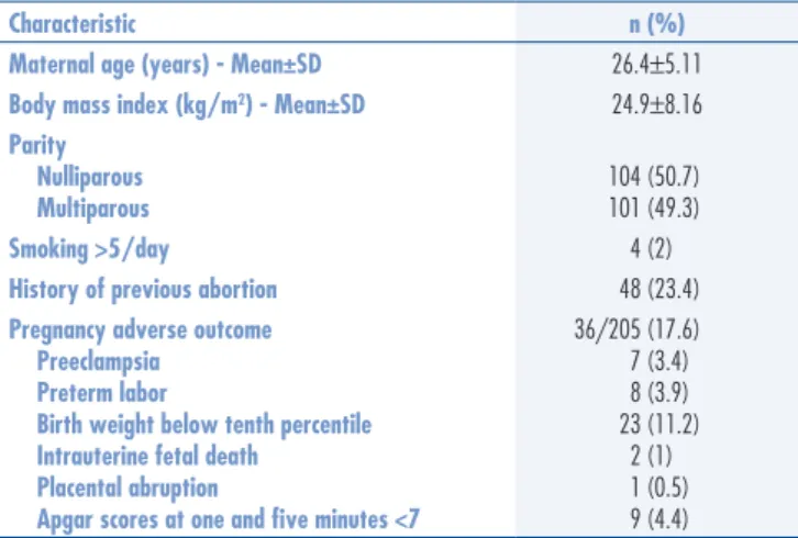
Two hundred and ive pregnant women were able to complete the study and their data were included in the inal analyses. The mean age of the entire group was 26.4±5.11 (range 16 to 42 years). At the time of birth, the mean±SD of gestational age and birth weight were 268.2±22.3 days and 3051.5±657.0 g, respectively. The complete demographic characteristic and outcome data are shown in Table 1.
Mean PI and RI in our population was 1.2 and 0.6 respectively at second trimester and it was 0.85 and 0.5 at third trimester. The values of uterine artery PI and RI for both second and third trimesters evaluations were signiicantly higher in patients who developed pregnancy complications than in normal women (Table 2). Also, the prevalence of bilateral notches in patients with sub-sequent complications is signiicantly higher than those with normal pregnancies (55.5 versus 36.7% for second trimester and 35 versus 15% for third trimester evaluation). Mean PI>95th percentile for gestational age at 15–20 weeks’ gestation and at 30–34 weeks’ gestation were present in 43 (21%) and 19 (9.5%) patients, respectively. The uterine artery 90th, 95th and 97.5th RI percentiles were calculated to be 0.76, 0.80 and 0.82 at second trimester and (0.66, 0.7 and 0.72) at third trimester, respectively.
Sensitivity, speciicity, PPV and NPV of uterine artery Doppler ultrasonography parameters for predict-ing the pregnancies adverse outcomes are summarized in
Table 1. Demographic characteristics and outcome data of the study population
Characteristic n (%)
Maternal age (years) - Mean±SD 26.4±5.11
Body mass index (kg/m2) - Mean±SD 24.9±8.16
Parity Nulliparous Multiparous
104 (50.7) 101 (49.3)
Smoking >5/day 4 (2)
History of previous abortion 48 (23.4)
Pregnancy adverse outcome Preeclampsia
Preterm labor
Birth weight below tenth percentile Intrauterine fetal death
Placental abruption
Apgar scores at one and ive minutes <7
36/205 (17.6) 7 (3.4) 8 (3.9) 23 (11.2)
2 (1) 1 (0.5) 9 (4.4)
Table 2. Demographic characteristics and description of Doppler ultrasound indings in
normal pregnancies versus pregnancies with adverse outcome
Parameter
Normal pregnancies
Mean±SD
Complicated pregnancies
Mean±SD
p-value
n 169 36
Demographic data Maternal age (years) Body mass index (kg/m2)
Parity (%) Nulliparous - n (%) Multiparous - n (%)
25.5±3.1 24.1±3.2
85 (50.3) 84 (49.7)
26.9±8.5 25.2±8.8
19 (53) 17 (47)
Second trimester PI
RI
Bilateral notches (%)
1.1±0.42 0.55±0.09 36.7
1.53±0.59 0.72±0.13 55.5
0.002* 0.000* 0.041**
Third trimester PI RI
Bilateral notches (%)
0.77±0.31 0.46±0.10
15
1.09±0.46 0.60±0.14
35
0.000* 0.010* 0.011**
Table 3. The sensitivity for predicting adverse outcomes for mean RI>95th percentile in third trimester was 58%. It increased to 77.2% at second trimester. The sensitivity of RI to predict complicated pregnancy in both trimes-ters was higher than sensitivity of PI and combined RI, PI and bilateral notch. On the other hand, the value of speciicity to detect adverse outcome was higher when RI, PI and bilateral notch values were combined.
Discussion
This study evaluated clinical usefulness of uterine artery color Doppler ultrasound as a predictor of adverse outcomes in pregnancy. In line with previous indings1,16, our results also showed that uterine artery PI and RI values were signiicantly higher in patients who eventually developed pregnancy complications than in women with normal outcome. This inding was encountered for both second and third trimesters. The sensitivity of mean RI>95th percentile in second trimester was higher than third tri-mester (77.2 versus 58.0%), which suggests the valuable role of second trimester’s uterine Doppler sonography in screening complicated pregnancy. After the combination of uterine indices, the value of sensitivity did not differ from each index alone in third trimester (57.5 versus 58.0%), but decreased in second trimester (36 versus 77%). With regards to speciicity, although each parameter (PI, RI or presence of bilateral notches) may provide a good diagnostic yield for the adverse pregnancy outcome, combination of all parameters together may result in a great speciicity. As it is evident in Table 3, a mean PI and mean RI>95th percentile along with presence of bilateral notches may produce a positive predictive value of 88%. This positive predictive value is acceptable since our subjects were not selected from high-risk population.
We realize that the study could have some weak-nesses. The main bias is that the study has low number of patients. To decrease the sonographic measurement bias, all women were assessed by one experienced radiologist. Participants in this study were not conined to high-risk population, which could enhance the external validity of our research; however, the results of the study could not be generalized to all population.
Screening abilities of color Doppler ultrasound in assessing single certain outcomes such as stillbirth18, pre-eclampsia and fetal growth restriction19 were extensively addressed. In a study by Singh and colleagues, which showed elevated second-trimester Doppler indices, a proxy for impaired placentation, they are more strongly associated with stillbirth than conventional risk fac-tors18. This is while in that study; stillbirth was the only primary outcome. In another study by Coleman et al.20 adverse outcomes were deined as development of pre-eclampsia and Small for Gestational Age (SGA). In that study, just high-risk women were included and it has been shown that uterine artery Doppler waveform analysis performed best in the prediction of severe adverse outcome and was better than clinical risk assessment in the pre-diction of preeclampsia and SGA babies. The deinition of adverse pregnancy outcome in the current study was similar to Jamal et al., including preeclampsia, IUGR, preterm delivery, placental abruption and fetal death1. The combination of several different adverse outcomes is more clinically relevant and may help obstetrician for a better clinical judgment. Jamal and colleagues reported a sensitivity of 60% for the mean PI>95th percentile for gestational age and/or bilateral notches in predicting a severe adverse outcome1. In another study by Harrington et al. the sensitivity for an adverse outcome in screen-positive women of low-risk group was 33.3% for a speciicity of 92.8% and a positive predictive value of 24.4%16.
In the present study, the presence of bilateral uterine artery notches in complicated and normal pregnancies was compared. The prevalence of bilateral notches in patients with subsequent complications was signiicantly higher than those with normal pregnancies. Jamal et al. showed a prevalence of bilateral notch of 20% in com-plicated pregnancies compared to 4.5% in normal ones1. This was in agreement with ours. Evaluation of bilateral notch presence is usually performed along with the RI and PI, but there exist studies that solely included this item to predict adverse pregnancy outcome. Mires et al.21 have shown that the presence of bilateral uterine arterial notching is associated with a signiicantly increased risk of adverse pregnancy outcome. In concordance with another study that showed a strong positive predictive value in high-risk populations22, in recent research, combination
Table 3. Screening characteristics of uterine artery resistance indices during second and third trimesters for adverse pregnancy outcome
Characteristic >95Mean RIth percentile
Mean PI >95th percentile
PI and mean RI >95th percentile
and bilateral notch Second trimester
Sensitivity (%) 77.2 48.8 36.1
Speciicity (%) 89.6 90.7 97
PPV (%) 47.2 58 72.2
NPV (%) 97 86.9 87.7
Third trimester
Sensitivity (%) 58 63.1 57.5
Speciicity (%) 95.8 89.5 98.2
PPV (%) 53.3 38.7 75
NPV (%) 87.6 95.8 87.3
of bilateral notch to other indices resulted in a good posi-tive predicposi-tive value in normal population.
The beneit of detecting such screen positive pregnancy is that they will form an ideal group for prophylactic trials such as low-dose aspirin. Also, it helps health care resources to target women with abnormal Doppler results for a better antenatal surveillance16,23.
The results of this study suggest that uterine artery Doppler may be a valuable screening tool for the pre-diction of a variety of adverse outcomes in second and third trimesters. Further studies with larger sample size will be necessary to determine whether this information could be incorporated into the routine antenatal care of pregnant women.
1. Jamal A, Abbasalizadeh F, Vafaei H, Marsoosi V, Eslamian L. Multicenter screening for adverse pregnancy outcomes by uterine artery Doppler in the second and third trimester of pregnancy. Med Ultrason. 2013;15(2):95-100.
2. Ghi T, Contro E, Youssef A, Giorgetta F, Farina A, Pilu G, et al. Persistence of increased uterine artery resistance in the third trimester and pregnancy outcome. Ultrasound Obstet Gynecol. 2010;36(5):577-81.
3. Shwarzman P, Waintraub AY, Frieger M, Bashiri A, Mazor M, Hershkovitz R. Third-trimester abnormal uterine artery Doppler indings are associated with adverse pregnancy outcomes. J Ultrasound Med. 2013;32(12):2107-13.
4. Kramer MS. The epidemiology of adverse pregnancy outcomes: an overview. J Nutr. 2003;133(5 Suppl 2):1592S-6S.
5. Afrakhteh M, Moeini A, Taheri MS, Haghighatkhah HR. Correlation between placental thickness in the second and third trimester and fetal weight. Rev Bras Ginecol Obstet. 2013;35(7):317-22. 6. Campbell S, Black RS, Lees CC, Armstrong V, Peacock JL. Doppler
ultrasound of the maternal uterine arteries: disappearance of abnormal waveforms and relation to birth weight and pregnancy outcome. Acta Obstet Gynecol Scand. 2000;79(8):631-4. 7. Papageorghiou AT, Yu CK, Cicero S, Bower S, Nicolaides KH.
Second-trimester uterine artery Doppler screening in unselected populations: a review. J Matern Fetal Neonatal Med. 2002;12(2):78-88. 8. Papageorghiou AT, Yu CK, Erasmus IE, Cuckle HS, Nicolaides
KH. Assessment of risk for the development of preeclampsia by maternal characteristics and uterine artery Doppler. BJOG. 2005; 112(6):703-9.
9. Yu CK, Smith GC, Papageorghiou AT, Cacho AM, Nicolaides KH; Fetal Medicine Foundation Second Trimester Screening Group. An integrated model for the prediction of preeclampsia using maternal factors and uterine artery Doppler velocimetry in unselected low-risk women. Am J Obstet Gynecol. 2005;193(2):429-36.
10. Gómez O, Martínez JM, Figueras F, Del Río M, Borobio V, Puerto B, et al. Uterine artery Doppler at 11–14 weeks of gestation to screen for hypertensive disorders and associated complications in an unselected population. Ultrasound Obstet Gynecol. 2005;26(5):490-4. 11. Campbell S. First-trimester screening for preeclampsia. Ultrasound
Obstet Gynecol. 2005;26(5):487-9.
12. Albaiges G, Missfelder-Lobos H, Lees C, Parra M, Nicolaides KH. One-stage screening for pregnancy complications by color Doppler assessment of the uterine arteries at 23 weeks’ gestation. Obstet Gynecol. 2000;96(4):559-64.
13. North RA, Ferrier C, Long D, Townend K, Kincaid-Smith P. Uterine artery Doppler low velocity waveforms in the second trimester for the prediction of preeclampsia and fetal growth retardation. Obstet Gynecol.1994;83(3):378-86.
14. Pariente G, Shwarzman P, Aricha-Tamir B, Weintraub AY, Hershkovitz R. Association between irst trimester vaginal bleeding and uterine artery Doppler measured at second and third trimesters of pregnancy. J Matern Fetal Neonatal Med. 2013;26(17):1724-7.
15. Severi FM, Bocchi C, Visentin A, Falco P, Cobellis L, Florio P, et al. Uterine and fetal cerebral Doppler predict the outcome of third-trimester small-for-gestational age fetuses with normal umbilical artery Doppler. Ultrasound Obstet Gynecol. 2002;19(3):225-8.
16. Harrington K, Fayyad A, Thakur V, Aquilina J. The value of uterine artery Doppler in the prediction of uteroplacental complications in multiparous women. Ultrasound Obstet Gynecol. 2004;23(1):50-5. 17. Brown MA, Lindheimer MD, de Swiet M, Van Assche A, Moutquin
JM. The classiication and diagnosis of the hypertensive disorders of pregnancy: statement from the International Society for the Study of Hypertension in Pregnancy (ISSHP). Hypertens Pregnancy. 2001; 20(1):IX-XIV.
18. Singh T, Leslie K, Bhide A, D’Antonio F, Thilaganathan B. Role of second-trimester uterine artery Doppler in assessing stillbirth risk. Obstet Gynecol. 2012;119(2 Pt 1):256-61.
19. Martin AM, Bindra R, Curcio P, Cicero S, Nicolaides KH. Screening for pre‐eclampsia and fetal growth restriction by uterine artery Doppler at 11-14 weeks of gestation. Ultrasound Obstet Gynecol. 2001;18(6): 583-6.
20. Coleman MA, McCowan LM, North RA. Mid‐trimester uterine artery Doppler screening as a predictor of adverse pregnancy outcome in high‐risk women. Ultrasound Obstet Gynecol. 2000; 15(1):7-12.
21. Mires GJ, Williams FL, Leslie J, Howie PW. Assessment of uterine arterial notching as a screening test for adverse pregnancy outcome. Am J Obstet Gynecol. 1998;179(5):1317-23.
22. Caruso A, Caforio L, Testa AC, Ferrazzani S, Mastromarino C, Mancuso S. Chronic hypertension in pregnancy: color Doppler investigation of uterine arteries as a predictive test for superimposed preeclampsia and adverse perinatal outcome. J Perinat Med. 1996;24(2):141-53.
23. Aalami-Harandi R, Karamali M, Moeini A. Induction of labor with titrated oral misoprostol solution versus oxytocin in term pregnancy: randomized controlled trial. Rev Bras Ginecol Obstet. 2013;35(2):60-5.