Association between segmental aplasia of great saphenous vein
and varicose veins of lower limbs, evaluated using color
Doppler ultrasonography
Associação entre aplasia segmentar de veia safena magna e varizes em membros
inferiores avaliada pelo ecocolor Doppler
Amélia Cristina Seidel1
*
, Pedro Cavalari Júnior1, Robson Marcelo Rossi1, Fausto Miranda Júnior2
Abstract
Background: here are individual diferences in the diameter of the great saphenous vein (GSV) in both normal and non-functional limbs and it is possible to identify these diferences using color Doppler ultrasonography. Objectives: To assess the association between segmental GSV aplasia and the presence of varicose veins and/or GSV insuiciency in lower limbs using color Doppler ultrasonography, in patients with chronic venous disease (CVD). Methods: A total of 1,408 patients with complaints compatible with CVD of lower limbs were examined using color Doppler ultrasonography. he age range of the sample was from 17 to 85 and 1,286 of the patients were female. People with clinical classiications (CEAP) ranging from C0 to C4 were included. On the basis of clinical examination, the sample was subdivided as follows: group A patients had varicose veins and group B patients were free from varicose veins. Color Doppler ultrasonography was used to determine whether there was GSV aplasia, by analysis of its route into the saphenous compartment, and the presence of varicose veins in diferent sites. Statistical analysis was conducted using the chi-square test or Fisher’s exact tests followed by an analysis of residuals in tables, with a 5% signiicance level.
Results: In group A, there were 479 (83.9%) patients with GSV insuiciency, 169 (38.2%) with aplasia and 71 (80.7%) with both insuiciency and aplasia. In group B, there were 92 (16.1%) patients with GSV insuiciency, 273 (61.8%) with aplasia and 17 (19.3%) with both insuiciency and aplasia. Conclusion: Segmental GSV aplasia was more common in lower limbs with no varicose veins and/or insuiciency, but there was a higher incidence of patients with both aplasia and insuiciency in the group with varicose veins.
Keywords: venous relux; saphenous vein (abnormalities); color Doppler ultrasonography; venous insuiciency; anatomy.
Resumo
Contexto: Há diferenças individuais no diâmetro da veia safena magna (VSM) em membros normais e doentes; sendo possível a identiicação dessas alterações pelo ecocolor Doppler. Objetivo: Avaliar a associação da aplasia segmentar da VSM com a presença de varizes e/ou insuiciência da mesma em membros inferiores, usando o ecocolor Doppler em pacientes com clínica de doença venosa crônica (DVC). Métodos: 1.408 pacientes com queixas compatíveis de DVC de membros inferiores, sendo 1.286 do sexo feminino, com idade entre 17 e 85 anos, examinados com ecocolor Doppler. Foram incluídos aqueles com classiicação CEAP clínica C0 a C4. Pela avaliação clínica, a amostra foi distribuída em grupo A, pacientes com varizes, e grupo B, aqueles sem varizes. O ecocolor Doppler determinou se havia aplasia da VSM pela análise do seu trajeto no compartimento safeno e presença de veias varicosas nos diferentes sítios. Para estatística, foram considerados os testes Qui-quadrado ou Exato de Fisher e uma análise de resíduos em tabelas, com nível de signiicância de 5%. Resultados: No grupo A houve 479 (83,9%) de VSM insuicientes, 169 (38,2%) com aplasia e 71 (80,7%) com insuiciência e aplasia associadas. No grupo B, houve 92 (16,1%) de VSM insuicientes, 273 (61,8%) com aplasia e 17 (19,3%) com insuiciência e aplasia associadas. Conclusão: A aplasia segmentar da VSM ocorre mais em membros inferiores que não apresentam varizes e/ou insuiciência da mesma, mas considerando-se a presença da associação de aplasia e insuiciência, houve maior incidência no grupo de membros que apresentavam varizes.
Palavras-chave: reluxo venoso; veia safena (anormalidades); ultrassonograia; Doppler em cores; insuiciência venosa; anatomia.
1Universidade Estadual de Maringá - UEM, Maringá, PR, Brazil. 2Universidade Federal de São Paulo - UNIFESP, São Paulo, SP, Brazil.
Financial support: None.
Conlicts of interest: No conlicts of interest declared concerning the publication of this article. Submitted: January 29, 2015. Accepted: May 05, 2015.
INTRODUCTION
A good understanding of the venous anatomy is very important to enable vascular ultrasonographers to identify diagnoses and contributes to indicating treatment for diseases of the venous system.1
The great saphenous vein (GSV) is easy to identify in the thigh because it is located within the compartment known as the “Egyptian eye” or “saphenous eye” which is delimited by the muscular fascia and the saphenous fascia and because of this any vein that is outside of this compartment is considered a tributary or accessory.1-5
There are differences between individuals in terms of the diameter of the GSV in normal limbs that are dependent on several different factors (age, exercise of muscles, thickness of subcutaneous cellular tissues) and diameters in diseased limbs also very, as has been demonstrated in microscopy studies.2,4,6 A smaller
than normal GSV diameter and a GSV that cannot be
seen within the saphenous compartment are deined,
respectively, as hypoplasia and aplasia and these
conditions generally affect speciic segments of the
vein. In such cases there is usually a vein outside of the compartment that courses in parallel and where the hypoplasia or aplasia returns to the compartment, connecting the segments of the saphenous vein.7
Although aplasia or hypoplasia of the GSV is very common, there are few studies dealing with this anatomic abnormality.4,7
This article deals with the term aplasia as used to describe a segment of the GSV that cannot be seen within the saphenous compartment using color Doppler ultrasonography.
The objective of the present study was to evaluate the association of aplasia of the GSV with varicose
veins and/or GSV insuficiency in the lower limbs
using color Doppler ultrasonography in a sample of patients with venous disease diagnosed clinically.
METHOD
This was a prospective, cross-sectional study of a sample of 1,408 consecutive patients with complaints compatible with venous disease of the lower limbs. The age range of the sample was from 17 to 85 years, 1,286 of the patients were female and 122 were male. They were examined using color Doppler ultrasonography over a 6-month period. Data collected when taking patient histories and during physical examinations were recorded on a specially designed form.
This sample does not represent the general population, but a group of patients with symptomatic chronic venous disease (CVD). Patients were excluded if
they had undergone a previous operation for varicose veins, had a history of deep venous thrombosis, were
expectant mothers or if they had been classiied as
CEAP classes 5 or 6, the majority of whom have a prior history of varicose vein operations or deep venous thrombosis. The sample therefore comprised
patients with CEAP clinical classiications C0 to C4.
On the basis of clinical assessment, the sample was subdivided into two groups, group A, comprising patients with varicose veins (C2 to C4a,b), and group B, comprising patients free from varicose veins (C0-C1). Color Doppler ultrasonography was conducted as recommended in the literature,8 with patients in an
orthostatic position (standing upright) for examination
of the supericial vein system, in particular the GSV of
all lower limbs analyzed, using linear transducers from 5 to 7 MHz and convex transducers from 2 to 3 MHz for obese patients. This evaluation determined whether there was aplasia of the GSV by analyzing its path within the saphenous compartment. Limbs were then analyzed for the presence of varicose veins in other
locations, irrespective of the source of relux, i.e., with
origins in the saphenous veins, irrespective of their anatomic path, or tributary branches. The criterion for
deinition of insuficiency was a relux time longer
than 500 ms.9
At the end of collection, data were stored using Microsoft® Excel® and the Statistical Analysis System (SAS) was used for analysis and interpretation of results.10 The chi-square test or Fisher’s exact test for
associations and homogeneity were used to investigate possible associations between certain variables of interest and the outcome variable: varicose veins (group with varicose veins versus group free from
varicose veins). For cases that were signiicant, an
analysis of residuals in a table was also performed.11
For all analyses, the signiicance cutoff was set at
5% (p<0.05).
This study was approved by the Ethics Committee at the Universidade Estadual de Maringá, PR, Brazil, under CAAE number 34386814.5.0000.0104.
RESULTS
A total of 2,665 lower limbs were examined from a sample of 1,408 patients, with a predominance of females (91.3%).
The classiication of GSV aplasia described by
Seidel et al.12 was adopted for the color Doppler
ultrasonography GSV examinations. This classiication
segment of the thigh and proximal segment of the leg; Type IV - vein in the saphenous compartment in the thigh and aplasia of the whole segment in the leg; Type V - vein in the saphenous compartment only in a short proximal segment in the thigh, outside of the compartment distally; and Type VI - vein with a short segment in the saphenous compartment only in the distal leg (Figures 1-3).
Table 1 shows the distribution of limbs in relation
to presence of insuficiency and/or aplasia of the
GSV in both groups, irrespective of the number of limbs in which the GSV was within the saphenous
compartment and of the absence of signs of relux
The chi-square test of homogeneity showed that
there is evidence that the groups classiied by presence
or absence of varicose veins differed (p<0.0001). Furthermore, the frequencies of “yes” and “no” for each type of GSV differed, with respective p-values
of (p=0) insuficiency, (p=0) aplasia and (p=0.001) insuficiency associated with aplasia.
Still using the chi-square test for analysis, a p-value of 0 shows that there is evidence of an association
between factor and outcome, to 5% signiicance,
and with an OR = 6.7 (4.04; 11.26), allowing for the
conclusion that exposure to insuficiency+aplasia
is associated with a 6.7 times greater likelihood of having varicose veins than exposure to aplasia alone.
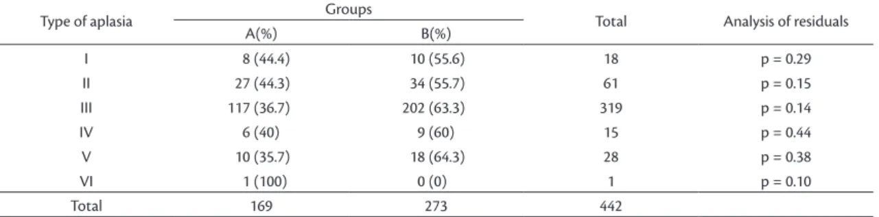
The types of aplasia found in the total sample of 2,665 limbs are detailed in Table 2.
Applying Fisher’s exact test (p=0.67) and the Williams test (p=0.67), the results indicate that the assumption of homogeneity between groups should not be rejected, i.e. the frequencies of “yes” and “no” are the same for all types of aplasia. This conclusion
is conirmed by the analysis of residuals in the table. The evaluation of presence of an insuficient
GSV segment against type of aplasia for limbs in each group was conducted as a descriptive analysis because, despite the size of the sample, there were nevertheless few limbs that exhibited this association, with multiple levels of aplasia types and segments with
relux, and because of the large number of “zeros”,
the statistical analysis was not conducted as a matter of course, i.e. for the whole of Table 3.
The results show that in group A there was a larger
number of limbs with both GSV insuficiency and
GSV aplasia than in group B and, furthermore, that among the overall number of limbs with aplasia in
both groups, there was a predominance of relux in the
proximal segment (within the saphenous compartment) and the epifascial branch (Table 3, Figure 4).
In summary, the totals for each segment with relux
enabled testing of the homogeneity between groups A and B using binomial tests. These results, shown in Table 4, provide evidence for homogeneity between groups A and B for all segments.
DISCUSSION
The international anatomic nomenclature serves as a basis for communication for research, treatment and exchange of information in phlebology and so
Figure 1. Classiication of the types of aplasia of the GSV described by Seidel et al.12
Figure 2. GSV exiting the saphenous compartment via the saphenous fascia in the proximal 1/3 of the thigh.
it is important that only veins located within the saphenous compartment should be considered to be true GSV,3,7,8,13-17 irrespective of possible reductions
in their diameter or when they cannot be located in certain segments, in which cases, the correct terms are segmental hypoplasia or aplasia, respectively.4,18
On color Doppler ultrasonography, the GSV is easily
identiied in the thigh within the saphenous compartment,
known as the “saphenous eye” or “Egyptian eye”1,7,19
which is not the case at the level of the knee, where
the reference used for identiication is the triangle
formed by the tibia, medial gastrocnemius muscle
and, supericially, the fascial sheath. The examinations
analyzed in the present study were conducted in accordance with the literature.8
Since there was no interest in making sex-distinction inferences in the statistical analysis of the results, the
Table 1. Number of limbs with GSV insuiciency and/or aplasia in groups A and B.
Insuiciency Aplasia Insuiciency + Aplasia
Group A (with varicose veins) 479 (83.9%) 169 (38.2%) 71 (80.7%)
Group B (free from varicose veins) 92 (16.1%) 273 (61.8%) 17 (19.3%)
Total 571 442 88
Chi-square test: p<0.0001.
Table 2. Distribution of the limbs by group, according to the type of GSV aplasia, and p values identiied by the analysis of residuals.
Type of aplasia Groups Total Analysis of residuals
A(%) B(%)
I 8 (44.4) 10 (55.6) 18 p = 0.29
II 27 (44.3) 34 (55.7) 61 p = 0.15
III 117 (36.7) 202 (63.3) 319 p = 0.14
IV 6 (40) 9 (60) 15 p = 0.44
V 10 (35.7) 18 (64.3) 28 p = 0.38
VI 1 (100) 0 (0) 1 p = 0.10
Total 169 273 442
Fisher’s exact test p=0.67; Test G (Williams) p=0.67.
Table 3. Distribution of insuicient segments with each type of GSV aplasia in limbs from groups A or B.
Type of Aplasia/ Groups
I II III IV V VI Total
Segment with relux A B A B A B A B A B A B A B
Proximal segment 0 0 1 0 1 2 2 0 2 0 0 0 6 2
Proximal segment + epifascial branch 3 0 3 0 25 9 1 1 1 0 0 0 33 10
Epifascial branch 0 0 1 0 7 2 0 0 0 0 0 0 8 2
Epifascial branch + distal segment 0 0 0 0 3 0 0 0 1 0 0 0 4 0
Distal segment 0 0 1 0 0 0 0 0 0 0 0 0 1 0
Entire vein 1 0 4 0 11 2 0 0 2 1 1 0 19 3
Total 4 0 10 0 47 15 3 1 6 1 1 0 71 17
Table 4. Binomial test applied to data shown in Table 3, considering totals for Groups A and B.
Segment with relux Total p-value
A B
Proximal segment 6 2 0.3352
Proximal segment + epifascial branch 33 10 0.1820
Epifascial branch 8 2 0.4773
Epifascial branch + distal segment 4 0 0.0704
Distal segment 1 0 0.3120
Entire vein 19 3 0.2189
Total 71 17
predominance of female patients in the sample did not affect the results.
The etiology of anatomic abnormalities of the GSV
has not been deined, but it is assumed that they are
the result of developmental defects by which vessels with favorable hemodynamic conditions prevail over others, which undergo atrophy.2,4,7
IN an analysis of limbs with hypoplastic segments, Caggiati and Mendoza4 studied 676 limbs with
normally functioning GSVs and found 86 limbs that had hypoplastic segments, while an analysis of
320 limbs with GSV insuficiency showed that 79 had
hypoplasia. In both groups, hypoplasia predominated in the segment between the proximal third of the leg and the distal third of the thigh, similar to what was observed in the present study.
Next, an analysis will be conducted of the aplastic
segments and presence or absence of GSV insuficiency
in the limbs of patients with or without clinical signs
of venous insuficiency.
The results of this study show that 61.8% of limbs
with aplasia were in group B, which is signiicantly
greater than in group A, which accounted for 38.2%. This result is different from the results for which
groups had insuficiency or insuficiency combined
with aplasia, where the percentage was greater for limbs in group A (Table 1).
Ricci and Caggiati17, Ricci and Cavezzi20 and
Caggiati and Mendoza4 analyzed samples that differed
from the sample analyzed here in that they compared groups of patients with primary varicose veins with people without varicose veins and competent GSV, concluding that segmental hypoplasia is more common among patients with varicose veins than among healthy people and they also referred to the presence of hypoplasia as a possible factor in pathogenesis of varicose veins. This hypothesis was not shared by
Oğuzkurt5 who, having listed as a study limitation
the lack of a control group containing normal people, stated that it was not possible to conclude whether the segment with aplasia had any clinical importance in the pathogenesis of varicose veins.
Comparing just the 1,255 limbs from group A of the present study with the 200 limbs that had varicose veins studied by Ricci and Cavezzi,20 the conclusions
contrast, since those authors concluded that there was absence or hypoplasia of the GSV in the knee, with a prevalence of tributaries, in almost 30%, whereas in the present study 128 of the 169 GSV with aplasia (75.7%) exhibited this abnormality at the medial surface of the knee. However, results were similar in terms of the low rate of complete GSV incompetence, with rates of 6% and 3.9%, respectively.
In a different study5 that analyzed a sample of
patients with clinical signs of CVD, one third of the patients exhibited segmental aplasia of the GSV with similar frequencies among patients with and without
GSV insuficiency. The results showed that segmental
aplasia was observed in 16.6% of the total and was more frequent, in percentage terms, in the group of limbs without varicose veins, but without statistical
signiicance (p>0.05).
In the conclusions of an article by Ricci and Cavezzi,20
the incidence of segmental hypoplasia of the GSV was higher among people with varicose veins than among those without varicose veins (43 vs. 30%),
but without statistical signiicance; which coincides
with a study by Caggiati and Mendoza,4 who observed
segmental hypoplasia in 25% of limbs with GSV relux
due to incompetence at the saphenofemoral junction
and in 12% of limbs without GSV relux, stating that hypoplastic segments do not allow venous relux, which is diverted into supericial branches. In our
study, 71 (14.8%) patients in group A, which included
479 GSV with insuficiency, exhibited some type
of aplasia, while in group B, 17 (18.5%) of 92 with
insuficiency had an aplastic segment.
The percentages of veins with segmental aplasia
in the subsets of the sample studied by Oğuzkurt5
are equivalent, with 34% among those with GSV
insuficiency and 31% among those with normal
GSV, with a predominance of type 1, corresponding to type III in this study.
According to Caggiati and Mendoza,4 segmental
hypoplasia of the GSV leads to hemodynamic overload of the accessory saphenous and the result is merely physiological compensatory dilation of the tributaries, but, in limbs with a predisposition to varicose disease, there is greater overload of the accessory saphenous vein which results in larger varicose veins, earlier, which are clinically more evident than in patients with GSV incompetence.
It is important to deine these anatomic abnormalities
because, if there is a drainage vein connecting the two segments of the vein then proximal incompetence is usually transferred to the distal segment, but if there is no connection between them, only one segment will develop incompetence.6
These indings could have an important role in routine practice and signiicance for the pathophysiology
of varicose disease. The presence of the wall of this compartment supplementing muscle contraction could modify the diameter of the vein and, consequently,
modulate its blood low, as happens in the deep vein
still preserve the GSV from excessive pathological dilation, providing mechanical protection.12,20
Knowledge of the presence of segmental hypoplasia or aplasia of the GSV is important because if there is
a reduction in diameter then there may be dificulty
in advancing endoluminal instruments such as vein
strippers and laser ibers or for thermoablation, and
thermal damage to the skin may also occur because of
the more supericial location of tributaries branches.
Notwithstanding, abnormalities in anatomic course do not invalidate its use, as long as it has been evaluated in advance using color Doppler ultrasonography and an adequate diameter is preserved.
CONCLUSIONS
It can be concluded that segmental aplasia of the GSV occurred more often in lower limbs that do not
exhibit varicose veins and/or GSV insuficiency, but that the combination of aplasia and insuficiency
had a higher incidence in the group of limbs that did exhibit varicose veins.
REFERENCES
1. Chen SSH, Prasad SK. Long saphenous vein and its anatomical variations. AJUM. 2009;12(1):28-31.
2. Caggiati A, Ricci S. The caliber of the human long saphenous vein and its congenital variations. Ann Anat. 2000;182(2):195-201. http:// dx.doi.org/10.1016/S0940-9602(00)80083-7. PMid:10755188. 3. Caggiati A, Bergan JJ. The saphenous vein: derivation of its name
and its relevant anatomy. J Vasc Surg. 2002;35(1):172-5. http:// dx.doi.org/10.1016/S0741-5214(02)52480-0. PMid:11802151. 4. Caggiati A, Mendoza E. Segmental hypoplasia of the great
saphenous vein and varicose disease. Eur J Vasc Endovasc Surg. 2004;28(3):257-61. http://dx.doi.org/10.1016/j.ejvs.2004.06.002. PMid:15288628.
5. Oğuzkurt L. Ultrasonography study on the segmental aplasia of the great saphenous vein. Phlebology. 2014;29(7):447-53. http:// dx.doi.org/10.1177/0268355513484016. PMid:23761865.
6. Seidel AC, Miranda F Jr, Juliano Y, Novo NF. Relationship between the diameter of great saphenous vein and body mass index. J Vasc Bras. 2005;4(3):265-9. http://dx.doi.org/10.1590/ S1677-54492005000300008.
7. Oğuzkurt L. Ultrasonographic anatomy of the lower extremity superficial veins. Diagn Interv Radiol. 2012;18(4):423-30. PMid:22427019.
8. Kalodiki E, Calahoras L, Nicolaides A. Make it easy: duplex examination of the venous system. Phlebology. 1993;8:17-21.
9. Labropoulos N, Tiongson J, Pryor L, et al. Definition of venous reflux in lower-extremity veins. J Vasc Surg. 2003;38(4):793-8. http://dx.doi.org/10.1016/S0741-5214(03)00424-5. PMid:14560232.
10. SAS Institute. Statistics Analysis System [software]. Version 9.3. Cary; 2013. DVD-ROM.
11. Pagano M, Gauvereau K. Princípios de bioestatística. 2. ed. São Paulo: Thomson; 2004. 506 p.
12. Seidel AC, Cavalari Jr P, Rossi RM, Miranda Jr F. Proposal for classification of the great saphenous vein aplasia by the echo-color
Doppler. In: VEITH Symposium; 2014; New York. Tema livre com apresentação oral.
13. Caggiati A. Fascial relationships of the long saphenous vein. Circulation. 1999;100(25):2547-9. http://dx.doi.org/10.1161/01. CIR.100.25.2547. PMid:10604894.
14. Caggiati A. Fascial relations and structure of the tributaries of the saphenous veins. Surg Radiol Anat. 2000;22(3-4):191-6. http:// dx.doi.org/10.1007/s00276-000-0191-3. PMid:11143312.
15. Caggiati A, Ricci S. The long saphenous vein compartment. Phlebology. 1997;12:107-11.
16. Caggiati A, Bergan JJ, Gloviczki P, Jantet G, Wendell-Smith CP, Partsch H. Nomenclature of the veins of the lower limbs: an international interdisciplinary consensus statement. J Vasc Surg. 2002;36(2):416-22. http://dx.doi.org/10.1067/mva.2002.125847. PMid:12170230.
17. Ricci S, Caggiati A. Echoanatomical patterns of the long saphenous vein in patients with primary varices and in healthy subjects. Phlebology. 1999;14(2):54-8. http://dx.doi.org/10.1007/ s005230050044.
18. Caggiati A, Bergan JJ, Gloviczki P, Eklof B, Allegra C, Partsch H. Nomenclature of the veins of the lower limb: extensions, refinements, and clinical application. J Vasc Surg. 2005;41(4):719-24. http:// dx.doi.org/10.1016/j.jvs.2005.01.018. PMid:15874941.
19. Ricci S. Comment to: ultrasonography study on the segmental aplasia of the great saphenous vein by Oguzkurt L. Phlebology 2013. Veins and Lymphatics. 2013. No prelo. [citado 2014 jul. 21]. http://www.pagepressjournals.org/index.php/vl/rt/printerFriendly/ ByblioLab.2013.13/1704.
20. Ricci S, Cavezzi A. Echo-anatomy of long saphenous vein in the knee region: proposal for a classification in five anatomical patterns. Phlebology. 2002;16(3):111-6. http://dx.doi.org/10.1007/ s005230200005.
*
Correspondence
Amélia Cristina Seidel Rua Dr. Gerardo Braga 118, Jardim Vila Rica CEP 87050-610 - Maringá (PR), Brazil Tel.: +55 (44) 3026-7590 E-mail: [email protected]
Author information
ACS - Associate professor of Angiology and Vascular Surgery, School of Medicine, Universidade Estadual de Maringá (UEM), Maringá, PR, Brazil; Vascular sonographer from SBACV and CBR. PCJ - Medical student (5th year), Universidade Estadual de Maringá (UEM). RMR - Adjunct professor at the Department of Statistics, Universidade Estadual de Maringá (UEM). FMJ - Full professor of Vascular Surgery, Department of Surgery, Universidade Federal de São Paulo (UNIFESP-EPM); vascular sonographer from SBACV and CBR.
Author contributions
Conception and design: ACS Analysis and interpretation: ACS, FMJ, RMR Data collection: ACS, PCJ Writing the article: ACS, FMJ, RMR Critical revision of the article: FMJ, ACS Final approval of the article*: ACS, FMJ, RMR, PCJ Statistical analysis: RMR Overall responsibility: ACS