Its E3 Subunit, Dihydrolipoamide Dehydrogenase Affects
Fertilization in Syrian Hamsters
Archana B. Siva*., Subbarayalu Panneerdoss., Purnima Sailasree, Durgesh K. Singh,
Duvurri B. Kameshwari, Sisinthy Shivaji*¤
Centre for Cellular and Molecular Biology (Council of Scientific and Industrial Research), Hyderabad, India
Abstract
Background/Aims:The importance of sperm capacitation for mammalian fertilization has been confirmed in the present study via sperm metabolism. Involvement of the metabolic enzymes pyruvate dehydrogenase complex (PDHc) and its E3 subunit, dihydrolipoamide dehydrogenase (DLD) in hamsterin vitrofertilization (IVF) viain vitrosperm capacitation is being
proposed through regulation of sperm intracellular lactate, pH and calcium.
Methodology and Principal Findings:Capacitated hamster spermatozoa were allowed to fertilize hamster oocytesin vitro
which were then assessed for fertilization, microscopically. PDHc/DLD was inhibited by the use of the specific DLD-inhibitor, MICA (5-methoxyindole-2-carboxylic acid). Oocytes fertilized with MICA-treated (MT) [and thus PDHc/DLD-inhibited] spermatozoa showed defective fertilization where 2nd polar body release and pronuclei formation were not observed. Defective fertilization was attributable to capacitation failure owing to high lactate and low intracellular pH and calcium in MT-spermatozoa during capacitation. Moreover, this defect could be overcome by alkalinizing spermatozoa, before fertilization. Increasing intracellular calcium in spermatozoa pre-IVF and in defectively-fertilized oocytes, post-fertilization rescued the arrest seen, suggesting the role of intracellular calcium from either of the gametes in fertilization. Parallel experiments carried out with control spermatozoa capacitated in medium with low extracellular pH or high lactate substantiated the necessity of optimal sperm intracellular lactate levels, intracellular pH and calcium during sperm capacitation, for proper fertilization.
Conclusions:This study confirms the importance of pyruvate/lactate metabolism in capacitating spermatozoa for successful fertilization, besides revealing for the first time the importance of sperm PDHc/ DLD in fertilization, via the modulation of sperm intracellular lactate, pH and calcium during capacitation. In addition, the observations made in the IVF studies in hamsters suggest that capacitation failures could be a plausible cause of unsuccessful fertilization encountered during human assisted reproductive technologies, like IVF and ICSI. Our studies indicate a role of sperm capacitation in the post-penetration events during fertilization.
Citation:Siva AB, Panneerdoss S, Sailasree P, Singh DK, Kameshwari DB, et al. (2014) Inhibiting Sperm Pyruvate Dehydrogenase Complex and Its E3 Subunit, Dihydrolipoamide Dehydrogenase Affects Fertilization in Syrian Hamsters. PLoS ONE 9(5): e97916. doi:10.1371/journal.pone.0097916
Editor:Suresh Yenugu, University of Hyderabad, India
ReceivedJune 4, 2012;AcceptedApril 26, 2014;PublishedMay 22, 2014
Copyright:ß2014 Siva et al. This is an open-access article distributed under the terms of the Creative Commons Attribution License, which permits unrestricted use, distribution, and reproduction in any medium, provided the original author and source are credited.
Funding:SP and DKS thank Council for Scientific and Industrial Research, Government of India, for the Research Fellowships. The funders had no role in study design, data collection and analysis, decision to publish, or preparation of the manuscript.
Competing Interests:The authors have declared that no competing interests exist. * E-mail: shivas@ccmb.res.in (SS); abs@ccmb.res.in (ABS)
.These authors contributed equally to this work.
¤ Current address: Jhaveri Microbiology Centre, L V Prasad Eye Institute, Banjara Hills, Hyderabad, India
Introduction
Fertilization is a complex biological process, for which many of the prerequisites are still poorly understood. Fertilization success or failure depends on several sperm and egg factors [1]. Sperm capacitation too is an obligatory phenomenon for successful fertilization in mammals [2,3]. Idiopathic fertilization failure in nature as well as during assisted reproductive practices such as
conventional in vitro fertilization (IVF) has been attributed to
problems of sperm capacitation [4,5]; warranting molecular studies on the contribution of sperm capacitation to fertilization success.
intracellular lactate, intracellular pH (pHi) and intracellular
calcium [Ca2+
]i[11–13]. Inhibition of PDHc/DLD was achieved
by the use of the DLD-specific inhibitor, 5-methoxyindole-2-carboxylic acid (MICA). Downregulation of the PDHc/DLD activity in these MICA-treated (MT) hamster spermatozoa inhibited capacitation and acrosome reaction, with no significant effects on hyperactivation and tyrosine phosphorylation [11]. The mechanism of inhibition of capacitation and acrosome reaction in the MT-spermatozoa was worked out in the laboratory [13]. It was demonstrated that MT-spermatozoa showed lactate accumu-lation (due to PDHc/DLD inhibition and thus, pyruvate non-consumption), which resulted in lowering of initially, the intracellular pH and eventually, the intracellular calcium in these cells, causing blocked capacitation and acrosome reaction.
Deviation in this regulation resulting in sperm capacitation failure; is likely to affect the fertilization-competence of these spermatozoa. To validate this premise and understand the
mechanism involved, we performed in vitro fertilization studies
with spermatozoa; in which PDHc/DLD was inhibited by the use of the specific DLD inhibitor, 5-methoxyindole-2-carboxylic acid (MICA).These MICA-treated (MT-), non-capacitated spermato-zoa, as anticipated, failed to fertilize the oocytes, thus, supporting the importance of sperm capacitation for successful fertilization. The results also substantiated the role of pyruvate/lactate metabolism in fertilization, in addition to establishing the requirement of a functional sperm PDHc/DLD in hamster fertilization.
Materials and Methods
Spermatozoa collection,in vitrocapacitation and
assessment of sperm hyperactivation
Male golden hamsters (Mesocricetus auratus) aged 6 months were
used for the in vitro capacitation studies that involved modified
TALP-PVA medium (Tyrode’s medium with albumin, lactate, pyruvate and polyvinyl alcohol) as described earlier [13]. Briefly, the caudae epididymidum were dissected out from anesthetized animals, rinsed in the medium, pierced with a fine needle and the released contents containing the spermatozoa was collected in the modified Tyrode’s medium. After a few minutes of incubation at
37uC, 5% CO2, a uniform suspension of spermatozoa was
obtained which was then taken for a sperm count in a Makler chamber and a HTM-CEROS (Hamilton Thorne, Beverly, MA)
computer assisted sperm analyzer (CASA). Forin vitrofertilization
(IVF) experiments; spermatozoa were collected after 3 h of capacitation in TALP-PVA medium and then used for insemi-nating the oocytes. MICA, the specific inhibitor of DLD, was dissolved in the TALP-PVA media as described earlier [11] and all the experiments were done with a 5 mM final concentration. The acrosome reaction was always assessed for MT- spermatozoa, to ensure that the inhibitor was working [13]. The present study was approved by the Institutional Animal Ethics Committee of the Centre for Cellular and Molecular Biology, Hyderabad, India.
Hamster sperm hyperactivation and the related motility kinematic parameters namely curvilinear velocity (VCL), linearity (LIN), amplitude of lateral head displacement (ALH) were assessed using CASA, according to the criteria described earlier [11]. The set up values of the CASA were as follows: frames acquired, 50; frame rate (Hz), 60; minimum contrast, 25; minimum cell size
(pixels), 3; low average path velocity cut off (mm/sec), 7.5; medium
average path velocity cut off (mm/sec), 12.5; low straight line
velocity cut off (mm/sec), 5; static head intensity limits, 0.2–1.47;
static head-size limits, 0.12–7.37;static elongation limits, 1–98; magnification, 1.43 (4x); video frequency (Hz), 60; bright field, off;
slide temperature, 37uC; field selection mode, manual [11]. Based
on these kinematic parameters, the non-hyperactivated spermato-zoa (exhibiting planar motility pattern) could be differentiated from the hyperactivated spermatozoa (exhibiting either circular or helical motility patterns) using the SORT facility of the CASA.
Spermatozoa with data points $15, VCL.300mm/sec, LIN,
40%, ALH.12mm were sorted as hyperactivated (those
exhib-iting either circular or helical motility pattern) and spermatozoa
with data points$15, VCL,300mm/sec, LIN.40% and ALH,
12mm were sorted as non-hyperactivated spermatozoa (exhibiting
planar motility pattern). A total of,100 individual spermatozoa
were sorted at each time point to establish whether the spermatozoa were hyperactivated or not.
Superovulation and oocyte collection
Three-month-old cyclic female hamsters were used in this investigation. On day 1 of the estrous cycle (confirmed by postovulatory discharge), before 10 a.m., ovarian hyperstimulation was induced by subcutaneous injection of 10 IU equine chorionic
gonadotrophin (eCG -FolligonH; Intervet, Boxmeer, The
Nether-lands) and ovulation was induced by 10 IU human chorionic
gonadotrophin (hCG-ChorulonH; Intervet, Boxmeer, The
Nether-lands) injected between 48–56 h after eCG injection [14]. Animals
were anesthetized at 1761 h after hCG injection. Oviducts were
collected in a 35 mm dish (Nunc, Roskilde, Denmark) containing 1 ml TALP-PVA medium. The cumulus–oocyte complexes (COCs) were collected by gently teasing the ampulla region of the oviducts, and the COC mass was digested using hyaluronidase (1 mg/ml) and the cumulus-free zona intact oocytes were washed
three times in TALP-PVA medium and incubated at 37uC in 5%
CO2, under mineral oil (embryo-tested, Sigma,, St. Louis, MO,
USA), until being used for IVF.
In vitrofertilization
Freshly collected oocytes (metaphase II-arrested, 10 oocytes per
drop) were placed in a 100ml fertilization drop of TALP-PVA
medium under mineral oil and an aliquot of spermatozoa (final
concentration of 10,000 – 20,000 spermatozoa, 2.5ml) previously
capacitated for 3 h (different capacitation conditions were used, as described under separate section) was added [14]. Co-incubation
was carried out for at least 3 h at 37uC in 5% CO2under mineral
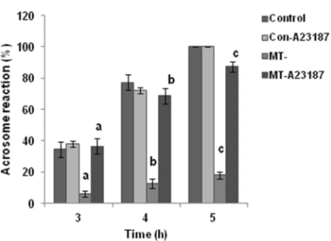
oil to prevent evaporation and pH changes. In all IVF experiments, spermatozoa were capacitated for 3 h under various conditions as indicated and then used for IVF, since in preliminary experiments it was established that in hamster spermatozoa, capacitation (as judged by the occurrence of acrosome reaction) begins at 3 h and reaches a peak by 5 h (Figure. 1).
After 3 h of co-incubation, the oocytes were washed in TALP-PVA medium to remove the excess bound spermatozoa, stained
with Hoechst 33342 (30mg/ml, Sigma, St. Louis, MO, USA) and
their fertilization status was confirmed in the Axiovert microscope (Carl Zeiss Inc, Germany), 40x objective. The various cellular events monitored included meiotic plate reorganization, second polar body release and formation of both pronuclei. Only those
oocytes that showed both 2ndpolar body release and pronuclei
formation were scored as ‘properly fertilized’. Thirty to 50 oocytes from at least 4–7 different females were used for each determi-nation. All experiments were repeated at least 4 times with spermatozoa from different males. All experiments were carried out with proper (solvent) control.
Irrespective of the media conditions for sperm capacitation, IVF was always done in the TALP-PVA medium. For alkalinization experiments, MT- spermatozoa were treated with 15 mM
and then used for IVF after 3 h. NH4Cl is routinely used for
increasing the pHi of spermatozoa [15]. For low pH studies,
control spermatozoa were capacitated for 3 h in TALP-PVA medium, the pH of which was lowered to 6.8 and 7.0. For G media (TALP-PVA medium without pyruvate-lactate) studies, MT- spermatozoa were capacitated in G medium (MT-G) with and without 5 mM MICA and then used for IVF. In media which had only pyruvate-lactate (PL medium) and no glucose, sperma-tozoa were incubated for 3 h in this media before IVF. For experiments involving treatment with calcium ionophore, both control and MT- spermatozoa were treated for 5 minutes with
0.2mM calcium ionophore, A23187 and then used for IVF.
Calcium ionophore (Sigma, St. Louis, MO, USA) was prepared as a stock solution in DMSO and working dilutions were made in the
TALP-PVA medium. 0.2mM A23187, did not affect sperm
motility drastically. It is known that a longer exposure of sperm to A23187 inhibits motility [16]. Acrosome reaction was assessed for control and MT-spermatozoa after the addition of A23187 at 3, 4 and 5 h of capacitation (Figure. 1).
Appropriate solvent and additives’ (NH4Cl, MICA, etc.)
controls were always done alongside to ensure that these did not have an effect on fertilization outcome via their direct effect on oocytes. In these, co-incubation of oocytes and control
sperma-tozoa was carried out in the presence of the additives (2.5ml of
5 mM MICA / 5 mM MICA+15 mM NH4Cl / 5 mM MICA-G /
5 mM MICA-G+5 mM NH4Cl / TL19 medium / pH 6.8 TALP
medium /pH 7.0 TALP medium / 0.2mM A23187 medium) and
these conditions were further used for experiments with MT-spermatozoa, as the fertilization was found to be 100% (Table S1A). Control experiments were set up to also rule out the effect of the
additives, NH4Cl and A23187 on the parthenogenetic activation of
the hamster oocytes (Table S1B).
Statistical analysis
ANOVA test with Tukey-Kramer multiple comparisons was performed to analyze the results statistically using the software
Graph Pad, Prism, version 3.02. P values,0.05 were considered
significant.
Results
Pyruvate/lactate alone in capacitating medium are sufficient to support fertilization
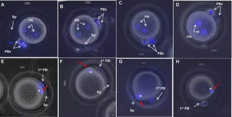
All the oocytes fertilized with hamster spermatozoa capacitated in TALP-PVA medium showed proper fertilization (Control), with both the polar bodies (PBs) and the pronuclei (PN) visible (Figure. 2A-D, Table 1). Hamster spermatozoa incubated in TALP medium devoid of glucose (PL medium) successfully fertilized all the oocytes (Control-PL), 100%, Table 1), indicating that the presence of pyruvate/lactate alone during capacitation is sufficient for hamster fertilization.
Inhibiting pyruvate/lactate metabolism in spermatozoa affects fertilization: MICA-treated spermatozoa are unable to fertilize oocytes properly
Inhibiting PDHc/DLD affects pyruvate/lactate metabolism [13], since PDHc metabolizes pyruvate to acetyl CoA. Aberrant pyruvate/lactate metabolism during capacitation in these
MT-spermatozoa resulted in only 6.264.6% oocytes showing proper
fertilization (Table 1). The majority of these oocytes (,90%)
showed only meiotic plate reorganization (MPR, asterisk, Figure. 2E-H). These oocytes were also observed after 8 and 18 h and they maintained the same condition of MPR (data not shown). All oocytes inseminated with control spermatozoa (Control) showed 100% fertilization (Table 1).
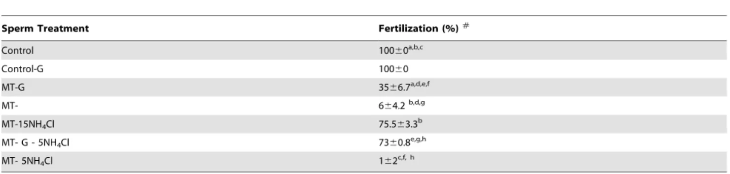
Reducing lactate load on the MICA-treated spermatozoa improves their fertilization ability
To ascertain if lactate accumulation in MT- spermatozoa [13] was responsible for the reduced fertilization, IVF was performed with MT- spermatozoa capacitated in G medium (MT-G). Reduced lactate load on these spermatozoa improved their
fertilizing potential. The IVF results indicated that 3566.7%
oocytes were fertilized with MT-G spermatozoa (Table 2) in contrast to 664.2 % oocytes fertilized by MT spermatozoa.
Alkalinization of the MICA-treated spermatozoa improves their fertilization potential
Increased lactate load decreases the pHiof the MT spermatozoa
[13] and thus, alkalinizing these spermatozoa was expected to improve the fertilization outcomes. Alkalinization of the male
gamete resulted in 75.563.3 % of the oocytes showing proper
fertilization, in contrast to 664.2% fertilization seen with
untreated MT spermatozoa (Table 2). IVF done with MT-G
spermatozoa alkalinized with only 5 mM NH4Cl (MTG
-5NH4Cl) revealed fertilization success (7362%) similar to
MT-15NH4Cl (75.563.3 %) [Table 2].
Control spermatozoa capacitated in TALP-PVA media with low pH or high lactate have compromised fertilizing ability
In order to assess if the accumulation of lactate and subsequent
lower pHiin the spermatozoa during capacitation [13] in general
had detrimental effects on the outcome of hamster IVF (similar to that seen with MT spermatozoa), we carried out IVF with control spermatozoa capacitated in TALP-PVA medium supplemented with 19 mM lactate (normally TALP has 12.8 mM lactate) or with lower pH of 6.8 and 7.0. IVF with MT-spermatozoa was also done in parallel as a control for defective fertilization. IVF results indicated that all the above sperm treatments resulted in low
success rate (4965.1% oocytes for TL19; 3866.9% for pH 6.8
and 4468% for pH 7.0 spermatozoa) [Table 3].
Figure 1. Acrosome reaction studies after induction with calcium ionophore, A23187.Induction of acrosome reaction was seen in MT- spermatozoa with 0.2mM A23187, when evaluated at 3, 4
Increasing [Ca2+]
iin MICA-treated spermatozoa during
capacitation improves their fertilizing ability
Since [Ca2+
]i level in MT-spermatozoa was low [13], we
envisaged that increasing calcium level in the MT spermatozoa would overcome the defective fertilization observed by these spermatozoa. Therefore, MT spermatozoa were treated at 2.55 h
briefly (for 5 min) with 0.2mM calcium ionophore, A23187
(MT-PreCa) before IVF. It was observed that oocytes fertilized with
MT-PreCa spermatozoa showed 3869.6% success as opposed to
664.6% in MT spermatozoa (Table 4). Acrosome reaction induction was also seen with this concentration of A23187 in MT-spermatozoa at all the 3 time points assessed (Figure. 1). A
correlation (Spearman correlation coefficient, r = 0.8503, p,0.05)
was seen between the sperm intracellular calcium levels [13] in the different treatments and the fertilization rates seen in this study (Figure. 3).
In another set of experiments with calcium ionophore, oocytes showing defective fertilization were treated with 40 nM A23187
for 10 minutes. It was seen that calcium ionophore treatment
resulted in 42.163.7% of oocytes showing both, PB release and
pronuclei formation, as compared to 6% fertilization seen in untreated MT-fertilized oocytes (Table 4).
Discussion
This study confirms the importance of sperm capacitation for fertilization, in general and of sperm pyruvate/lactate metabolism in fertilization, in particular [10,17]. Pyruvate/lactate are suffi-cient to support fertilization in hamsters, as demonstrated by IVF studies with spermatozoa capacitated in TALP medium devoid of glucose (Table 1). However, there is an optimum level of lactate/ pyruvate required by the spermatozoon for it to remain fertile, since increase in the lactate load on the spermatozoa reduces its fertilizing potential (Table 3). Inhibiting pyruvate/lactate metab-olism with PDHc/DLD inhibitor, MICA affected fertilization (Table 1). Since DLD mutant is embryonically lethal [18] making
Figure 2.In vitrofertilization results with control (A-D) and MT- spermatozoa (E-H).Oocytes fertilized using control spermatozoa showed proper fertilization (PF) as judged by the presence of both polar bodies (PBs) and both pronuclei [PN].(A-D). Oocytes fertilized with MT-spermatozoa showed defective fertilization. In these spermatozoa, only meiotic spindle reorganization was visible (asterisk, E-H) and the 2ndpolar body extrusion
had failed (red arrow in E-H). Oocytes were stained with Hoechst 33342 to visualize the polar bodies and pronuclei and the images presented are a merge of both brightfield and fluorescence. Magnification used was 400 x. Scale bars indicate 5mm.
doi:10.1371/journal.pone.0097916.g002
Table 1.Fertilization outcome with MICA-treated spermatozoa.
Sperm Treatment Fertilization (%)#
Control 10060a
Control-PL 10060b
MT- 6.264.6a, b
Control: Control spermatozoa capacitated in TALP-PVA medium; Control-PL: spermatozoa capacitated in PL medium; MT-: MICA-treated spermatozoa in TALP-PVA medium.
#
Values represent mean6SD.
the use of DLD specific inhibitor, MICA is the best possible approach. The importance of pyruvate metabolism has been highlighted in embryo development, by the use of PDHA1 knockout model as well, where it was seen that oocytes compromised in PDHc activity (PDHA1 is another subunit of
PDHc) fail to develop beyond the 1-cell zygote stagein vivo[19].
The authors hypothesized that this effect may be because of a ‘‘lactic acidosis-like condition’’, which is created in the oocyte thereby affecting normal development. This brings into view the fact that pyruvate metabolism (via PDHc activity, since both DLD and PDHA1 are subunits of this complex) is important for
fertilization and embryo development via pHiregulation
irrespec-tive of the cell type (spermatozoon or oocyte) or the approach used (i.e. either DLD inhibition in our study or PDHA1 knockout in the
study by Johnsonet al[19]).
Inhibition of sperm DLD results in defective fertilization due to lactate accumulation in the spermatozoa, which in turns adversely
affects sperm pHi and [Ca
2+]
i crucial for sperm capacitation
[8,20–25]. This high lactate, and low sperm pHiand [Ca2+]iaffects
capacitation and acrosome reaction [13], and eventually the fertilizing potential of the spermatozoa, as seen in this study [Table 2, 3]. Improvement in the fertilization rate after increasing
intracellular pH or calcium with NH4Cl [Table 2] and calcium
ionophore, A23187, respectively [Table 4] supports the hypothesis that increasing intracellular pH/calcium improves capacitation/ acrosome reaction in MT-spermatozoa, thereby eventually improving their fertilizing potential. This study on PDHc/DLD contributes to the knowledge available on the importance of sperm
pHiand [Ca2+]iin mammalian fertilization [26–32]. A correlation
(spearman correlation coefficient, r = 0.7683, p,0.05) was seen
between the sperm pHi[13] and the fertilization rate under the
various experimental conditions studied, where low sperm pHi
resulted in lower fertilization rates (Figure. 4). Alkalinized MT and MT-G spermatozoa, showed a deviation in this trend (Figure. 4, encircled), suggesting a likely post-fertilization effect of the added
NH4Cl to the fertilized oocyte. In fact, it was also seen that
treatment of defectively fertilized oocytes with 2.5 mM NH4Cl
post-fertilization resulted in 7163% fertilization (Table S2) when compared to 6.264.6 % fertilization with untreated MT-spermatozoa.
One another sperm characteristic, sperm hyperactivation (HA) is considered very important for mammalian fertilization [33,34]. To assess if the various conditions used in this study affected sperm HA, which ultimately would have a bearing on the fertilization outcome, we assessed sperm HA during capacitation for the various conditions used (Figure. 5). It was seen that the effect was evident until 2 h of incubation in VCL (Figure. 5A and B), ALH (Figure. 5C and D) and LIN (Figure. 5E and F) parameters, after which the spermatozoa recovered. This trend with MICA has been shown earlier from our laboratory by Mitra and Shivaji [11]. Since the spermatozoa were picked up at 3 h of capacitation for IVF studies, it is possible that effects on HA may not influence IVF outcomes.
The oocyte resumes meiosis and becomes competent to begin embryonic development upon activation. The mammalian oocyte is activated in a fertilization-dependent manner. Oocyte activation
Table 2.Fertilization with MICA-treated spermatozoa capacitated in G medium (MICA-treated-G) with or without ammonium chloride [NH4Cl].
Sperm Treatment Fertilization (%)#
Control 10060a,b,c
Control-G 10060
MT-G 3566.7a,d,e,f
MT- 664.2b,d,g
MT-15NH4Cl 75.563.3b
MT- G - 5NH4Cl 7360.8e,g,h
MT- 5NH4Cl 162c,f, h
Control: Control spermatozoa in TALP-PVA medium; Control-G: control spermatozoa in G medium; MT-: MICA-treated spermatozoa; MT-G: MT spermatozoa in G medium; MT-15NH4Cl: MT spermatozoa in TALP-PVA medium alkalinized with 15 mM ammonium chloride; MT-G-5NH4Cl: MT-G spermatozoa alkalinized with 5 mM
ammonium chloride; MT-5NH4Cl: MT- spermatozoa alkalinized with 5 mM ammonium chloride. #
Values represent mean6SD.
Values with the same superscript differ significantly at p,0.05. doi:10.1371/journal.pone.0097916.t002
Table 3.Fertilization with spermatozoa capacitated in TALP-PVA medium with high lactate and low pH (6.8 and 7.0).
Sperm Treatment Fertilization (%)#
Control 10060a, b, c
TL19 4965.1a
pH 6.8 3866.9b
pH 7.0 4468.0c
Control: Control spermatozoa in TALP-PVA medium with pH 7.5 and 12.8 mM lactate; TL19: spermatozoa in TALP-PVA medium having 19 mM lactate; pH 6.8: spermatozoa in TALP-PVA medium having pH 6.8; pH 7.0: spermatozoa in TALP-PVA medium having pH 7.0.
#
Values represent mean6SD.
is characterized by many events including changes in membrane to prevent polyspermy, release of the second meiotic arrest and completion of meiosis, posttranscriptional modifications of mater-nal mRNAs, and cytoskeletal rearrangements [35]. In this context, a careful observation of the oocytes fertilized with MT-sperma-tozoa revealed that the defective oocytes were arrested at the meiotic resumption step during oocyte activation. Subsequent to sperm penetration, initial occurrence of meiotic plate
reorganiza-tion was observed; but the subsequent release of 2ndpolar body
and pronuclei formation were not seen. Post-fertilization treatment of defectively-fertilized oocyte with calcium ionophore (Table 4)
and NH4Cl (Table S2) resulted in 2nd polar body release and
pronuclei formation (both male and female), confirming sperm-penetration and ruling out the possibility of parthenogenetic activation. Oocytes activated parthenogenetically using ethanol showed 2 polar bodies and a single pronucleus (data not shown). Asch et al[36] report such observations of fertilization arrest in human IVF and in this context our study supports the hypothesis that such arrests could arise owing to defects in the spermatozoa and sperm capacitation, in particular. Although our study is in rodents [fertilization was inhibited in mouse species as well (unpublished results)], these results would help in understanding
the clinical dilemma faced in unsuccessful human assisted reproductive technologies (ARTs).
Although sperm capacitation is an indispensable part of sperm fertility and is being studied for more than 60 years now, it is still accepted that ‘‘this process is not clearly understood’’. The definition of capacitation has evolved over time (summarized by Ruffenach, 2009) [37] and in 1984, Chang [38] suggested that all processes leading up to the acrosome reaction should be referred to as the first part of sperm capacitation or in his original words ‘‘definition of capacitation should include all the events that lead to the development of the capacity of mammalian spermatozoa to ‘penetrate’eggs.’’ It now seems essential to re-discuss the process of capacitation in the light of the interesting findings from this study. We observe sperm penetration but not fertilization, (i.e. the
subsequent events of activation, pronucleus formation, 2ndpolar
body extrusion, etc.) in the case of MICA- treated/low pH spermatozoa. This reveals that these spermatozoa with improper capacitation have compromised fertility, in the post-penetration window. This is interesting because this highlights the importance of capacitation beyond penetration; thus, strengthening the hypothesis that fertilization failures can be due to paternal effects
Figure 3. Graph showing the sperm intracellular calcium levels (nM) under different sperm treatments and the fertilization outcomes (%) [secondary axis].
doi:10.1371/journal.pone.0097916.g003
Table 4.Fertilization with MT-spermatozoa pretreated with calcium ionophore, A23187.
Sperm Treatment Fertilization (%)
Control 10060a
MT- 664.6a,b,c
MT-PreCa 3869.6b
Post-fertilization 42.163.9c
Control: Control spermatozoa in TALP-PVA medium; MT-: MICA treated spermatozoa; MT-PreCa: MT-spermatozoa pretreated with 0.2mM calcium ionophore A23187;
Post-fertilization: treatment of defective oocytes with 40 nM A23187, post-fertilization. Values represent mean6SD.
Values with the same superscript differ significantly at p,0.05. doi:10.1371/journal.pone.0097916.t004
as highlighted in literature [39–42]. These studies point out that the failure to complete the fertilization process, syngamy or early cleavage could be the result of an early paternal effect. To validate and resolve this further in the context of humans, research on understanding the functional role of the male gamete beyond penetration, and unraveling the underlying causes of sperm pathology need to be carried out extensively [43,44].
Adverse paternal effects on fertilization and embryo develop-ment could be due to centrosomal dysfunction, deficiency of oocyte-activating factors, failure of sperm head decondensation/ damaged chromatin packaging, etc. [45–47]. These altered steps arising due to capacitation anomalies, cannot be ruled out. In the
case of PDHc inhibition, sperm have reduced pHiand elevated
ROS [13,48], which are likely to have a role in these altered sperm events, especially sperm chromatin packaging [49] and eventually fertilization. Besides these capacitation-associated anomalies, direct effects of ‘‘sperm acidification’ on oocyte activation/zygote development cannot be ruled out, although this hypothesis would require additional study.
Defectively fertilized oocytes showed completion of oocyte activation after treatment with calcium ionophore A23187,
revealing that low [Ca2+
]i is responsible for the arrest seen and
the MT- spermatozoa presumably fail to induce the calcium influx required for successful oocyte activation. Calcium signaling is crucial for fertilization [50] and it has been shown recently that calcium influx across the plasma membrane is mandatory for completion of meiosis; especially the extrusion of polar body in the metaphase II arrested oocytes [51] which are in accordance with our observations. It is evident that the molecular changes occurring during sperm capacitation pertinent for calcium influx and eventual oocyte-activation is compromised in the MT-spermatozoa, which are not capacitated properly. The mecha-nism, however, by which this happens, is not clear yet, but could be manifold as suggested by Barrosso et al [52], such as an improper localization of the oocyte activating factor PLC zeta due to improper capacitation [53]; untimely entry of spermatozoa into the oocyte due to delayed hyperactivation and penetration [54], compromised centriolar function, etc. Experiments to investigate these possibilities in the human and hamster spermatozoa are essential to understand how failed sperm capacitation due to low pHiand [Ca2+]icauses low calcium levels in fertilized oocytes and
oocyte activation/fertilization failure.
In conclusion, this study has been an attempt to understand
metabolic activities that regulate pHiand calcium in sperm and
Figure 5. Assessment of sperm hyperactivation of spermatozoa under the various experimental conditions used.VCL (A and B), ALH (C and D) and LIN (E and F) were considered for assessing hyperactivation in the spermatozoa. Values with same superscript indicate statistically significant changes at p,0.05.
modulate the capacitation-associated changes required for fertility. It highlights the role of the capacitation-associated, sperm metabolic proteins, PDHc/DLD in fertilization. Inhibition of sperm PDHc/DLD results in a ‘‘lactic acidosis- like condition’’ in the spermatozoa, where lactate, a common energy source turns unfavorable, upon exceeding its optimal limits and also affects sperm intracellular pH and calcium; thereby also highlighting the importance of pyruvate metabolism and lactate-pyruvate
equilib-rium during capacitation in the maintenance of sperm pHi,
calcium and fertility. To the best of our knowledge, this appears to be for the first time that essentiality of sperm capacitation in the phenomenon of fertilization/ oocyte activation via pyruvate/ lactate metabolism has been suggested. This observation would help in understanding the fertilization failure in human ARTs.
Supporting Information
Table S1 A: Control IVF experiments set up with various additives. B: Control experiments done to study parthenogentic activation of oocytes.
(DOCX)
Table S2 Fertilization outcome on alkalinization of MT-fertilized oocytes with NH4Cl, post-fertilization.
(DOCX)
Author Contributions
Conceived and designed the experiments: SPD ABS DKS DBK SS PS. Performed the experiments: SPD ABS DKS DBK SS PS. Analyzed the data: SPD ABS DKS DBK SS PS. Contributed reagents/materials/ analysis tools: SPD ABS DKS DBK SS PS. Wrote the paper: SPD ABS DBK SS.
References
1. Kashir J, Heindryckx B, Jones C, De Sutter P, Parrington J, et al. (2010) Oocyte activation, phospholipase C zeta and human infertility. Hum Reprod Update 16: 690–703
2. Austin CR (1951) Observations on the penetration of the sperm into the mammalian egg. Aust J Biol Sci 4: 581–596.
3. Chang MC (1951) Fertilizing capacity of spermatozoa deposited into fallopian tubes. Nature 168: 697–698.
4. Tucker MJ, Leong MKHC, Leung KM, Wong CJY, Chan HHY (1987) Is delayed capacitation a complicating factor in the treatment of idiopathic infertility by intrauterine insemination? J Assist Reprod Genet 4: 245–247. 5. Ambrosini A, Zolese AG, Wozniak M, Genga D, Boscaro M, et al. (2003)
Idiopathic infertility: susceptibility of spermatozoa to in-vitro capacitation, in the presence and the absence of palmitylethanolamide (a homologue of ananda-mide), is strongly correlated with membrane polarity studied by Laurdan fluorescence. Mol Hum Reprod 9: 381–388.
6. de Lamirande E, Leclerc P, Gagnon C (1997) Capacitation as a regulatory event that primes spermatozoa for the acrosome reaction and fertilization. Mol Hum Reprod; 3: 175–194.
7. Jha KN, Kameshwari DB, Shivaji S (2003) Role of signaling pathways in regulating the capacitation of mammalian spermatozoa. Cell Mol Biol (Noisy-le-grand. 49: 329–340.
8. Visconti PE, Westbrook VA, Chertihin O, Demarco I, Sleight S, et al. (2002) Novel signaling pathways involved in sperm acquisition of fertilizing capacity. J Reprod Immunol 53: 133–150.
9. Fraser LR, Ahuja KK (1988) Metabolic and surface events in fertilization. Gamete Res 20: 491–519.
10. Hereng TH, Elgstøen KBP, Cederkvist FH, Eide L, Jahnsen T, et al. (2011) Exogenous pyruvate accelerates glycolysis and promotes capacitation in human spermatozoa. Hum Reprod 26: 3249–3263.
11. Mitra K, Shivaji S (2004) Novel tyrosine-phosphorylated post-pyruvate metabolic enzyme, dihydrolipoamide dehydrogenase, involved in capacitation of hamster spermatozoa. Biol Reprod 70: 887–899.
12. Mitra K, Rangaraj N, Shivaji S (2005) Novelty of the pyruvate metabolic enzyme dihydrolipoamide dehydrogenase in spermatozoa: correlation of its localization, tyrosine phosphorylation, and activity during sperm capacitation. J Biol Chem 280: 25743–25753.
13. Panneerdoss S, Siva AB, Kameshwari DB, Rangaraj N, Shivaji S (2012) Association of Lactate, Intracellular pH and Intracellular Calcium During Capacitation and Acrosome Reaction: Contribution of Hamster Sperm Dihydrolipoamide Dehydrogenase, the E3 Subunit of Pyruvate Dehydrogenase Complex. J Androl. 33:699–710.
14. Bavister BD (1989) A consistently successful procedure for in vitro fertilization of golden hamster eggs. Gamete Res 23:139–158.
15. Fraire-Zamora JJ, Gonzalez-Martinez MT (2004) Effect of intracellular pH on depolarization-evoked calcium influx in human sperm. Am J Physiol Cell Physiol 287:C1688–C1696.
16. Liu DY, Baker HWG (1998) Calcium ionophore-induced acrosome reaction correlates with fertilization rates in vitro in patients with teratozoospermic semen. Hum Reprod 13: 905–910.
17. Odet F, Duan C, Willis WD, Goulding EH Kung, A, et al. (2008) Expression of the gene for mouse Lactate Dehydrogenase C (Ldhc) is required for male fertility. Biol Reprod 79: 26–34.
18. Johnson MT, Yang HS, Magnuson T, Patel MS (1997) Targeted disruption of the murine dihydrolipoamide dehydrogenase gene (Dld) results in perigastrula-tion lethality. Proc Natl Acad Sci USA 94: 14512–14517.
19. Johnson MT, Freeman EA, Gardner DK, Hunt PA (2007) Oxidative metabolism of pyruvate is required for meiotic maturation of murine oocytes in vivo. Biol Reprod 77: 2–8.
20. Working PK, Meizel S (1993) Correlation of increased intraacrosomal pH with the hamster sperm acrosome reaction. J Exp Zool 227: 97–107.
21. Acott TS, Carr DW (1984) Inhibition of bovine spermatozoa by caudal epididymal fluid: II. Interaction of pH and a quiescence factor. Biol Reprod 30: 926–935.
22. Yanagimachi R (1994) Mammalian fertilization. In: Knobil, E, Neill JD, editors, The Physiology of Reproduction. 2nd
edn. New York: Raven Press Ltd; pp. 189– 317.
23. Vredenburgh-Wilberg WL, Parrish JJ (1995) Intracellular pH of bovine sperm increases during capacitation. Mol Reprod Dev 40: 490–502
24. Harrison RAP, Gadella BM (2005) Bicarbonate-induced membrane processing in sperm capacitation. Theriogenology 63: 342–351.
25. Pons-Rejraji H, Bailey JL, Leclerc P (2009) Modulation of bovine sperm signalling pathways: correlation between intracellular parameters and sperm capacitation and acrosome exocytosis. Reprod Fertil Dev 21: 511–524. 26. Ren D, Navarro B, Perez G, Jackson AC, Hsu S, et al. (2001) A sperm ion
channel required for sperm motility and male fertility. Nature 413: 603–609 27. Wang D, King SM, Quill TA, Doolittle LK, Garbers DL (2003) A new
sperm-specific Na+ /H+
exchanger required for sperm motility and fertility. Nature Cell Biol 5: 1117–1122.
28. Kirichok Y, Navarro B, Clapham DE (2006) Whole-cell patch-clamp measurements of spermatozoa reveal an alkaline-activated Ca2+
channel. Nature 439: 737–740.
29. Navarro B, Kirichok Y, Clapham DE (2007) KSper, a pH-sensitive K- current that controls sperm membrane potential. Proc Natl Acad Sci USA 104:7688– 7692.
30. Liu T, Huang JC, Zuo WL, Lu CL, Chen M, Zhang XS, et al. (2010) A novel testis-specific Na+/H+exchanger is involved in sperm motility and fertility. Front Biosci (Elite Ed) 2: 566–581.
31. Ren D, Xia J (2010) Calcium Signaling Through CatSper Channels in Mammalian Fertilization. Physiology 25: 165–175.
32. Santi CM, Santos T, Herna´ndez-Cruz A, Darszon C (1998) Properties of a novel pH-dependent Ca2+
permeation pathway present in male germ cells with possible roles in spermatogenesis and mature sperm function. J Gen Physiol 112: 33–53.
33. Suarez SS, Katz DF, Owen DH, Andrew JB, Powell RL (1991). Evidence for the function of hyperactivated motility in sperm. Biol Reprod 44, 375–381. 34. McPartlin LA, Suarez SS, Czaya CA, Hinrichs K, Bedford-Guaus SJ (2009)
Hyperactivation of stallion sperm is required for successful in vitro fertilization of equine oocytes. Biol Reprod. 81:199–206.
35. Horner VL, Wolfner MF (2008) Transitioning from egg to embryo: Triggers and mechanisms of egg activation. Dev Dynamics 527–544.
36. Asch R, Simerly C, Ord T, Ord VA, Schatten G (1995) The stages at which human fertilization arrests: microtubule and chromosome configurations in inseminated oocytes which failed to complete fertilization and development in humans. Hum Reprod 10: 1897–1906.
37. Ruffenach S (2009) "Sperm Capacitation". Embryo Project Encyclopedia (2009-07-07). ISSN: 1940-5030. http://embryo.asu.edu/handle/10776/1938. 38. Chang MC (1984) The Meaning of Sperm Capacitation. J Androl 5:45–50. 39. Tesarik J, Greco E, Mendoza C (2004) Late, but not early, paternal effect on
human embryo development is related to sperm DNA fragmentation. Hum Reprod 19: 611–615.
41. Paternal Influences on Human Reproductive Success (2013) Edited by Douglas T. Carrell Andrology and IVF Laboratories, University of Utah School of Medicine Publisher: Cambridge University Press.
42. Kumar M, Kumar K, Jain S, Hassan T, Dada R (2013) Novel insights into the genetic and epigenetic paternal contribution to the human embryo. Clinics (Sao Paulo). 68: 5–14.
43. Oehninger S (2011) Clinical management of male infertility in assisted reproduction: ICSI and beyond. Int J Androl 34: e319.
44. Mehta A1, Sigman M. Identification and preparation of sperm for ART. Urol Clin North Am. 2014 41:169–180.
45. Fissore RA, Reis MM, Palermo GD (1999) Isolation of the Ca2+ releasing component(s) of mammalian sperm extracts: the search continues. Mol. Hum. Reprod.,5, 189–192.
46. Tesarik J, Mendoza C, Greco E (2002) Paternal effects acting during the first cell cycle of human preimplantation development after ICSI. Hum Reprod 17: 184– 189.
47. Gawecka JE, Marh J, Ortega M, Yamauchi Y, Ward MA, et al. (2013) Mouse Zygotes Respond to Severe Sperm DNA Damage by Delaying Paternal DNA Replication and Embryonic Development. PLoS ONE 8: e56385.
48. Kumar V, Kota V, Shivaji S (2008) Hamster sperm capacitation: role of pyruvate dehydrogenase A and dihydrolipoamide dehydrogenase. Biol Reprod. 79:190–199.
49. Irvine DS, Twigg JP, Gordon EL, Fulton N, Milne PA, Aitken RJ (2000) DNA integrity in human spermatozoa: relationships with semen quality. J Androl. 21:33–44.
50. Wakai T, Vanderheyden V, Fissore RA (2011) Ca2+
signaling during mammalian fertilization: requirements, players, and adaptations. Cold Spring Harb Perspect Biol. 3: pii: a006767.
51. Miao Y-L, Stein P, Jefferson WN, Padilla-Banks E, Williams CJ (2012) Calcium influx-mediated signaling is required for complete mouse egg activation. PNAS 109: 4169–4174.
52. Barroso G, Valdespin C, Vega E, Kershenovich R, Avila R, et al. (2009) Developmental sperm contributions: fertilization and beyond. Fertil Steril 92: 835–848.
53. Young C, Grasa P, Coward K, Davis LC, Parrington J (2009) Phospholipase C zeta undergoes dynamic changes in its pattern of localization in sperm during capacitation and the acrosome reaction. Fertil Steril 91: 2230–2242. 54. Quill TA, Sugden SA, Rossi KL, Doolittle LK, Hammer RE, et al. (2003)
![Figure 4. Graph showing the intracellular pH of spermatozoa (primary axis) under various sperm treatments and their corresponding fertilization outcomes (%) [secondary axis].](https://thumb-eu.123doks.com/thumbv2/123dok_br/16478896.199681/6.918.89.445.93.371/figure-intracellular-spermatozoa-treatments-corresponding-fertilization-outcomes-secondary.webp)
