Mycoplasma bovis
and Establishment of an Indirect
ELISA Based on Recombinant E1 Beta Subunit of the
Pyruvate Dehydrogenase Complex
Zhenhong Sun., Ping Fu., Kai Wei, Haiyan Zhang, Yuewei Zhang, Jian Xu, Fei Jiang, Xu Liu, Wei Xu,
Wenxue Wu*
Key Laboratory of Animal Epidemiology and Zoonosis, Ministry of Agriculture, College of Veterinary Medicine, China Agricultural University, Beijing, P. R. China
Abstract
The pathogenMycoplasma bovis(M. bovis) is a major cause of respiratory disease, mastitis, and arthritis in cattle. Screening the key immunogenic proteins and updating rapid diagnostic techniques are necessary to the prevention and control ofM. bovisinfection. In this study, 19 highly immunogenic proteins fromM. bovisstrain PD were identified using 2-dimensional gel electrophoresis, immunoblotting and MALDI-TOF/TOF MS. Of these 19 proteins, pyruvate dehydrogenase E1 component beta subunit (PDHB) showed excellent immune reactivity and repeatability. PDHB was found to be conserved in differentM. bovisisolates, as indicated by Western blot analysis. On the basis of these results, a rPDHB-based indirect ELISA (iELISA) was established for the detection of serum antibodies using prokaryotically expressed recombinant PDHB protein as the coating antigen. The specificity analysis result showed that rPDHB-based iELISA did not react with other pathogens assessed in our study exceptM. agalactiae(which infects sheep and goats). Moreover, 358 serum samples from several disease-affected cattle feedlots were tested using this iELISA system and a commercial kit, which gave positive rates of 50.8% and 39.9%, respectively. The estimated Kappa agreement coefficient between the two methods was 0.783. Notably, 39 positive serum samples that had been missed by the commercial kit were all found to be positive by Western blot analysis. The detection rate of rPDHB-based iELISA was significantly higher than that of the commercial kit at a serum dilution ratio of 1:5120 to 1:10,240 (P,0.05). Taken together, these results provide important information regarding the novel immunogenic proteins ofM. bovis. The established rPDHB-based iELISA may be suitable for use as a new method of antibody detection inM. bovis.
Citation:Sun Z, Fu P, Wei K, Zhang H, Zhang Y, et al. (2014) Identification of Novel Immunogenic Proteins fromMycoplasma bovisand Establishment of an Indirect ELISA Based on Recombinant E1 Beta Subunit of the Pyruvate Dehydrogenase Complex. PLoS ONE 9(2): e88328. doi:10.1371/journal.pone.0088328
Editor:Mo´nica V. Cunha, INIAV, I.P.- National Institute of Agriculture and Veterinary Research, Portugal
ReceivedAugust 29, 2013;AcceptedJanuary 7, 2014;PublishedFebruary 10, 2014
Copyright:ß2014 Sun et al. This is an open-access article distributed under the terms of the Creative Commons Attribution License, which permits unrestricted use, distribution, and reproduction in any medium, provided the original author and source are credited.
Funding:This work was supported by Agricultural Finance Program, Ministry of Agriculture of China and Program for New Century Excellent Talents in University of Ministry of Education of China. The funders had no role in study design, data collection and analysis, decision to publish, or preparation of the manuscript.
Competing Interests:The authors have declared that no competing interests exist. * E-mail: [email protected]
.These authors contributed equally to this work.
Introduction
Mycoplasma bovis (M. bovis) is a major but often overlooked pathogen. It mainly causes respiratory disease, mastitis, arthritis, keratoconjunctivitis, and otitis. M. boviswas first isolated from a case of severe mastitis in cattle in 1961 [1]. It has since been reported to be connected with bovine respiratory disease [2]. In China, it was first isolated in 2008, from the lungs of calves infected with pneumonia [3]. This disease exists worldwide today. In Europe, about 25–33% of cases of calf pneumonia are caused by or associated withM. bovis. In the U.S.,M. bovisis responsible for annual losses of USD 140 million resulting from bovine respiratory disease and breast disease, with a maximum infection rate of up to 70% per cattle feedlot [4–6].
Under natural conditions, M. bovis infection is difficult to identify and easy to confuse with contagious pleuropneumonia because their clinical symptoms and pathologic changes are very similar. This leaves laboratory differential diagnosis as the best available way to identifyM. bovisinfection. Generally, serological
diagnosis is more sensitive thanM. bovisisolation, especially for the chronic cases or animals treated with antibiotics [5]. Currently, a few commercial indirect ELISA kits have been used for this purpose. The commonly used are theMycoplasma bovisAntibody Test Kit which is produced by Canada’s Biovet Company and Bio-XMycoplasma bovis ELISA Kit produced by Belgium’s Bio-X Diagnostics Company. Most kits are based on whole-cell proteins, and the effects with respect to the detection ofM. bovisinfection in different geographic regions have yet to be verified. However, the use of specific, highly pure antigens with high affinity to antibodies as coating antigens may render the diagnosis more accurate.
[11], and glyceraldehyde-3-phosphate dehydrogenase (GAPDH) [12]. These proteins may be suitable for use as candidate antigens for diagnosis and subunit vaccines againstM. bovis. Currently, only a few of the immunogenic proteins ofM. bovisare well understood. More immunogenic proteins must be identified to facilitate development of more effective approaches to both the diagnosis and prevention ofM. bovis.
Proteome analysis is a useful complementary method of studying pathogens. It facilitates genome annotation and protein identification [13,14]. Immunoproteomics, which combines con-ventional proteomics with serology, is a powerful method of identifying immunodominant antigens that have diagnostic and protective value [15]. In this study, 19 immunogenic proteins were identified in a strain ofM. bovisthat had been isolated in China. These proteins were identified using immunoproteomics with four positive sera (Table S1) collected from the disease-affected cattle feedlots in different provinces. An iELISA method of detecting serum antibodies was established based on prokaryotically expressed antigen protein E1 beta subunit of the pyruvate dehydrogenase complex (PDHB). It was found to be highly sensitive and specific.
Results
Two-dimensional gel electrophoresis (2-DE) and immunoblotting
To separate the whole-cell proteins of M. bovis, IPG strips of different pH ranges (pH 3–10 and pH 4–7) were used for 2-DE (Fig. 1A). This 2-DE process was shown to be reproducible by running different batches of protein samples to each IPG strip three times (data not shown). About 570 prominent protein spots, with molecular weights ranging from 15 to 130 kDa, were detected in Coomassie brilliant blue R-350 stained 2-DE gels with pH 3-10 IPG strips (17 cm, NL) (Fig. 2A). The isoelectric points (pI) of most proteins were concentrated within the range of 4–7. Taking into account that there are 765 proteins encoded by the genome ofM. bovisreference strain PG45, the rate of coverage of proteins separated in the present study was found to be 74.5%. According to the isoelectric points of most proteins, IPG strips of pH 4–7 were selected for use in the following tests to facilitate better separation of the proteins (Fig. 2C).
The proteins separated in 2-DE gels with pH 3–10 and 4–7 IPG strips were blotted onto PVDF membranes, respectively. Then, each immunoblot assay was performed using the strongly positive sera A, B, C, and D (Table S1), collected from naturally infected cattle. About 60 and 35 spots were found to be positive on the two PVDF membranes, as shown in Figs. 2B and 2D. These immunoreactive protein spots showing good reproducibility were identified using each strongly positive serum. No positive spot was detected in the test using the negative sera (data not shown).
Immunogenic proteins identified using MALDI-TOF/TOF MS and bioinformatics analysis
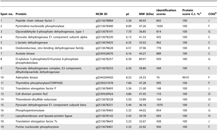
At first, 21 and 11 immunoreactive protein spots with good reproducibility were excised from 2-DE gels stained with Coomassie with pH 3–10 and 4–7 IPG strips, respectively. MALDI-TOF/TOF MS analysis indicated the presence of 31 protein spots, corresponding to a total of 19 different proteins. Of these, 12 proteins corresponded to single spots and 7 proteins were represented by multiple immunoreactive spots, indicating post-translational modifications such as chemical modification and proteolytic cleavage. The information regarding the identity, isoelectric point, molecular weight, identification score, and protein score of these immunogenic proteins are shown in Table 1.
PSORTb analysis predicted that 18 of the 19 immunogenic proteins would be located in the cytoplasm, and the cell division protein ftsZ might be located at multiple sites, including the cytoplasm and cytoplasmic membrane. Most of the proteins identified in this way are enzymes involved in cell metabolism, cell structure and host cell invasion. According to the clusters of orthologous groups (COG) functional classification system, these proteins were assigned to the metabolism, cellular processing and signaling, and information storage and processing groups. Specifically, these proteins were found to be involved in energy production and conversion (spots 4, 5, 7, 9, and 15); carbohydrate transport and metabolism (spots 3, 8, and 16); amino acid transport and metabolism (spot 6); nucleotide transport and metabolism (spots 2, 10, 11, and 19); coenzyme transport and metabolism (spot 17); cell cycle control, cell division, and chromosome partitioning (spot 13); posttranslational modification, protein turnover, and chaperoning (spot 14), and translation, ribosomal structure and biogenesis (spots 1, 12, and 18). Pyruvate dehydrogenase E1 component alpha subunit (PDHA, spot 4), pyruvate dehydrogenase E1 component beta subunit (PDHB, spot 15), and GAPDH (spot 3) have also been reported to participate in the process of adhesion to host cells.
PDHB is usually considered a cytoplasmic protein, but it has been detected on the surfaces of some bacteria, and the antigenicity of PDHB in other mycoplasma species has also been demonstrated [16–18]. In the present study, the immunogenicity of PDHB was proved to be reproducible by Western blot analysis using strongly positive sera. Specific primers have been designed to amplify PDHB genes from differentM. bovisisolates. The amino acid sequence of PDHB of theM. bovis strain PD showed over 99.7% homology with PDHBs from otherM. bovisisolates, about 98.2% homology with the PDHB fromM. agalactiae(which infects sheep and goats) and 66.2% homology with those of other
Figure 1. Extraction of the whole-cell proteins ofM. bovisstrain PD and expression of rPDHB protein.(A1) The extracted whole-cell proteins were separated by SDS-PAGE. (A2) Purity analysis of the recombinant His-tagged PDHB protein by SDS-PAGE. (B) Purified rPDHB protein was subjected to Western blot analysis using an anti-His-tag antibody.
mycoplasma species (isolated from cattle, sheep and goats) (Table 2). These data showed the protein PDHB to be structurally conserved within theM. boviscluster.
Expression ofM. bovisPDHB
The 987 bp gene encoding PDHB was amplified from the genome ofM. bovisstrain PD and cloned into a prokaryotic vector. Then recombinant PDHB (rPDHB) containing six histidine residues was expressed in E. coli, as illustrated in Fig. 1A. The Western blot analysis result indicated the presence of an immunoreactive band of about 37 kDa corresponding to the rPDHB (Fig. 1B), with a slightly higher molecular mass than the native protein ofM. bovis.
Antigenicity analysis ofM. bovisPDHB
In order to examine whether the antigenicity ofM. bovisPDHB was species-specific, Western blot analysis was performed to assess
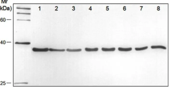
the reactivity of the prepared rabbit anti-rPDHB polyclonal antibody and the whole-cell proteins of eightM. bovisisolates. As shown in Fig. 3, all eightM. bovisstrains isolated from different regions reacted with the rabbit anti-rPDHB polyclonal antibody. For the reaction withM. bovirhinis,M. ovipneumoniae, M. agalactiae, bovine viral diarrhea virus (BVDV), bovine parainfluenza virus type 3 (BPIV3), and infectious bovine rhinotracheitis virus (IBRV), all these pathogens failed to be recognized by the anti-rPDHB polyclonal antibody exceptM. agalactiae(Fig. S1). All pathogens in the present study were identified using specific PCR, as shown in Fig. S2. The results indicated that the antigenicity of M. bovis
PDHB was similar to that of M. agalactiae PDHB but markedly different from that of other pathogens.
Establishment of rPDHB-based iELISA
According to the immunogenicity and antigenicity ofM. bovis
PDHB, a rPDHB-based iELISA was established to confirm the
Figure 2. Two-dimensional gel electrophoresis (2-DE) and immunoblotting of the whole-cell proteins ofM. bovisstrain PD.First, 350mg and 100mg of protein were separated by IEF using (A) a pH 3–10 IPG strip and (C) a pH 4–7 IPG strip, respectively. This was followed by
SDS-PAGE on 12% gels and staining with Coomassie brilliant blue R-350. Immunoblotting was performed using each of the four antisera (Table S1) from naturally infected cattle with three replicates. The immunoreactive protein spots on PVDF membranes B and D contained all spots with good reproducibility as identified by each of the four positive sera. These corresponded to the proteins separated by 2-D gels A and C, respectively. pI values are shown on top, and the standard molecular weights are shown to the left of the gels. The spot numbers correspond to those identified by MS and listed in Table 1.
feasibility of rPDHB as a diagnostic antigen. Standard positive and negative sera were used to optimize the reaction conditions. Eventually, the best reaction conditions were selected as follows: the concentration of rPDHB used for coating was 100 ng/well, blocking buffer was 10% sheep serum, the best dilutions of serum
sample and secondary antibody were 1:160 and 1:2000 (v/v), respectively, and the cutoff value was 0.316.
To examine the specificity of this iELISA, the prepared positive sera of other pathogens, including Mycoplasma mycoides subsp. mycoidesSC (MmmSC),M. agalactiae,M. bovirhinis,M. ovipneumoniae, BVDV, BPIV3, and IBRV, were tested. According to the
Table 1.M. bovisproteins identified by mass spectrometry and reactions with sera from naturally infected cattle in immunoblotting experiments.
Spot no. Protein NCBI ID pI MW (kDa)
Identification scores
Protein
score C.I. %a COGb
1 Peptide chain release factor 1 gi|313678884 5.30 40.43 842 100 J
2 Pyrimidine-nucleoside phosphorylase gi|313678400 8.09 47.26 1020 100 F
3 Glyceraldehyde-3-phosphate dehydrogenase, type 1 gi|313678191 7.70 36.85 814 100 G
4 Pyruvate dehydrogenase E1 component subunit alpha gi|313678230 6.13 41.33 435 100 C
5 L-lactate dehydrogenase gi|313678425 6.35 35.02 962 100 C
6 Oxidoreductase, zinc-binding dehydrogenase family gi|313678628 6.51 37.96 778 100 E
7 Acetate kinase gi|339320670 6.16 44.27 889 100 C
8 D-xylulose 5-phosphate/D-fructose 6-phosphate phosphoketolase
gi|313678257 6.30 89.91 929 100 G
9 Pyruvate dehydrogenase complex, E3 component, dihydrolipoamide dehydrogenase
gi|313678233 6.50 58.80 569 100 C
10 Adenylate kinase gi|344204920 8.53 24.33 76 99.91 F
11 Thymidine phosphorylase(TDRPASE) gi|339321078 7.60 47.28 995 100 F
12 Translation elongation factor P gi|313678493 5.36 21.00 148 100 J
13 Cell division protein ftsZ gi|339320924 4.86 41.95 110 100 D
14 Thioredoxin-disulfide reductase gi|313678228 5.50 33.89 164 100 O
15 Pyruvate dehydrogenase E1 component subunit beta gi|313678231 5.44 36.18 1070 100 C
16 Phosphopentomutase gi|313678465 5.32 44.02 854 100 G
17 Lipoyltransferase and lipoate-protein ligase gi|313678142 5.43 39.78 693 100 H
18 Translation elongation factor Ts gi|313678643 5.23 32.67 438 100 J
19 Purine nucleoside phosphorylase gi|313678401 5.33 25.92 956 100 F
Protein spots from 2-DE were sequenced using MALDI-TOF/TOF MS and identified by searching mycoplasma databases using the MASCOT search engine 2.2. A GPS explorer protein confidence index$95% were used for further manual validation.
aC.I. %: the confidence interval for the protein score.
bCOG database functional classes: (C) energy production and conversion, (D) cell cycle control, cell division, chromosome partitioning, (E) amino acid transport and metabolism, (F) nucleotide transport and metabolism, (G) carbohydrate transport and metabolism, (H) coenzyme transport and metabolism, (J) translation, ribosomal structure and biogenesis, (O) post-translational modification, protein turnover, chaperones.
doi:10.1371/journal.pone.0088328.t001
Table 2.Homology of PDHB from theM. bovisstrain PD and other mycoplasmas.
Mycoplasmas NCBI Reference sequence Homology
Mycoplasma bovisPG45 YP_004055971.1 100.0%
Mycoplasma bovisHubei-1 YP_004683146.1 99.7%
Mycoplasma bovisHB0801 YP_006470739.1 99.7%
Mycoplasma agalactiaePG2 YP_001256238.1 98.2%
Mycoplasma bovigenitalium WP_004420033.1 66.2%
Mycoplasma ovipneumoniae WP_010321080.1 58.5%
Mycoplasma mycoides subsp. capristr. GM12 ACU79371.1 47.9%
Mycoplasma mycoides subsp. mycoidesSC str. PG1 NP_975265.1 47.9%
Mycoplasma capricolum subsp. capricolumATCC 27343 YP_424213.1 47.3%
Mycoplasmas shown on the list were selected using the NCBI BLAST server, basing on the principle of recent homology. Then the amino acid sequences of these PDHBs were downloaded from NCBI, and DNAstar software (version 5.0) was used to analyze the homology.
detection results of all the positive sera listed above, only the sera againstM. agalactiaereacted withM. bovisrPDHB (mean OD was 0.578).
Performance of rPDHB-based iELISA and a commercial kit
rPDHB-based iELISA and the commercial kit were both used to test 358 serum samples collected from cattle feedlots in different provinces. As shown in Table 3, 182 and 143 sera were confirmed to be positive for anti-M. bovis antibodies by iELISA and the commercial kit. The estimated Kappa agreement coefficient between the two detection methods was 0.783. Then Western blot analysis was performed to confirm the incompatible sera by both methods using the whole-cell proteins ofM. bovisstrain PG45. Notably, 39 positive serum samples that had been missed by the commercial kit were all correctly found to be positive by Western blot analysis. All negative samples detected by rPDHB-based iELISA were also found to be negative by the commercial kit. The results suggested that the detection rate of rPDHB-based iELISA was higher than that of the commercial kit currently used forM. bovisantibody detection.
To compare the sensitivity of two methods, 140 positive and 20 negative sera were selected, diluted, and detected with the commercial kit and rPDHB-based iELISA. As shown in Table 4, the highest dilution ratios of iELISA and commercial kit for detecting all 140 positive serum samples were both 1:640. However, iELISA was able to detect 127/140 positive sera at dilution ratio of 1:2560, and the detection rate of commercial kit was only 51/140. The difference was statistically significant (P,
0.05). Moreover, the detection rate of rPDHB-based iELISA was significantly higher than that of the commercial kit at a serum dilution ratio of 1:5120 to 1:10,240 (P,0.05). In addition, iELISA was still competent to detect positive sera at dilution ratio as high as 1:10,240 and showed a detection rate of 12/140, but the highest dilution ratio for the commercial kit was 1:5120. The data suggested that the sensitivity of rPDHB-based iELISA was higher than that of the commercial kit.
Discussion
M. bovis has caused severe losses to the worldwide cattle industry. Currently, only a few M. bovis vaccines, including bacterin and autogenous vaccines, have been approved in the United States. However, most bacterin-based vaccines cannot
provide complete protection. In some cases, vaccination did not bring down the morbidity and mortality but even aggravated the symptoms instead [19–21]. For this reason, laboratory diagnosis is of great significance to the prevention and treatment ofM. bovis
infection. Because M. bovis infection is often latent and the bacterium is seldom shed from healthy cattle, serological detection ofM. bovisantibody, which can last for several months and can be detected at high levels by ELISA, is considered a more reliable method of diagnosis ofM. bovisinfection [5]. However, commer-cial kits based on the whole-cell proteins currently used for serological diagnosis of M. bovis infection cannot ensure the detection of the variegatedM. bovisisolates, and the development of ELISA using the specific conserved antigen with higher specificity and sensitivity is considered a promising alternative. Therefore, the exploitation of M. bovis proteins with excellent immunogenicity is necessary to the renewal and improvement of
M. bovisdiagnostic techniques. In the present study, immunopro-teomics was used to screen the immunogenic proteins from aM. bovisstrain isolated in China. Information regarding the immuno-genic proteins identified in this way can provide a reference for the diagnosis ofM. bovisinfection.
Immunoproteomics involves combining 2-DE, immunoblotting, and mass spectrometry for analysis of functional proteins. In recent years, the immunoproteomics approach has seen increasingly widespread use in the diagnosis and vaccine research of important livestock pathogenic mycoplasmas, such asMycoplasma capricolum subsp. capripneumoniae (Mccp), M. hyopneumoniae, and MmmSC [15,18,22]. However, it has seen less use in research intoM. bovis. Thomas et al. performed 2-DE and MS to analyze the protein expression differences between the 7th and 116th generation ofM. bovisstrains [23]. Then an undiscovered adhesive protein as a new member of the Vsps family was identified. The study also showed that the phenotype of M. bovis may change after a long-term culturein vitro. For this reason, only the first three generations of
M. bovisfield-isolate strain PD were used for the preparation of protein samples in the present study. It was to prevent any change inM. bovis antigen proteins caused by long-term culture in vitro
from affecting the results. Moreover, whole-cell proteins served as the protein sample for 2-DE instead of membrane proteins to
Figure 3. Antigenicity of PDHBs from differentM. bovisisolates. The whole-cell proteins of eight M. bovisisolates were separated by SDS-PAGE, blotted onto a PVDF membrane, and subjected to Western blot analysis with a rabbit anti-rPDHB polyclonal antibody. The positions of molecular mass markers are indicated at the left in kDa. Lanes 1–8:M. bovisstrain PD, PG45, SD-2, Hubei-1, HRB-1, GY-7, GY-14, and WF-3, respectively.
doi:10.1371/journal.pone.0088328.g003
Table 3.Detection rates of rPDHB-based iELISA and the commercial kit.
No. of sera Detection results Commercial kita
rPDHB-based
iELISA Western blotb
19 4+ Positive NTc
71 3+ Positive NT
38 2+ Positive NT
15 1+ Positive NT
176 Negative Negative NT
39 Negative Positive Positive
Detection rate (%)d39.9 (143/358) 50.8 (182/358)
Kappa 0.783
aPositive results were classified from 1
+to 4+according to the kit protocol. bWestern blot analysis was performed to confirm that the sera were incompatible. It was performed using the whole-cell proteins ofM. bovisstrain PG45.
c
NT, not tested.
dDetection rate = (number of positive samples/total number of samples)
6100.
prevent the loss of important cytoplasmic antigen proteins [12]. Joerg et al. demonstrated the importance of antiserum selection in the immunogenic protein screening process [15]. They screened out 24 immunogenic proteins using serum obtained from the cattle that had been experimentally infected withMmmSC B237, but 13 additional proteins were detected when serum from the cattle with clinical acute onset was used. It indicated that the antibody induced in the naturally infected cattle is indispensable to the screening of immunogenic proteins of M. bovis. For this reason, strongly positive antisera from the cattle with natural onset were used in the present study for Western blot analysis to ensure that as many immunogenic proteins as possible would be recognized.
In the present 2-DE assay, about 570 and 338 prominent protein spots were separated in the gels with pH 3–10 and 4–7 IPG strips, respectively. According to the information provided by NCBI, there are 765 proteins encoded byM. bovisgenome, which means that the present study acquired a proteome coverage of over 70%. It is similar to that observed in proteomic studies of other mycoplasma species [14,15,24]. Later, 19 immunogenic proteins were identified by Western blot and MALDI-TOF/TOF MS analysis. Most of these proteins were cytoplasmic proteins that were mainly involved in cell metabolism, cellular processes and signaling, and information storage and processing. Some of them were also related to adherence and invasion of host cells. Remarkably, some similar immunogenic proteins from other mycoplasma species have already been reported. These included PDHA, PDHB, GAPDH, L-lactate dehydrogenase (LDH), and translation elongation factors [15,22,25]. GAPDH is a highly conserved protein, and one of the features of GAPDH is its presence on the cell surface of several prokaryotic and eukaryotic cells, where it is able to bind mucin perhaps contributing to adherence to the epithelia [26]. In 2007, a study performed by Perez-Casal and Prysliak suggested thatM. bovis GAPDH is an important immunogenic protein and might be a good candidate for diagnosis and vaccines [27]. However, they subsequently found that although cattle vaccinated with a subunit vaccine based on GAPDH produced high titers of IgG1 antibodies, there were few differences in the number of lung lesions and survival rate after challenge with a combination of three M. bovisisolates [12]. It indicates that screening and exploiting new immunogenic proteins are very necessary to the diagnosis and prevention of M. bovis
infection. EF-Tu, EF-Ts, and EF-G are three factors usually involved in the protein translation process in prokaryotic cells. Elongation factor thermo stable proteins (EF-Ts) serve as guanine nucleotide exchange factors for elongation factor thermo unstable proteins (EF-Tu), catalyzing the release of guanosine diphosphate from EF-Tu. It allows EF-Tu to bind to a new guanosine triphosphate molecules, release EF-Ts, and go on to catalyze another aminoacyl tRNA addition. The EF-Tu proteins of many pathogens, including mycoplasmas, bacteria, and parasites have
been reported to be immunoreactive [25,28,29]. EF-Tu has also been described as surface-localized, which allows it to mediate binding ofM. pneumoniaeto fibronectin [16]. In the present study,
M. bovis EF-Ts was found to be immunoreactive. The issues of whether EF-Ts is antigenic conserved or involved in the adherence to host cells merit further study.
Pyruvate dehydrogenase (E1), dihydrolipoyl transacetylase (E2), and dihydrolipoyl dehydrogenase (E3) catalyze the conversion of pyruvic acid to acetyl CoA. With other cofactors, they form pyruvate dehydrogenase complex (PDHC). This conversion is a bond linking glycolysis and the tricarboxylic acid cycle. E1 is an
a2b2 tetramer. It mainly catalyzes decarboxylation of pyruvate. Some studies have demonstrated that mycoplasma PDHB (beta subunit of E1) is a phosphoprotein with a cytoskeleton-like structure, and it can also be expressed on the surfaces of mycoplasma cells. Its primary roles are biosynthetic and metabolic and take place in the cytoplasm, but it is also involved in binding to the surface of the host cell. Previous studies have suggested that the conformation that PDHB assumes on the surface of the membrane may confer biological and virulence-related functions [16,17]. Zhao et al. identified nine proteins, including PDHB, in the membrane protein fraction of Mycoplasma capricolum subsp. capripneumoniaeusing a MS system that reacted with convalescent sera in the immunoblots [22]. Pinto et al. identified five highly immunoreactive antigens, including the PDHB of the M. hyopneumoniae pathogenic strain 7448, using immunoproteomics [18].
The present study is the first to findM. bovisPDHB to exhibit excellent immunogenicity and repeatability. Homology analysis indicated that the amino acid sequences of PDHB of differentM. bovisisolates were nearly identical ($99.7%). With one exception,
M. bovisPDHB had less than or equal to 66.2% homology with any other mycoplasma species isolated from cattle, sheep, or goats. It exhibited 98.2% homology with M. agalactiae PDHB. We examined the antigenicity of M. bovis PDHB by Western blot assay, the presence of specific straps observed in all eightM. bovis
field isolates probed with the rabbit anti-rPDHB polyclonal antibody demonstrated that this protein is structurally and antigenically conserved within the M. bovis cluster. For other species of pathogens from ruminants, cross-reactivity was detected solely withM. agalactiaewhich infects sheep and goats.M. agalactiae
is considered to be the classical agent of contagious agalactia, which occurs worldwide and is one of the principal mycoplasmoses of sheep and goats.M. bovisand M. agalactiae are closely related both phenotypically and genotypically.M. boviswas once calledM. agalactiaesubsp. bovis [5]. They share a 16S rDNA similarity of 99.8% and an unusually high number of related antigens and common epitopes. The existence of cross reactivity is not unexpected between M. bovis and M. agalactiae. However, epidemiological investigations have shownM. bovisto be generally
Table 4.Sensitivity of rPDHB-based iELISA and the commercial kit.
Detection methods Dilutions of the sera
1:320 1:640 1:1280 1:2560 1:5120 1:10240 1:20480
Commercial kit (no. of positive/total no.)
140/140a 140/140a 124/140b 51/140c 7/140d 0/140e 0/140e
rPDHB-based iELISA (no. of positive/total no.)
140/140a 140/140a 131/140b 127/140b 49/140c 12/140d 0/140e
host-specific. AlthoughM. agalactiaehas been isolated from cattle on extremely rare occasions, the roles ofM. bovisandM. agalactiae
as pathogens outside cattle and small ruminants, respectively, have yet to be defined [30]. Most studies indicated that the probability of cross-reaction betweenM. bovisandM. agalactiaeis very low in clinical situations. Therefore, this cross-reactivity is expected to have a negligible effect on the serologic diagnosis of M. bovis
infection in most of the countries in which farms that raise both cattle and small ruminants are rare [10].
Based on these considerations, an iELISA with rPDHB as a diagnostic antigen was established to detect anti-M. bovisantibody. A commercial ELISA kit was used to assess the detection rate and sensitivity of the rPDHB-based iELISA. The comparative analysis between the two methods was carried out by testing 358 serum samples, the detection rate of the iELISA (50.8%) was higher than that of the commercial kit (39.9%), even though the two methods showed a good consistency (kappa = 0.783, where 1.0 is perfect consistency). Furthermore, rPDHB-based iELISA showed a significantly higher detection rate than the commercial kit at a serum dilution ratio of 1:2560 to 1:10,240, suggesting that rPDHB-based iELISA would be a better method for clinical detection with the higher sensitivity. In terms of the specificity of rPDHB-based iELISA, no cross-reaction was detected with the positive sera of other pathogens exceptM. agalactiae. As described above, the cross-reaction betweenM. bovisandM. agalactiaeshould not limit the establishment of this new ELISA detection method forM. bovis.
In conclusion, 19 immunogenic proteins of M. bovis were identified for the first time using 2-DE and MALDI-TOF/TOF MS approaches. An iELISA used to detect serum antibodies ofM. bovis was established with rPDHB expressed in a prokaryotic system. It can provide the supplementary diagnosis to the infectious Mycoplasma bovispneumonia in cattle and advance the associated epidemiological investigation and quarantine.
Materials and Methods
Ethics statement
All animal research was approved by the Beijing Association for Science and Technology (approval ID SYXK (Beijing) 2007-0023) and was in compliance with Beijing Laboratory Animal Welfare and Ethics guidelines as issued by the Beijing Administration Committee of Laboratory Animals. All animal studies were also performed in accordance with the China Agricultural University Institutional Animal Care and Use Committee guidelines (ID: SKLAB-B-2010-003) and approved by animal welfare committee of China Agricultural University.
Mycoplasmas and culture conditions
M. bovis strain PD was used in this study. It was isolated in Shandong Province and stored in the laboratory. It was cultivated at 37uC in PPLO broth (BD, U.S.) containing 2.5% yeast extract (w/v), 20% horse serum (v/v), 0.5% phenol red (v/v) and 2000 IU/l penicillin. pH was maintained at 7.4–7.6.
Preparation of serum samples
The 358 serum samples and the corresponding nasal swabs were collected from cattle feedlots in Beijing, Shandong, Hebei, Tianjin, Hubei, Hunan, and Heilongjiang, all of which are in China. Some of these samples were taken from cattle that showed symptoms of M. bovis infection, such as fever, arthritis, mastitis, conjunctivitis, and pneumonia. PCR amplification and commer-cial ELISA (Mycoplasma bovisAntibody Test Kit, BioVet, Canada) were carried out separately on all samples [31,32]. The serum
samples were kept at –70uC for further study of screening immunogenic protein and establishment of a method of detection.
Extraction of the whole-cell proteins
A modified version of a previously described method was used to extractM. bovisproteins [33]. Briefly, cells were harvested at a density of 108CFU/ml by centrifugation at 4uC and 12,000 g for 20 min and then resuspended in lysis buffer (7 M urea, 2 M thiourea, 4% (w/v) CHAPS, 1% (w/v) DTT, 1% (v/v) cocktail, 0.5% (v/v) IPG buffer and 40 mM Tris-base, pH 9.6). Then cells were sonicated for 10 min on ice with a Sonifier 750 (Branson Ultrasonics Corp., U.S.). The proteins were collected by centri-fugation at 4uC, 100,000 g for 1 h and lysed in the same lysis buffer at room temperature (RT) for 1 h. The protein concentra-tion was determined using a 2-D Quant Kit (GE Healthcare, U.S.).
2-DE
Isoelectric focusing (IEF): IEF was performed as described previously [34]. Briefly, each pH 3–10 IPG strip (17 cm, NL) or pH 4–7 IPG strip (7 cm, NL) was rehydrated at RT for 12 h with 350mg or 100mg M. bovis protein sample in 400ml or 150ml
rehydration sample buffer (6 M urea, 2 M thiourea, 4% (w/v) CHAPS, 65 mM DTT, 0.5% (v/v) IPG, 0.04% (w/v) Bromo-phenol blue and 40 mM Tris-base, pH 9.6). IEF was performed in a PROTEANHIEF System (Bio-Rad, U.S.). The parameters used for IEF were as follows: 50 V for 2 h, 500 V for 30 min, 1000 V for 30 min, 8000 V for 4 h. The final phase of 8000 V was terminated after 50,000 Vh.
SDS-PAGE: After IEF, the IPG strips were successively equilibrated for 15 min in equilibration buffer I containing 64.8 mM DTT and buffer II containing 135 mM iodoacetamide. Two equilibrated IPG strips were subjected to 12% polyacryl-amide gel electrophoresis and sealed with 0.5% agarose solution. The second dimension was carried out at 50 V for 3 h followed by 100 V for 15 h. One strip gel was stained with Coomassie brilliant blue R-350, and the other was subjected to immunoblotting. The stained gel was scanned with an Image Scanner (Amersham Biosciences, U.S.) and analyzed using the PDQuest Basic 8.0 program (Bio-Rad). Three replicates were performed.
Immunoblotting
The separated protein spots from 2-DE gels were electroblotted onto PVDF membranes (Amersham Biosciences) using a trans-blot semi-dry transfer cell (Bio-Rad, U.S.). Blotted membranes were blocked with 10% (v/v) sheep serum (Macgene, China) in PBST (0.5% v/v Tween). Then the membranes were incubated with four anti-M. bovispositive sera tested with both a commercial kit and PCR (Table S1) diluted 1:800 at 4uC overnight, respectively. Three replicates were performed with each positive serum. HRP-conjugated sheep anti-bovine IgG (Sigma, USA; 1:8000 dilution) was used as the secondary antibody at RT for 30 min. Finally, the membranes were treated with Super Enhanced Chemiluminescent Substrate (ECL) Plus (Applygen, China) in accordance with the manufacturer’s instructions. Sera from healthy cattle served as negative controls.
MALDI-TOF/TOF MS and bioinformatic analysis
Immunoreactive protein spots were manually excised from Coomassie stained gels and in-gel digested with trypsin. Briefly, gel pieces were destained with 30% acetonitrile (ACN) in 100 mM NH4HCO3 and dried in vacuum centrifuge at RT. Then they
NH4HCO3at 37uC. The dry peptide samples were reconstituted
in 2ml standard diluent (20:80 ACN:water) and spotted on a 384-well Opti-TOF stainless steel plate. They were then covered with 0.5ml oversaturation cyano-4-hydroxy-cinnamic acid (CHCA) in
50% ACN and 0.1% trifluoroacetic acid (TFA). MS and MS/MS were performed using a MALDI-TOF/TOF instrument (4800 proteomics analyzer, Applied Biosystems). The parameters were set using the 4000 Series Explorer software (Applied Biosystems). The MS spectra were recorded in reflector mode at masses ranging from 800 to 4000 with a focal mass of 2000. MS involved a CalMix5 standard to calibrate the instrument (ABI 4700 Calibration Mixture). Combined peptide mass fingerprinting (PMF) and MS/MS queries were performed using the MASCOT search engine 2.2 (Matrix Science, Ltd., U.S.) embedded into GPS-Explorer Software 3.6 (Applied Biosystems), which had been downloaded from NCBI database (Taxonomy: Mycoplasma (7442 sequences), Dec. 10, 2010). The peptide mass tolerance was 100 ppm. MS/MS fragment tolerance was set to 0.4 Da. A GPS Explorer protein confidence index of$95% was used for further manual validation.
The corresponding protein sequence was downloaded from NCBI. Then the protein function classification was determined by comparison to COGs database with RPS-BLAST program, and the subcellular localization was predicted by PSORTb version 3.0 software.
Expression of rPDHB
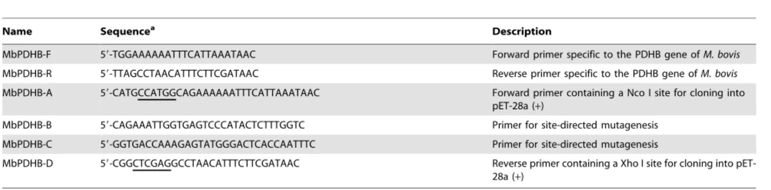
Sequence analysis showedM. bovis PDHB to contain a UGA codon. This codon is translated as tryptophan according to mycoplasma genetic code but translated as a stop codon in theE. coliexpression system. To avoid the production of truncated gene products, this UGA codon was mutated to UGG using two PCR runs by standard overlap extension PCR (39). Two restriction enzyme cutting sites, Nco I and Xho I, were added to the flanks of the PDHB gene sequence. The mutagenic primers and flanking primers are as shown in Table 5.
Then PDHB gene fragment was cloned into a pET-28a(+) prokaryotic expression vector to construct a pET-28a(+)-PDHB plasmid. Transetta (DE3) chemically competent cell (TransGen Biotech, China) was used to express the recombinant protein. Briefly, the competent cells carrying pET-28a(+)-PDHB plasmids were grown in Luria-Bertani (LB) media at 37uC for 3 h and induced with 1 mM isopropylb-D-thiogalactoside (IPTG) for 4 h. Then the harvested cells were disrupted by sonication and recombinant proteins were purified by affinity chromatography to Ni-NTA columns as directed by the manufacturer (Qiagen, Germany).
Preparation of polyclonal antibody against rPDHB
Three New Zealand white rabbits aged 6–7 weeks were injected subcutaneously with purified rPDHB (0.5 mg/kg) three times at 2-week intervals. rPDHB was blended with the same volume of complete Freund’s adjuvant (CFA, Sigma) in the first immuniza-tion and with incomplete Freund’s adjuvant (IFA) in the following immunization. A rabbit injected with adjuvants alone served as a negative control. Sera were collected two weeks after the last immunization and the antibody titers were tested using ELISA. The positive sera were either tested for specificity by Western blot analysis or stored at –70uC.
Establishment of rPDHB-based iELISA
Indirect ELISA was performed as described previously [10]. Briefly, 96-well ELISA plates were coated with 25, 50, 100, 200, or 400 ng/well rPDHB and allowed to incubate at 4uC overnight. Sheep serum, swine serum, horse serum, protein-free blocking buffer, and gelatin served as blocking buffers. Sera (primary antibodies) were diluted to 1:40, 1:80, and 1:160–1:2560 (v/v). HRP-conjugated sheep anti-bovine IgG secondary antibody was diluted to 1:1000, 1:2000, 1:4000, and 1:8000 (v/v). Finally the substrate TMB and 2 M H2SO4 was added for coloration and
termination of the reaction, respectively. The plates were read at an optical density of 450 nm (OD450) with a reference filter of 630
nm in an ELISA plate reader (Pharmacia, U.S.).
Fifty OD450 values of negative sera presenting normal
distribution were used to calculate the mean optical density (OD) and standard deviation (SD). The cutoff value between positive and negative sera was calculated as the mean OD of the fifty negative sera plus 3 SDs of the mean. This calculation provides 99% confidence that all negative values fell within the defined range [35,36].
Specificity
Two methods were used to confirm the specificity ofM. bovis
PDHB: i) Whole-cell proteins fromM. agalactiae(CVCC 344),M. bovirhinis(ATCC 27748),M. ovipneumoniae(ATCC 29419), BVDV (CVCC 69), BPIV3 (ATCC VR-281), IBRV (CVCC AV-346), and eightM. bovisstrains were used to assess cross-reactivity with anti-rPDHB polyclonal antibodies in Western blot assays. The eightM. bovisstrains wereM. bovisPG45 (ATCC 25523),M. bovisHubei-1 (donated by China Animal Health and Epidemiol-ogy Center),M. bovisSD-2 (donated by China Animal Health and Epidemiology Center) and five strains preserved in the lab, specificallyM. bovis PD,M. bovis HRB-1,M. bovisGY-7,M. bovis
GY-14, andM. bovis WF-3. ii) rPDHB-based iELISA was used to detect three rabbit polyclonal antibodies againstM. bovirhinis,M.
Table 5.Primers used for expression of rPDHB in the present study.
Name Sequencea Description
MbPDHB-F 59-TGGAAAAAATTTCATTAAATAAC Forward primer specific to the PDHB gene ofM. bovis
MbPDHB-R 59-TTAGCCTAACATTTCTTCGATAAC Reverse primer specific to the PDHB gene ofM. bovis
MbPDHB-A 59-CATGCCATGGCAGAAAAAATTTCATTAAATAAC Forward primer containing a Nco I site for cloning into pET-28a (+)
MbPDHB-B 59-CAGAAATTGGTGAGTCCCATACTCTTTGGTC Primer for site-directed mutagenesis MbPDHB-C 59-GGTGACCAAAGAGTATGGGACTCACCAATTTC Primer for site-directed mutagenesis
MbPDHB-D 59-CGGCTCGAGGCCTAACATTTCTTCGATAAC Reverse primer containing a Xho I site for cloning into pET-28a (+)
agalactiae, and M. ovipneumoniae (prepared using the method described above), and four positive sera of MmmSC, BVDV, BPIV3, and IBRV that had been purchased from Real Bio-technology (China). All protein samples were boiled before being loaded onto the SDS-PAGE gel. The parameters used to transfer the gel to the PVDF membrane included 60 V for 2 h. All the pathogens described above were identified using specific PCR. The primer sequences and the references are given in Table S2.
rPDHB-based iELISA and commercial kit
The 358 serum samples were tested using rPDHB-based iELISA under optimized conditions, and the degree of agreement and sensitivity between the iELISA and the commercial kit were determined.
Sensitivity: 140 positive serum samples and 20 negative controls were randomly selected. Twofold serial dilutions of the sera from 1:80 to 1:20,480 were employed in the test. The commercial kit and rPDHB-based iELISA were used separately for detection. Samples with OD450values greater than or equal to twice that of
the negative serum were considered positive (A 630 nm filter served as a reference filter).
Statistical analysis
The degree of agreement between the commercial kit and rPDHB-based iELISA was measured using kappa statistics [37]. The sensitivity of the detection of differences between the rPDHB-based iELISA and commercial kit were analyzed using the Chi-square or Fisher’s Exact test with SPSS v19.0 software.P values below 0.05 were considered statistically significant.
Supporting Information
Figure S1 Antigenicity analysis of M. bovisPDHB. M. bovisrPDHB (lane 1) and the whole-cell proteins ofM. bovis(lane
2),M. agalactiae(lane 3),M. bovirhinis(lane 4),M. ovipneumoniae(lane 5), BVDV (lane 6), BPIV3 (lane 7) and IBRV (lane 8) were separated by SDS-PAGE, blotted onto a PVDF membrane and subjected to the following Western blot analysis with rabbit
anti-M. bovisrPDHB polyclonal antibody. (TIF)
Figure S2 PCR identification of the pathogens used in the present study.Photograph of a 1% agarose gel loaded with the PCR or RT-PCR (for RNA viruses: BVDV and BPIV3) products. M: molecular weight marker. Lane 1-7: M. bovis, M. agalactiae, M. bovirhinis, M. ovipneumoniae, BVDV, BPIV3, and IBRV, respectively. The specific primers are listed in Table S2. (TIF)
Table S1 Positive bovine sera used in the immunoblot assays.
(DOC)
Table S2 Primer sequences used for PCR identification.
(DOC)
Acknowledgments
We would like to thank Huiling Chen and Wenqiang Gan for providing the bovine sera. We would also like to thank Xiaolin Zhu and Ming Zhou for improving the English of the manuscript.
Author Contributions
Conceived and designed the experiments: ZHS PF KW WXW. Performed the experiments: ZHS PF HYZ YWZ JX FJ XL WX. Analyzed the data: ZHS PF KW HYZ WXW. Contributed reagents/materials/analysis tools: WXW. Wrote the paper: ZHS PF KW WXW.
References
1. Hale HH, Helmboldt CF, Plastridge WN, Stula EF (1962) Bovine mastitis caused by a Mycoplasma species. Cornell Vet 52: 582–591.
2. Pfutzner H, Sachse K (1996)Mycoplasma bovisas an agent of mastitis, pneumonia, arthritis and genital disorders in cattle. Rev Sci Tech 15: 1477–1494. 3. Xin JQ, Li Y, Guo D, Song NH, Hu SP, et al. (2008) First isolation ofMycoplasma
bovisfrom calf lung with pneumoniae in China. Chinese Journal of Preventive 30: 661–664.
4. Tschopp R, Bonnemain P, Nicolet J, Bumens A (2001) Epidemiological study of risk factors forMycoplasma bovisinfections in fattening calves. Schweiz Arch Tierheilkd 143: 461–467.
5. Nicholas RA, Ayling RD (2003)Mycoplasma bovis: disease, diagnosis, and control. Res Vet Sci 74: 105–112.
6. Caswel JL. Arehambauh M (2007)Mycoplasma bovispneumonia in cattle. Anim Health Res Rev 8: 161–186.
7. Lysnyansky I, Rosengarten R, Yogev D (1996) Phenotypic switching of variable surface lipoproteins inMycoplasma bovisinvolves high-frequency chromosomal rearrangements. J Bacteriol 178: 5395–5401.
8. Nussbaum S, Lysnyansky I, Sachse K, Levisohn S, Yogev D (2002) Extended repertoire of genes encoding variable surface lipoproteins inMycoplasma bovis strains. Infect Immun 70: 2220–2225.
9. Sachse K, Pfutzner H, Heller M, Hanel I (1993) Inhibition ofMycoplasma bovis cytadherence by a monoclonal antibody and various carbohydrate substances. Vet Microbiol 36: 307–316.
10. Robino P, Alberti A, Pittau M, Chessa B, Miciletta M, et al. (2005) Genetic and antigenic characterization of the surface lipoprotein P48 ofMycoplasma bovis. Vet Microbiol 109: 201–209.
11. Scherm B, Gerlach GF, Runge M (2002) Analysis of heat shock protein 60 encoding genes of mycoplasmas and investigations concerning their role in immunity and infection. Vet Microbiol 89: 141–150.
12. Prysliak T, van der Merwe F, Perez-Casal J (2013) Vaccination with recombinantMycoplasma bovisGAPDH results in a strong humoral immune response but does not protect feedlot cattle from an experimental challenge with M. bovis. Microb Pathog 12: 1–8.
13. Jaffe JD, Berg HC, Church GM (2004) Proteogenomic mapping as a complementary method to perform genome annotation. Proteomics 4: 59–77.
14. Ueberle B, Frank R, Herrmann R (2002) The proteome of the bacterium Mycoplasma pneumoniae: comparing predicted open reading frames to identified gene products. Proteomics 2: 754–764.
15. Jores J, Meens J, Buettner FF, Linz B, Naessens J, et al. (2009) Analysis of the immunoproteome ofMycoplasma mycoides subsp. mycoidessmall colony type reveals immunogenic homologues to other known virulence traits in related Mycoplas-ma species. Vet Immunol Immunopathol 131: 238–245.
16. Dallo SF, Kannan TR, Blaylock MW, Baseman JB (2002) Elongation factor Tu and E1bsubunit of pyruvate dehydrogenase complex act as fibronectin binding proteins inMycoplasma pneumoniae. Mol Microbiol 46: 1041–1051.
17. Su HC, Hutchison CA 3rd, Giddings MC (2007) Mapping phosphoproteins in Mycoplasma genitaliumandMycoplasma pneumoniae. BMC Microbiol 7: 47–63. 18. Pinto PM, Chemale G, de Castro LA, Costa AP, Kich JD, et al. (2007)
Proteomic survey of the pathogenicMycoplasma hyopneumoniaestrain 7448 and identification of novel post-translationally modified and antigenic proteins. Vet Microbiol 121: 83–93.
19. Soehnlen MK, Aydin A, Lengerich EJ, Houser BA, Fenton GD, et al. (2011) Blinded, controlled field trial of two commercially availableMycoplasma bovis bacterin vaccines in veal calves. Vaccine 29: 5347–5354.
20. Maunsell FP, Woolums AR, Francoz D, Rosenbusch RF, Step DL, et al. (2011) Mycoplasma bovisinfections in cattle. J Vet Intern Med 25: 772–783. 21. Maunsell FP, Donovan GA (2009)Mycoplasma bovisinfections in young calves.
Vet Clin North Am Food Anim Pract 25: 139–177.
22. Zhao P, He Y, Chu YF, Gao PC, Zhang X, et al. (2012) Identification of novel immunogenic proteins in Mycoplasma capricolum subsp. Capripneumoniae strain M1601. J Vet Med Sci 74: 1109–1115.
23. Thomas A, Leprince P, Dizier I, Ball H, Gevaert K, et al. Identification by two-dimensional electrophoresis of a new adhesin expressed by a low-passaged strain ofMycoplasma bovis. Res Microbiol 156: 713–718.
24. Wasinger VC, Pollack JD, Humphery-Smith I (2000) The proteome of Mycoplasma genitalium. Chaps-soluble component. Eur J Biochem 267: 1571– 1582.
26. Alvarez RA, Blaylock MW, Baseman JB (2003) Surface localized glyceralde-hyde-3-phosphate dehydrogenase ofMycoplasma genitalium binds mucin. Mol Microbiol 48: 1417–1425.
27. Perez-Casal J, Prysliak T (2007) Detection of antibodies against theMycoplasma bovisglyceraldehyde-3-phosphate dehydrogenase protein in beef cattle. Microb Pathog 43: 189–197.
28. Sun XM, Ji YS, Elashrama SA, Lu ZM, Liu XY, et al. (2012) Identification of antigenic proteins of Toxoplasma gondii RH strain recognized by human immunoglobulin G using immunoproteomics. J proteomics 77: 423–432. 29. Yang YL, Wang L, Yin JG, Wang XL, Cheng SP, et al. (2011)
Immunoproteomic analysis of Brucella melitensis and identification of a new immunogenic candidate protein for the development of brucellosis subunit vaccine. Mol Immunol 49: 175–184.
30. Bashiruddin JB, Frey J, Konigsson MH, Johansson KE, Hotzel H, et al. (2005) Evaluation of PCR systems for the identification and differentiation of Mycoplasma agalactiae andMycoplasma bovis: a collaborative trial. Vet J 169: 268–275.
31. Hou X, Fu P, Zhang HY, Zhang YW, Wu WX (2012) Development of loop-mediated isothermal amplification for rapid detection of Mycoplasma bovis. Journal of Agricultural Biotechnology 20: 218–224.
32. Tenk M, Ba´lint A, Stipkovits L, Biro´ J, Dencso L (2006) Detection ofMycoplasma boviswith an improved pcr assay. Acta Vet Hung 54: 427–435.
33. Regula J, Ueberle B, Boguth G, Gorg A, Schnolzer M, et al. (2000) Towards a two-dimensional proteome map ofMycoplasma pneumoniae. Electrophoresis 21: 3765–3780.
34. Go¨rg A, Weiss W, Dunn MJ (2004) Current two-dimensional electrophoresis technology for proteomics. Proteomics 4: 3665–3685.
35. Webster KA, Giles M, Dawson C (1997) A competitive ELISA for the serodiagnosis of hypodermosis. Vet Parasitol 68: 155–164.
36. Tiwari S, Kumar A, Thavaselvam D, Mangalgi S, Rathod V, et al. (2013) Development and comparative evaluation of a plate enzyme-linked immuno-sorbent assay based on recombinant outer membrane antigens Omp28 and Omp31 for diagnosis of human brucellosis. Clin Vaccine Immunol 20: 1217– 1222.