CASE REPORT
Neonatal pyruvate dehydrogenase deficiency due to a R302H
mutation in the PDHA1 gene: MRI findings
João P. Soares-Fernandes&Roseli Teixeira-Gomes&
Romeu Cruz&Manuel Ribeiro&Zita Magalhães&
Jaime F. Rocha&Lara M. Leijser
Received: 4 September 2007 / Revised: 13 November 2007 / Accepted: 18 November 2007 / Published online: 16 January 2008
#Springer-Verlag 2007
Abstract Pyruvate dehydrogenase (PDH) deficiency is one of the most common causes of congenital lactic acidosis. Correlations between the genetic defect and neuroimaging findings are lacking. We present conventional and diffu-sion-weighted MRI findings in a 7-day-old male neonate with PDH deficiency due to a mosaicism for the R302H mutation in the PDHA1 gene. Corpus callosum dysgenesis, widespread increased diffusion in the white matter, and bilateral subependymal cysts were the main features. Although confirmation of PDH deficiency depends on specialized biochemical analyses, neonatal MRI plays a role in evaluating the pattern and extent of brain damage, and potentially in early diagnosis and clinical decision making.
Keywords Pyruvate dehydrogenase deficiency . Neonate . MRI . DWI
Introduction
Pyruvate dehydrogenase (PDH) deficiency results from mutations in the mitochondrial PDH complex, an important enzyme for mitochondrial energy metabolism. In 90% of patients the abnormality occurs in the X-linked gene for the E1-α subunit (PDHA1 gene) [1]. Neonatal presentation with lactic acidosis is most common [2]. Although confirmation of the diagnosis depends on specialized biochemical analyses, neonatal neuroimaging plays a role in the evaluation of brain damage and abnormalities. We describe the conventional and diffusion-weighted (DW) MRI findings in a 7-day-old male infant with PDH deficiency due to mosaicism for the R302H mutation in the PDHA1 gene. Imaging correlations at 18 months of age are provided. Potential injury mechanisms, and the diag-nostic and clinical decision-making value of the neuro-imaging findings are discussed.
Case report
The patient was a male infant, born at 38 weeks’gestation to a 16-year-old primigravid Caucasian mother; the parents were nonconsanguineous. Prenatal US screening was not performed. His birth weight was 3,180 g and his head circumference was on the 25th centile. He was admitted to the neonatal intensive care unit after birth because of respiratory distress. His face was mildly dysmorphic, with hypertelorism, a long nasal philtrum, and anteverted nostrils. Initial neurological examination revealed absent swallowing reflex, exaggerated Moro reflex, and axial hypotonia.
On admission, elevated plasma lactate (8.09 mmol/l; normal range, NR, 0.63–2.44 mmol/l) and pyruvate
Pediatr Radiol (2008) 38:559–562
DOI 10.1007/s00247-007-0721-9
J. P. Soares-Fernandes (*)
:
M. Ribeiro:
Z. Magalhães:
J. F. Rocha
Department of Neuroradiology, Hospital de S. Marcos, Largo Engenheiro Carlos Amarante,
Braga 4710-965, Portugal e-mail: [email protected]
R. Teixeira-Gomes
Division of Neuropediatrics, Hospital Pedro Hispano, Matosinhos, Portugal
R. Cruz
Department of Neuroradiology, Hospital Geral de Sto. António, Porto, Portugal
L. M. Leijser
Department of Paediatrics, Division of Neonatology, Leiden University Medical Center,
(339μmol/l; NR 54.1–119.9μmol/l) levels, with a normal lactate/pyruvate ratio, were found. Subsequently, elevated cerebrospinal fluid lactate (6.6 mmol/l; NR 1.2–2.1 mmol/l) and alanine (95μmol/l; NR 13.8–32.6μmol/l) levels were also demonstrated.
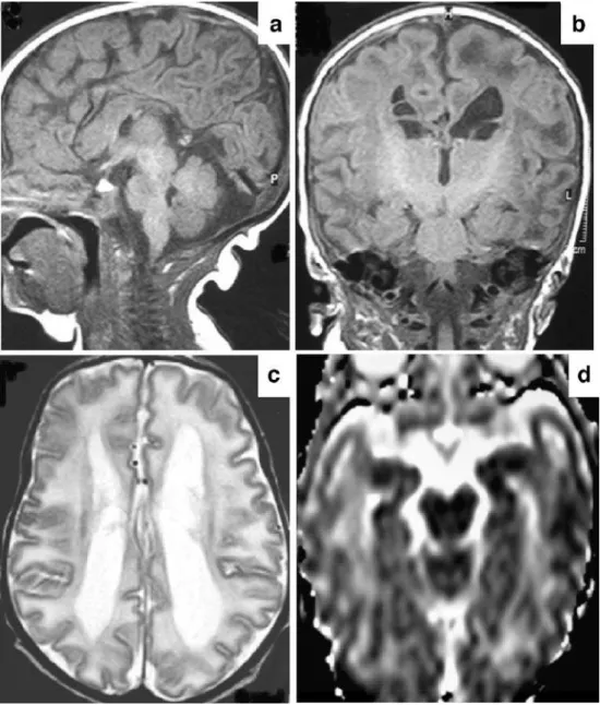
MRI at 7 days of age showed colpocephaly, dysgenesis of the corpus callosum, prominent bilateral germinolytic cysts in the caudothalamic groove and lateral ventricles, and widespread T1 and T2 prolongation of the white matter with cystic change in the anterior temporal lobes (Fig. 1). A few punctate high-signal lesions were found bilaterally in the basal ganglia on T2-weighted (T2-W) images. No cortical malformations were detected and myelination was appropriate for gestational age. The cerebellum appeared normal. In addition, widespread
increased diffusion was detected in the white matter on DW images and ADC maps. MR spectroscopy (MRS; region of interest positioned in the left centrum semiovale, TE 135 ms; not shown) demonstrated a lactate doublet at 1.33 ppm, but failed to reveal a pyruvate peak at 2.37 ppm. Fibroblast biochemical analysis confirmed significantly decreased PDH activity. Mutation analysis detected mosa-icism for the R302H mutation in the PDHA1 gene. No specific treatment was started.
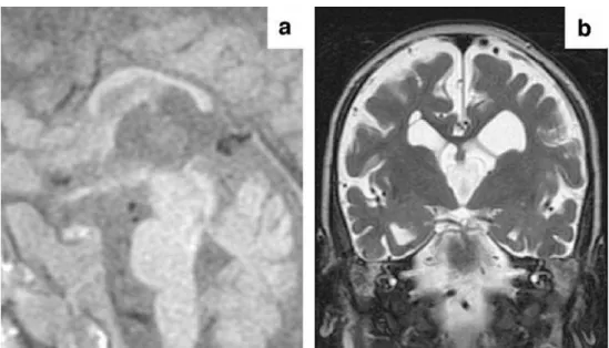
At 18 months of age, the child demonstrated microceph-aly and severe neurodevelopmental delay with a total absence of response to visual or auditory stimuli. MR images showed severe atrophy of the white matter and corpus callosum (Fig.2). White matter signal intensity was normal on T1-W and T2-W images.
Fig. 1 Brain MRI at 7 days of
age.aMid-sagittal T1-W
spin-echo (TR/TE 624/15 ms) image demonstrates dysgenesis of the corpus callosum. Note thicken-ing of the genu and anterior body while the mid- and poste-rior body are very thin. There is agenesis of the splenium and
rostrum.bCoronal T1-W
spin-echo image demonstrates subependymal cysts in the right lateral ventricle and left caudo-thalamic groove. Also note nor-mal myelination (hyperintensity) in the posterior limb of the internal capsule and widespread hypointensity of the white
mat-ter.cAxial T2-W turbo
spin-echo (TR/TE 3,000/120 ms) image shows enlarged and mildly colpocephalic lateral ventricles and widespread white
matter hyperintensity.dADC
map reveals bilaterally increased water diffusion in the white matter, particularly in the tem-poral lobes
Discussion
PDH is a key mitochondrial enzyme complex in the oxidative carboxylation of pyruvate to acetyl-CoA, control-ling the flow of substrate entering the Krebs cycle [1,2]. In neonates, recognition of PDH deficiency is difficult due to the wide variety of clinical presentations that depend on the particular mutation and the residual PDH complex activity. Poor feeding, lethargy, hypotonia, and respiratory distress are nonspecific signs that may point in the direction of a metabolic disorder. Lactic acidosis and normal lactate/ pyruvate ratios are more characteristic findings. Decreased PDH complex activity confirms the diagnosis, but the bio-chemical studies may take weeks to months to complete.
Brain MRI forms part of the investigation of metabolic disorders, including neonatal PDH deficiency. Convention-al MRI features of PDH deficiency have been previously described [1–5], and include enlarged ventricles, cerebral and cerebellar white matter involvement (including cystic change), brainstem atrophy, absence of medullary pyra-mids, abnormal inferior olives, cerebellar dysplasia, partial or complete agenesis of the corpus callosum, and basal ganglia hyperintensities on T2-W images. To our knowl-edge, this is the first report of conventional MRI and DW imaging findings in a male neonate. The gender differen-tiation is of importance because E1-α deficiency is associated with various types of brain damage and significant differences in symptoms have been described between female and male patients; for example, infantile spasms have been encountered almost exclusively in females [3].
In the infant presented here, diffuse and widespread white matter involvement presented with T1 hypointensity and T2 hyperintensity on conventional images, and
in-creased signal intensity on ADC maps. Elevated ADC values (i.e. an increase in water content and a decrease in restriction of water diffusion) are suggestive of diffuse white matter abnormality (vasogenic oedema or encepha-lomalacia), and may be predictive of progressive atrophy. Occasionally, infants with a metabolic disorder may be suspected of hypoxic-ischaemic encephalopathy (HIE). In HIE, white matter injury, occurring as a consequence of a hypoxic-ischaemic insult, may present with similar con-ventional and DW imaging findings and evolution [6]. In metabolic disorders, however, the basal ganglia abnormal-ities tend to be more focal and punctate and mostly do not evolve [7]. HIE is an important differential diagnosis to consider since implications for medicolegal proceedings and genetic counselling can be considerable. Thus, al-though nonspecific, diffuse white matter involvement, demonstrated by conventional MRI and DW imaging, may be of prognostic value.
Corpus callosum dysgenesis can be identified in prenatal life by US and fetal MRI, and is relevant for the differential diagnosis with HIE, indicating a disease onset during the first half of gestation. Callosal malformations have been previously reported in PDH deficiency [1, 4] and several other metabolic disorders, such as other mitochondrial disorders, peroxisomal biogenesis disorders, amino acid metabolism disorders and urea cycle defects [7].
Germinolytic cysts are more often seen in preterm than term-born infants and are probably the result of haemor-rhage and subsequent lysis of the germinal matrix. In our term-born infant the cysts were already demonstrated at 7 days of age and, therefore, were probably congenital [3,
4]. Their location in the frontal horns of the lateral ventricles, as well as in the caudothalamic grooves, was atypical for the evolution of germinal matrix haemorrhage. Fig. 2 Brain MRI at 18 months
of age. Mid-sagittal T1-W
spin-echo image (a) and coronal
T2-W turbo spin-echo image (b)
demonstrate a thin callosal body and marked atrophy of the hemispheric white matter. Intra-ventricular septations are also
seen inb
Subependymal cysts have also been described in congenital heart disease, congenital infections and several metabolic disorders and cause primary lactic acidosis, such as Zellweger syndrome, holocarboxylase synthetase deficien-cy, pyruvate carboxylase deficiendeficien-cy, and glutaryl CoA dehydrogenase deficiency [4]. To our knowledge, only a few prior MRI observations of germinolytic cysts in term-born neonates with PDH deficiency have been reported [1,
4]. In one of these infants, PDH deficiency was due to a rarer mutation in the PDX1 gene that encodes for the E3BP subunit of the PDH complex [4]. In another, a different mutation in the E1-α subunit was detected, and the subependymal cysts were interpreted as having evolved from germinal matrix haemorrhages [1]. It is interesting to note that different mutations leading to PDH deficiency are associated with germinolytic cysts on neonatal neuroimag-ing, suggesting a noncausal relationship. Although nonspe-cific, the cysts, in combination with other neuroimaging and clinical findings, can be a useful finding to suggest a metabolic disorder, and specifically PDH deficiency, espe-cially as they are mostly easily detected by cranial US or MRI. Intraventricular septations, as seen in our patient on follow-up imaging, have been previously reported in older patients [3], and probably reflect the evolution of sub-ependymal cysts.
MRS in PDH deficiency, performed with a long TE (135 ms), usually shows an inverted lactate doublet at 1.33 ppm that is also a nonspecific finding. In our patient, a narrow singlet at 2.37 ppm, which corresponds to pyruvate, was not seen. This peak has been described previously in a female neonate with PDH deficiency who died during the neonatal period [2]. It probably reflects severe disease with very low PDH activity levels.
The imaging differential diagnosis of the combination of widespread white matter injury, germinolytic cysts and dysgenesis of the corpus callosum includes oxidative phosphorylation disorders, amino acid metabolism and urea cycle disorders, and peroxisomal biogenesis disorders. In these disorders findings are often associated with delayed or absent myelination and cortical malformations [7], which we were unable to demonstrate in our patient.
The exact mechanisms of the cerebral lesions associated with PDH deficiency are largely unexplained. The white
matter injury may be due to reduced energy generation. Astrocytic glutamate uptake, a protective mechanism against excitotoxic neuronal injury, is coupled with glycol-ysis and cellular ATP generation [8]. Impairment of cell energy metabolism, which occurs in PDH deficiency, can lead to irreversible excitotoxicity. However, the white matter lesions may also be attributable to selective susceptibility of immature oligodendrocytes to injury from free radicals and glutamate [8].
In conclusion, we present the first description of early MRI and MRS findings in a male neonate with PDH deficiency. The combination of clinical findings, increased lactate and pyruvate levels, and neuroimaging findings, including ventriculomegaly, widespread white matter in-volvement, callosal malformations and germinolytic cysts, is suggestive of PDH deficiency and may lead to early treatment and clinical decision making, before biochemical confirmation.
References
1. Blanco-Barca O, Gomez-Lado C, Rodrigo-Saez E et al (2006) Pyruvate dehydrogenase deficit associated to the C515T mutation
in exon 6 of the E1alpha gene. Rev Neurol 43:341–345
2. Zand DJ, Simon EM, Pulitzer SB et al (2003) In vivo pyruvate detected by MR spectroscopy in neonatal pyruvate dehydrogenase
deficiency. AJNR 24:1471–1474
3. Wada N, Matsuishi T, Nonaka M et al (2004) Pyruvate dehydro-genase E1alpha subunit deficiency in a female patient: evidence of antenatal origin of brain damage and possible etiology of infantile
spasms. Brain Dev 26:57–60
4. Dey R, Mine M, Desguerre I et al (2003) A new case of pyruvate dehydrogenase deficiency due to a novel mutation in the PDX1
gene. Ann Neurol 53:273–277
5. Willemsen M, Rodenburg RJ, Teszas A et al (2006) Females with PDHA1 gene mutations: a diagnostic challenge. Mitochondrion
6:155–159
6. Rutherford MA, Ward P, Malamatentiou C (2005) Advanced MR techniques in the term-born neonate with perinatal brain injury.
Semin Fetal Neonatal Med 10:445–460
7. Leijser LM, de Vries LS, Rutherford MA et al (2007) Cranial ultrasound in metabolic disorders presenting in the neonatal period: characteristic features and comparison with MR imaging. AJNR
28:1223–1231
8. Yoshioka A, Bacskai B, Pleasure D (1996) Pathophysiology of
oligodendroglial excitotoxicity. J Neurosci Res 46:427–437