Plasminogen Activator Inhibitor-2 Plays a
Leading Prognostic Role among Protease
Families in Non-Small Cell Lung Cancer
Chia-Yi Su1, Yu-Peng Liu2, Chih-Jen Yang3, Yuan-Feng Lin4, Jean Chiou1,5, Li-Hsing Chi6, Jih-Jong Lee7, Alex T. H. Wu6, Pei-Jung Lu8, Ming-Shyan Huang3*, Michael Hsiao1*
1Genomics Research Center, Academia Sinica, Taipei, Taiwan,2Department of Genome Medicine, Kaohsiung Medical University, Kaohsiung, Taiwan,3Department of Internal Medicine, Kaohsiung Medical University Hospital, Kaohsiung Medical University, Kaohsiung, Taiwan,4Graduate Institute of Clinical Medicine, College of Medicine, Taipei Medical University, Taipei, Taiwan,5The Ph.D. Program for Cancer Biology and Drug Discovery, China Medical University and Academia Sinica, Taichung, Taiwan,6The Ph.D. Program for Translational Medicine, Taipei Medical University and Academia Sinica, Taipei, Taiwan,
7Institute of Veterinary Clinical Science, School of Veterinary Medicine, National Taiwan University, Taipei, Taiwan,8Institute of Clinical Medicine, National Cheng-Kung University, Tainan, Taiwan
*mhsiao@gate.sinica.edu.tw(MH);shyang@kmu.edu.tw(MSH)
Abstract
Background
In lung cancer, uPA, its receptor (uPAR), and the inhibitors PAI-1 and PAI-2 of the plasmino-gen activator family interact with MMP-2 and MMP-9 of the MMP family to promote cancer progression. However, it remains undetermined which of these markers plays the most important role and may be the most useful indicator to stratify the patients by risk.
Methods
We determined the individual prognostic value of these 6 markers by analyzing a derivation cohort with 98 non-small cell lung cancer patients by immunohistochemical staining. The correlation between the IHC expression levels of these markers and disease prognosis was investigated, and an immunohistochemical panel for prognostic prediction was subse-quently generated through prognostic model analysis. The value of the immunohistochemi-cal panel was then verified by a validation cohort with 91 lung cancer patients.
Results
In derivation cohort, PAI-2 is the most powerful prognostic factor (HR = 2.30;P= 0.001), fol-lowed by MMP-9 (HR = 2.09;P= 0.019) according to multivariate analysis. When combining PAI-2 and MMP-9, the most unfavorable prognostic group (low PAI-2 and high MMP-9 IHC expression levels) showed a 6.40-fold increased risk of a poor prognosis compared to the most favorable prognostic group (high PAI-2 and low MMP-9 IHC expression levels). PAI-2 and MMP-9 IHC panel could more precisely identify high risk patients in both derivation and validation cohort.
a11111
OPEN ACCESS
Citation:Su C-Y, Liu Y-P, Yang C-J, Lin Y-F, Chiou J, Chi L-H, et al. (2015) Plasminogen Activator Inhibitor-2 Plays a Leading Prognostic Role among Protease Families in Non-Small Cell Lung Cancer. PLoS ONE 10(7): e0133411. doi:10.1371/journal.pone.0133411
Editor:Joseph Najbauer, University of Pécs Medical School, HUNGARY
Received:February 12, 2015
Accepted:June 26, 2015
Published:July 31, 2015
Copyright:© 2015 Su et al. This is an open access article distributed under the terms of theCreative Commons Attribution License, which permits unrestricted use, distribution, and reproduction in any medium, provided the original author and source are credited.
Data Availability Statement:All relevant data are within the paper and its Supporting Information files.
Funding:The authors have no support or funding to report.
Conclusions
We revealed PAI-2 as the most powerful prognostic marker among PA and MMP protease family even after considering their close relationships with each other. By utilizing a combi-nation of PAI-2 and MMP-9, more precise prognostic information than merely using patho-logical stage alone can be obtained for lung cancer patients.
Introduction
Lung cancer remains the most dominant cause of cancer-related death in spite of advances in
treatment strategies [1]. Stage-based management were generally used as the guide to decide
which patients should receive surgery, chemotherapy, or radiotherapy, or targeted therapy [2].
However, different prognosis was not uncommonly seen in patients within the same stage. Although high risk factors such as poorly differentiated tumors and vascular or pleural involve-ment were recommended to be used as additional indicators for more aggressive treatinvolve-ment,
these risk factors alone are still not enough to precisely stratify the patients by risk [3,4].
There-fore, more novel prognostic predictive markers are urgently needed to guide therapeutic deci-sion-making. Recently, studies that compare and combine markers of cancer regulatory
pathways, such as the pathways controlling tumor proliferation [5] and the
epithelial-mesen-chymal transition [6], with the aim of generating greater prognostic efficacy to identify high
risk patients have received increasing attention.
In lung cancer, the high incidence of local aggressiveness and metastatic behavior is one of
the main causes of cancer-related mortality and may lead to treatment failure [7]. Extracellular
matrix (ECM) degradation is one of the most crucial steps involved in local invasion and
dis-tant metastasis [8]. The plasminogen activator (PA) family and the matrix metalloproteinase
(MMP) family are two well-known protease families that play essential roles in ECM
degrada-tion during cancer progression [9,10], and their members have been reported to be useful
prog-nostic markers in lung cancer. High expression levels of urokinase-type plasminogen activator (uPA), its receptor (uPAR), and plasminogen activator inhibitor type 1 (PAI-1) in the PA fam-ily and matrix metalloproteinase 2 (MMP-2) and matrix metalloproteinase 9 (MMP-9) in the MMP family have been correlated with a poor prognosis and unfavorable clinicopathological
parameters [11–18]. In contrast, patients with high plasminogen activator inhibitor type 2
(PAI-2) expression levels tend to have a favorable prognosis [19–21]. Although in some cancer
types such as endometrial and colorectal cancers, high PAI-2 expression was associated with
poor prognosis [22,23], high PAI-2 expression was also shown to be correlated with better
prognosis in breast and ovarian cancer [24,25].
In addition to acting alone, molecules in the PA and MMP families also interact with each
other to further modulate the process of ECM degradation in a complicated manner (S1 Fig).
Plasmin, which is activated from plasminogen by the binding of uPA to uPAR, can degrade the
ECM directly or indirectly through the proteolytic activation of MMP-2 and MMP-9 [26].
When either PAI-1 or PAI-2 is present, the ability of uPA to activate plasmin is inhibited, and in turn, ECM degradation is also inhibited. However, uPA-PAI-1 complexes have also been reported to increase MMP-2 and MMP-9 expression level through downstream signaling
path-way [27], whereas the uPA-PAI-2 complex has been shown to facilitate the clearance of uPA
without activating downstream signaling [28]. Moreover, MMP-9 can enhance uPA activity by
regulating protease nexin-1 cleavage [29]. These markers form a complex network that
Therefore, identification of the most important markers within the signaling network for prognostic and therapeutic decision will increase the clinical value. Rather than focusing on a single marker, our primary interest in this study was to assess and compare the prognostic value of markers in PA and MMP families and to evaluate their combined effects. After immu-nohistochemical staining of clinical non-small cell lung cancer (NSCLC) specimens to deter-mine the expression levels of the components of this network, a three-step approach was used to integrate the prognostic values of uPA, uPAR, PAI-1, PAI-2, MMP-2, and MMP-9. We first verified the correlation between these 6 markers and then carried out a multi-marker assess-ment, whereby we identified PAI-2 as the marker with the greatest prognostic value and MMP-9 as the second most powerful prognostic indicator. Finally, a practical IHC panel composed of PAI-2 and MMP-9 was generated for improved prognostic utility.
Materials and Methods
Ethics statement and Patients
The study was carried out with the approval of the Institutional Review Boards and with the permission from the ethics committees of the institution involved (KMUH-IRB-20110286), Institutional Review Board of Kaohsiung Medical University Chung-Ho Memorial Hospital. No informed consent was required because the data were analyzed anonymously. No patients' treatments were modified for the purpose of this study.
A total of 98 NSCLC patients, including 61 cases of adenocarcinoma, 31 cases of squamous cell carcinoma, and 6 cases of large cell carcinoma, from Kaohsiung Medical University
Hospi-tal of Taiwan from 1991 to 2007 were enrolled as derivation cohort (S1 Table). All patients
received standard treatment protocols according to hospital guidelines. Patients with operable stage I-III NSCLC underwent lobectomy or pneumonectomy with mediastinal lymphadenect-omy. Patients with resectable stage II and III NSCLC were treated with postoperative adjuvant platinum-based chemotherapy. Patients with nonresectable locally advanced or metastatic dis-ease received chemotherapy with or without radiotherapy. Representative 1-mm-diameter cores from each tumor with morphological features that were typical of the diagnosis were obtained from formalin-fixed paraffin embedded tissue to generate a tissue microarray (TMA). Overall survival (OS) and disease-free survival (DFS) were defined as the intervals from the ini-tial treatment time until death or until recurrence, metastasis, or death, respectively. All cases were staged according to the American Joint Committee on Cancer staging manual, and the histological cancer types were classified according to the criteria of the World Health Organiza-tion classificaOrganiza-tion. Another non-small cell lung cancer TMA with 91 cases was purchased from SuperBioChips (CC5 and CCA4, SuperBioChips Laboratories, Seoul, Korea) as the validation
cohort (S1 Table).
Immunohistochemical (IHC) staining and interpretation
polyclonal rabbit anti-uPA antibody (sc-14019, 1:50; Santa Cruz Biotechnology, Santa Cruz, CA), a polyclonal rabbit anti-uPAR antibody (GTX100466, 1:750; GeneTex, Taipei, Taiwan), and a polyclonal rabbit anti-MMP-9 antibody (#2270, 1:50; Cell Signaling Technology, Dan-vers, MA, USA). Manual IHC staining was performed for MMP-2. Briefly, the slides were sub-jected to heat-induced antigen retrieval for 10 minutes with DAKO antigen retrieval buffer (pH 6) and incubated at 4°C overnight with a polyclonal rabbit anti-MMP-2 antibody (10373-2-AP, 1:50; Proteintech, Chicago, USA) and then visualized using a 3, 3’-diaminobenzidine (DAB) peroxidase substrate kit (Vector Laboratories, USA). Negative control was performed by replacing the primary antibody by a rabbit polyclonal IgG isotype control (ab27478, 1:100;
Abcam, Cambridge, UK) (S2 Fig).
IHC staining was independently analyzed by 2 pathologists (Chia-Yi Su and Michael Hsiao) who were blinded to the patient’s outcome. We adapted the IHC scoring methods from previ-ous studies to establish a uniform IHC scoring criteria for all 6 markers. Like the scoring system
used by previous research focused on PA family [20] and MMP-2 and MMP-9 [17], both the
cytoplasmic staining intensity and the percentage (0–100%) of tumor cells stained were recorded in present study. The staining intensity was scored using a four-tier scale: 0, negative; 1+, weak; 2+, moderate; 3+, strong. High IHC expression level was defined as a staining inten-sity of 2+ or 3+ in more than 25% of the tumor cells, and the other cases were considered as
having low IHC expression levels (S3 Fig).
Statistical analysis
The statistical analysis was performed using SPSS 17.0 software (SPSS, USA). Correlations for IHC expression level of all 6 markers were evaluated using Spearman’s rank correlation analy-sis. The survival rates were estimated using the Kaplan-Meier survival analysis and univariate and multivariate Cox regression analyses. For the analysis with multiple comparisons,
Benja-mini and Hochberg false discovery rate (FDR)-correctedPvalue was used, and a
FDR-cor-rectedPvalue0.05 was considered significant [30]. For the prognostic model analysis, a
more complex model was generated by adding each marker sequentially into the former prog-nostic model to determine whether the added marker significantly improved the progprog-nostic prediction. The difference between models was also compared by the area under the curve (AUC) of ROC (receiver operative characteristic) curve, sensitivity, and specificity. For all
anal-yses, aPvalue<0.05 was regarded as statistically significant.
Results
Verification of the correlation between IHC expression levels of uPA,
uPAR, PAI-1, PAI-2, MMP-2, and MMP-9 in NSCLC
First, in order to verify whether there are significant correlations between the IHC expression status of uPA, uPAR, PAI-1, PAI-2, MMP-2, and MMP-9, Spearman’s rank correlation analy-sis was performed in our derivation cohort. The results identified significant correlations
(P<0.01) between PAI-1 and uPA and uPAR IHC expression levels (ρ= 0.616 and 0.431,
respectively), between uPA and uPAR IHC expression levels (ρ= 0.571), between MMP-9 and
uPA and uPAR IHC expression levels (ρ= 0.354 and 0.322, respectively), and between MMP-2
and uPA IHC expression levels (ρ= 0.261). A significant inverse correlation was observed
between IHC expression levels of PAI-2 and uPA (ρ= -0.246;P= 0.015), and PAI-2 IHC
expression level also tended to be negatively correlated with IHC expression levels of other
adenocarcinoma with low PAI-2 staining intensity and high staining intensity of all 5 of the other markers.
Determination of the greatest prognostic significance of PAI-2 and
MMP-9 among PA and MMP family markers
To determine the individual prognostic power of these 6 markers, we first evaluated the asso-ciation between their IHC expression levels and patient outcomes by Kaplan-Meier survival analysis in the derivation cohort. Low PAI-2 IHC expression level was significantly correlated
with a poor prognosis (P<0.001 for both overall survival (OS) and disease-free survival
(DFS)) (Fig 1B and 1C). High IHC expression level of PAI-1, uPA, and MMP-9 also
corre-lated with a poor prognosis. High MMP-2 IHC expression level was associated with an unfavorable OS but was not associated with DFS. The uPAR IHC expression level was not correlated with either OS or DFS. Besides, through clinicopathological analysis, a signifi-cant correlation between low PAI-2 IHC expression level and higher pathological stage
(P= 0.027) was found (S1 TableandS4 Fig). On the contrary, high IHC expression levels of
other markers tended to have a correlation with higher pathological stage. The correlations between each marker and other clinicopathological characteristics are summarized in
S1 Table.
Due to the significant correlation between each marker in the correlation analysis, a
multi-variate analysis was performed to determine their respective prognostic roles (Table 2). For
DFS, low PAI-2 IHC expression level (hazard ratio [HR] = 2.30; 95% confidence interval [CI]
= 1.40–3.79;P= 0.001), high MMP-9 IHC expression level (HR = 2.09; 95% CI = 1.13–3.88;
P= 0.019), and a higher pathological stage (HR = 3.44; 95% CI = 2.03–5.84;P<0.001)
remained independently correlated with an unfavorable prognosis. With regard to OS, only
PAI-2 IHC expression level (HR = 2.52; 95% CI = 1.50–4.22;P<0.001) and pathological stage
(HR = 4.14; 95% CI = 2.39–7.16;P<0.001) retained a significant influence. After false
discov-ery rate correction for multiple testing, PAI-2 (P= 0.01) and MMP-9 (P= 0.05) still had
prog-nostic significance in DFS. These results indicated that PAI-2 has the leading progprog-nostic value and MMP-9 is the second most significant prognostic marker among multiple markers in PA and MMP families.
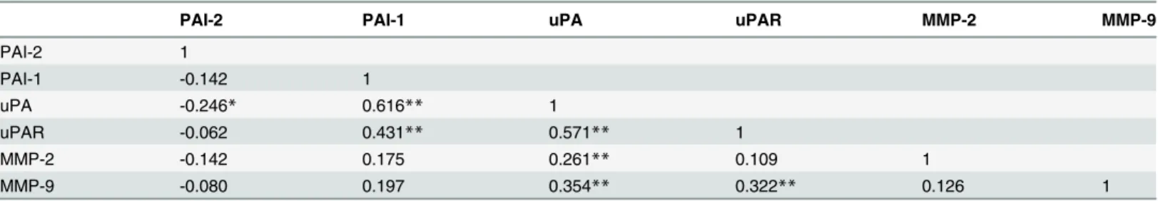
Table 1. The correlations between IHC expression levels of PAI-1, PAI-2, uPA, uPAR, MMP-2, and MMP-9 analyzed by Spearman’s rank correlation analysis in derivation cohort with 98 NSCLC cases.
PAI-2 PAI-1 uPA uPAR MMP-2 MMP-9
PAI-2 1
PAI-1 -0.142 1
uPA -0.246* 0.616** 1
uPAR -0.062 0.431** 0.571** 1
MMP-2 -0.142 0.175 0.261** 0.109 1
MMP-9 -0.080 0.197 0.354** 0.322** 0.126 1
**Correlation is significant at the 0.01 level (2-tailed).
*Correlation is significant at the 0.05 level (2-tailed).
PAI-1: plasminogen activator inhibitor-1; PAI-2: plasminogen activator inhibitor-2; uPA: urokinase-type plasminogen activator; uPAR: urokinase-type plasminogen activator receptor; MMP-2: matrix metalloproteinase 2; MMP-9: matrix metalloproteinase 9.
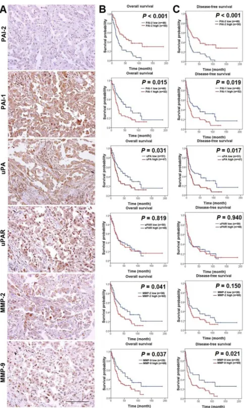
Fig 1. The prognostic value of markers in PA and MMP families in derivation cohort with 98 NSCLC cases.(A) A representative lung adenocarcinoma with low IHC expression level of PAI-2 and high IHC expression levels of PAI-1, uPA, uPAR, MMP-2, and MMP-9, as detected by IHC. Photographs were taken at a magnification of 400×. Scale bars represent 100μm. (B) Kaplan-Meier plots of overall survival relative to
Establishment of an IHC panel comprising PAI-2 and MMP-9 for more
precise prognostic prediction in NSCLC patients
Based on the results that PAI-2 had the greatest prognostic value and that MMP-9 was the sec-ond most powerful prognostic marker in the multivariate analysis, a prognostic model analysis, survival analysis, and ROC curve for testing the area under the curve (AUC), sensitivity, and specificity were performed to examine the appropriateness of combining PAI-2 and MMP-9 as
an IHC panel. In the prognostic model analysis (S3 Table), adding PAI-2 and MMP-9 to the
pathological stage variable resulted in the highest predictive power for DFS, yet adding more markers did not further improve the predictive power. Through combining PAI-2 and MMP-9 as an IHC panel, the patients could be separated into three groups. Patients with high PAI-2 and low MMP-9 expression levels had the most favorable prognosis, and patients with low
PAI-2 and high MMP-9 expression levels had the most unfavorable prognosis (P<0.001 for
both OS and DFS) (Fig 2A). In the ROC curve, increase of area under curve (AUC) was seen in
disease-free survival after combining PAI-2 and MMP-9 as a panel (Fig 2AandS4 Table). The
heat map displayed the IHC expression levels of PAI-2 and MMP-9 in individual patients survival (P<0.001); high IHC expression levels of PAI-1, uPA, and MMP-9 was also significantly associated
with shorter disease-free survival (P= 0.019,P= 0.017,P= 0.021, respectively).
doi:10.1371/journal.pone.0133411.g001
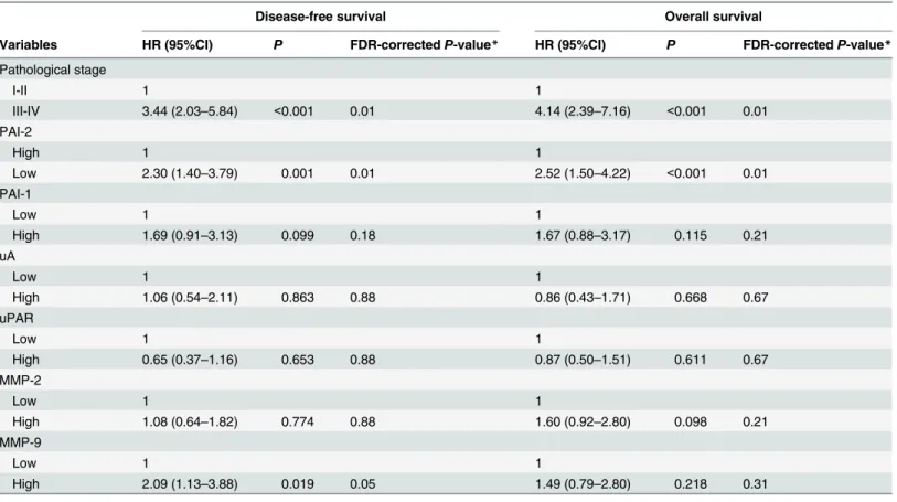
Table 2. Cox multivariate analysis with false discovery rate correction of PAI-1, PAI-2, uPA, uPAR, MMP-2 and MMP-9 IHC expression levels and pathological stage in derivation cohort with 98 NSCLC cases.
Disease-free survival Overall survival
Variables HR (95%CI) P FDR-correctedP-value* HR (95%CI) P FDR-correctedP-value*
Pathological stage
I-II 1 1
III-IV 3.44 (2.03–5.84) <0.001 0.01 4.14 (2.39–7.16) <0.001 0.01
PAI-2
High 1 1
Low 2.30 (1.40–3.79) 0.001 0.01 2.52 (1.50–4.22) <0.001 0.01
PAI-1
Low 1 1
High 1.69 (0.91–3.13) 0.099 0.18 1.67 (0.88–3.17) 0.115 0.21
uA
Low 1 1
High 1.06 (0.54–2.11) 0.863 0.88 0.86 (0.43–1.71) 0.668 0.67
uPAR
Low 1 1
High 0.65 (0.37–1.16) 0.653 0.88 0.87 (0.50–1.51) 0.611 0.67
MMP-2
Low 1 1
High 1.08 (0.64–1.82) 0.774 0.88 1.60 (0.92–2.80) 0.098 0.21
MMP-9
Low 1 1
High 2.09 (1.13–3.88) 0.019 0.05 1.49 (0.79–2.80) 0.218 0.31
*Pvalue after Benjamini and Hochberg false discovery rate (FDR) procedure.
PAI-1: plasminogen activator inhibitor-1; PAI-2: plasminogen activator inhibitor-2; uPA: urokinase-type plasminogen activator; uPAR: urokinase-type plasminogen activator receptor; MMP-2: matrix metalloproteinase 2; MMP-9: matrix metalloproteinase 9.
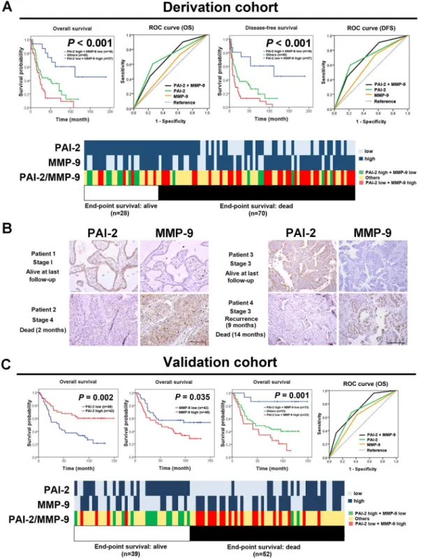
Fig 2. An IHC panel comprising PAI-2 and MMP-9 provides a more precise prognostic predictive power for 98 NSCLC patients in derivation cohort and 91 NSCLC patients in validation cohort.(A) Kaplan-Meier plots and ROC curve of overall survival and disease-free survival for the combination of PAI-2 and MMP-9 as an IHC panel. Patients with high PAI-2 and low MMP-9 IHC expression levels had the most favorable prognosis, whereas patients with low PAI-2 and high MMP-9 IHC expression levels had the most unfavorable prognosis (P<0.001 for both OS and DFS). Increase of area under curve (AUC)
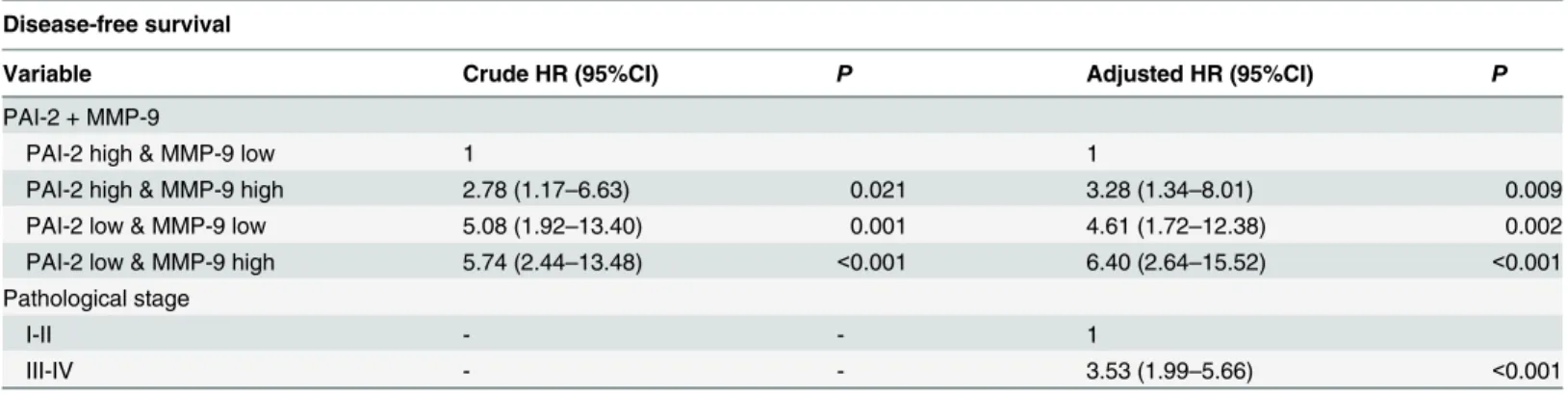
clustered by end-point survival status (Fig 2A). Patients who were dead at the end-point tended to have low PAI-2 and high MMP-9 IHC expression levels. Furthermore, the cumulative nostic effect of PAI-2 and MMP-9 was also investigated. Compared to the most favorable prog-nostic group (high PAI-2 and low MMP-9 IHC expression levels), the most unfavorable prognostic group (low PAI-2 and high MMP-9 IHC expression levels) showed a 6.40-fold
(95% CI = 2.64–15.52;P<0.001) increased risk of a poor prognosis after adjustment for
path-ological stage (Table 3).Fig 2Bshow representative examples of the usage of the PAI-2 and
MMP-9 IHC panel in NSCLC samples. Patient 1, who had stage I adenocarcinoma with high PAI-2 and low MMP-9 IHC expression levels, was alive at the final follow up. In contrast, patient 2 had stage IV adenocarcinoma with low PAI-2 and high MMP-9 IHC expression levels and died 2 months after treatment commenced. Patient 3 and patient 4 were both diagnosed with stage III adenocarcinoma. Patient 3 had high PAI-2 and low MMP-9 IHC expression lev-els and was alive and had no tumor recurrence at the final follow up. In contrast, patient 4, who had low PAI-2 and high MMP-9 IHC expression levels, experienced tumor recurrence within 9 months and died 14 months after treatment commenced.
Confirmation of the prognostic value of the IHC panel comprising PAI-2
and MMP-9 in a validation cohort
To validate the prognostic power of the IHC panel comprising PAI-2 and MMP-9 discovered in our derivation cohort, we further conducted the survival analysis and ROC curve analysis in a validation cohort with 91 NSCLC patients. In Kaplan-Meier survival analysis, patients with low PAI-2 IHC expression level had a poor prognosis, and patients with high MMP-9 IHC
expression level had a poor prognosis (Fig 2C). When combining PAI-2 and MMP-9 as an
IHC panel, patients with high PAI-2 and low MMP-9 IHC expression levels had the best prog-nosis, and patients with low PAI-2 and high MMP-9 IHC expression levels had the worst
out-come (P= 0.001). Increase of area under curve (AUC) of ROC was also noted after combining
PAI-2 and MMP-9 as a panel (Fig 2CandS4 Table). As shown in the heat map (Fig 2C),
patients who were dead at the end-point tended to have low PAI-2 and high MMP-9 IHC expression levels. Therefore, the PAI-2 and MMP-9 IHC panel we identified from markers of PA and MMP families could be used to stratify lung cancer patients by risk and select patients for more aggressive treatment.
Discussion
To our knowledge, this is the first study to integrate the PA and MMP families using IHC anal-ysis in lung cancer. We provide a novel insight that PAI-2 is the most powerful prognostic indi-cator among markers in PA and MMP families and, more importantly, PAI-2 remained its leading prognostic role even after taking its close relationship with other PA and MMP family markers into consideration. Research on both PA and MMP families revealed that they are an
appropriate model for examining prognostic value by multi-marker assessment (S5 Table)
[26–29]. With regard to the interconnected signaling pathway level, a recent study provides
information about the respective contributions of each PA family member in airway epithelial recurrence within 9 months and died 14 months after treatment commenced. Photographs were taken at a magnification of 400×. Scale bars represent 100
μm. (C) Kaplan-Meier plots and ROC curve of overall survival in validation cohort showed that patients with low PAI-2 IHC expression level (P= 0.002) or high MMP-9 IHC expression level (P= 0.035) had poor overall survival. When stratified by 2 and MMP-9 IHC panel, high risk group patients had low PAI-2 and high MMP-9 IHC expression levels and low risk group patients had high PAI-PAI-2 and low MMP-9 IHC expression level (P<0.001). Increase of area under curve (AUC) of ROC was observed after combining PAI-2 and MMP-9 as a panel. The heat map showed that patients who were dead at the end-point tended to have low PAI-2 and high MMP-9 IHC expression level.
cells [31]. Membrane-bound uPAR (muPAR) has been considered the key molecule because it
is the only member of the PA family shown to accelerate cell migrationin vitro. In comparison,
our IHC data demonstrated no correlation between uPAR IHC expression level and patient prognosis. This discrepancy may result from several factors. First, the limitation of the previous study was that the overexpression of each marker may not accurately mimic the coordinated
signaling networkin vivo[31]. Although our data may not be able to demonstrate the cause
and effect relationship between these markers, they do directly reflect the prognostic role of each marker in clinical patients in whom these markers are coexpressed and interact with each other. Moreover, the expression of ECM degradation-related enzymes, such as the PA and
MMP families, may also be regulated by tumor-associated stromal cells [32]. Using a tissue
microarray (TMA), we demonstrated the predictive value of these markers by considering both the tumor cells and their microenvironment. Detecting PA family markers level from tissue extraction by enzyme-linked immunosorbent assay (ELISA) was also a frequently used method
to analyze their roles in lung cancer (S6 Table) [33–37]. Although ELISA could more accurately
quantify the expression levels of these markers, the tumor samples may not be completely com-posed of tumor tissue due to not being able to see the morphology of the tissue. In contrast, IHC analysis used in present study could more avoid this limitation by directly observe the expression in situ. This may explain that the prognostic significance of PA family markers was
not seen in previous study by using ELISA [33], but was seen in our study by using IHC
analy-sis. Therefore, our results provide a complementary illustration of the roles of PA and MMP family members in lung cancer.
Our data reveal that PAI-2 plays a leading role in PAI-1, PAI-2, uPA, uPAR, MMP-2, and MMP-9. However, few studies have focused on the pathophysiological function of PAI-2 in cancer. PAI-2 overexpression was reported to reduce metastasis in xenograft models and
decrease the level of uPA and migrationin vitro[38,39]. It has recently been demonstrated that
the low binding affinity of PAI-2 for endocytic receptors facilitates the clearance of uPA with-out evoking downstream signaling events, providing a possible explanation for the inhibitory
role of PAI-2 in cancer progression [28,40]. The physiological function of PAI-2 may further
provide clues regarding its role in cancer progression. As a protein expressed in stress
condi-tion such as inflammacondi-tion and infeccondi-tion [41], elevated PAI-2 plasma level was found in
patients with sepsis or leukemia, and the expression level was increased during active or relapse
disease and was undetectable in remission [42,43]. In present study, we also analyzed the
PAI-Table 3. Cox univariate and multivariate analyses of disease-free survival in association with PAI-2 and MMP-9 IHC expression levels and patho-logical stage in derivation cohort with 98 NSCLC cases.
Disease-free survival
Variable Crude HR (95%CI) P Adjusted HR (95%CI) P
PAI-2 + MMP-9
PAI-2 high & MMP-9 low 1 1
PAI-2 high & MMP-9 high 2.78 (1.17–6.63) 0.021 3.28 (1.34–8.01) 0.009
PAI-2 low & MMP-9 low 5.08 (1.92–13.40) 0.001 4.61 (1.72–12.38) 0.002
PAI-2 low & MMP-9 high 5.74 (2.44–13.48) <0.001 6.40 (2.64–15.52) <0.001
Pathological stage
I-II - - 1
III-IV - - 3.53 (1.99–5.66) <0.001
PAI-1: plasminogen activator inhibitor-1; PAI-2: plasminogen activator inhibitor-2; uPA: urokinase-type plasminogen activator; uPAR: urokinase-type plasminogen activator receptor; MMP-2: matrix metalloproteinase 2; MMP-9: matrix metalloproteinase 9.
2 plasma level in NSCLC lung cancer patients through ELISA (S5 Fig). The finding showed ele-vated PAI-2 plasma level in lung cancer patients compared to normal control. Interestingly, a recent research revealed that PAI-2 which is able to inhibit uPA-mediated tumor cell migration
and invasion is secreted by tumor cells itself on microparticles, not by host cells [44]. Taken
together, a mechanism may be proposed that the elevated PAI-2 plasma level in lung cancer patients reflects PAI-2 positive microparticles in circulation secreted by cancer cells. And fur-ther research is clearly required to determine whefur-ther PAI-2 plasma level could be used as a surrogate for disease status in lung cancer patients.
Although the functional role of PAI-2 remains to be fully understood, by taking advantage of its inhibitory function, PAI-2 has been investigated as a vector targeting uPA in targeted
alpha radioimmunotherapy for various cancer types [45]. In our study, evaluation of the
expression level of PA and MMP family members by IHC analysis demonstrated provided a potential method to select patients with high uPA expression level and low PAI-2 expression level, who may obtain a greater benefit from PAI-2-targeted therapy. Moreover, the failure of broad-spectrum MMP inhibitors in clinical trials of cancer treatment has led to the
develop-ment of selective MMP inhibitors [46], and inhibitors that target MMP-9 have been indicated
as potential treatments for cancer [47]. Consequently, the clinical utility of PAI-2 and MMP-9
as prognostic markers identified in our study further suggests their potential for therapeutic application in NSCLC.
In conclusion, considering markers from PA and MMP protease families that is critical in regulating tumor progression, our study reveals the greatest prognostic significance of PAI-2 among uPA, uPAR, PAI-1 and PAI-2 in the PA family and MMP-2 and MMP-9 in the MMP family. By prioritizing the prognostic power of each marker, we generated an IHC panel composed of PAI-2 and MMP-9, which provides greater prognostic value than the pathological stage or a single marker alone. Integration of the prognostic values of the markers from protease families determined in the present study will allow their application in the clinical setting for predicting patient outcome and determining treatment direction in lung cancer.
Supporting Information
S1 Materials and Methods. ELISAs.Plasma levels of PAI-2 and MMP-9 were detected by commercial PAI-2 (LS-F5568, Lifespan Biosciences, USA) and MMP-9 (ab100610, Abcam, Cambridge, UK) ELISA kit. The plasma samples of 36 NSCLC patients and 6 normal controls were obtained from Kaohsiung Medical University Hospital of Taiwan.
(DOCX)
S1 Fig. The PA family and MMP family interact with each other and form a complex net-work that regulates ECM degradation.(A and B) Both PAI-1 and PAI-2 inhibit the proteo-lytic ability of uPA and in turn inhibit the formation of plasmin, which can degrade the ECM alone or through the activation of MMP-2 and MMP-9. Moreover, MMP-9 can enhance uPA activity by degrading protease nexin-1. (A) The uPA-PAI-1 complex can further increase the expression of MMP-2 and MMP-9 through downstream signaling. (B) However, the uPA--PAI-2 complex facilitates the clearance of uPA without increasing MMP-2 and MMP-9 expres-sion through downstream signaling.
(TIF)
was seen on debrides. Photographs were taken at a magnification of 200×. Scale bars represent
200μm.
(TIF)
S3 Fig. Representative images of PAI-1, PAI-2, uPA, uPAR, MMP-2, and MMP-9 immunoexpression in lung cancer patients.Representative images showing the intensity of immunostaining for PAI-1, PAI-2, uPA, uPAR, MMP-2, and MMP-9 in lung cancer tissue microarrays. The images were taken at a magnification of 400×. Scale bars represent
200μm.
(TIF)
S4 Fig. The heat map with IHC expression level of individual patient in derivation cohort clustered by stage.Patients with higher stage tend to have low PAI-2 and high MMP-9 IHC expression levels.
(TIF)
S5 Fig. Plasma level of PAI-2 and MMP-9 of NSCLC patients and normal controls. Signifi-cant elevated plasma level of PAI-2 and MMP-9 were seen in NSCLC patients compared to normal controls.
(TIF)
S1 Table. Clinicopathological and demographic characteristics of derivation and validation cohort lung cancer patients.
(DOCX)
S2 Table. Clinicopathological analysis of the correlation between clinicopathological fea-tures and PAI-1, PAI-2, uPA, uPAR, MMP-2 and MMP-9 IHC expression in derivation cohort with 98 NSCLC cases.
(DOC)
S3 Table. Prognostic model analysis evaluates prognostic values of PAI-1, PAI-2, uPA, uPAR, MMP-2 and MMP-9 as IHC panels added to pathological stage in derivation cohort with 98 NSCLC cases.
(DOC)
S4 Table. The AUR (area under curve) of ROC (receiver operating characteristic) curve, sensitivity, and specificity of the combined and separated PAI-2 and MMP-9 IHC expres-sion in derivation and validation cohort.
(DOCX)
S5 Table. Comparison of published clinicopathological studies of plasminogen activator family and matrix metalloproteinase family via immunohistochemical analysis in lung cancer.
(DOCX)
S6 Table. Published clinicopathological studies of plasminogen activator family markers analyzed by enzyme-linked immunosorbent assay (ELISA) in lung cancer.
(DOCX)
Acknowledgments
Author Contributions
Conceived and designed the experiments: CYS MSH MH. Performed the experiments: CYS MH. Analyzed the data: CYS MSH MH. Contributed reagents/materials/analysis tools: YPL CJY YFL JC LHC JJL AW PJL MSH MH. Wrote the paper: CYS MSH MH.
References
1. Siegel RL, Miller KD, Jemal A. Cancer statistics, 2015. CA Cancer J Clin. 2015; 65: 5–29. doi:10.3322/
caac.21254PMID:25559415
2. Johnson DH, Schiller JH, Bunn PA Jr. Recent clinical advances in lung cancer management. J Clin Oncol. 2014; 32: 973–982. doi:10.1200/JCO.2013.53.1228PMID:24567433
3. Strauss GM, Herndon JE 2nd, Maddaus MA, Johnstone DW, Johnson EA, Harpole DH, et al. Adjuvant paclitaxel plus carboplatin compared with observation in stage IB non-small-cell lung cancer: CALGB 9633 with the Cancer and Leukemia Group B, Radiation Therapy Oncology Group, and North Central Cancer Treatment Group Study Groups. J Clin Oncol. 2008; 26: 5043–5051. doi:10.1200/JCO.2008.
16.4855PMID:18809614
4. Higgins KA, Chino JP, Ready N, D'Amico TA, Berry MF, Sporn T, et al. Lymphovascular invasion in non-small-cell lung cancer: implications for staging and adjuvant therapy. J Thorac Oncol. 2012; 7: 1141–1147. doi:10.1097/JTO.0b013e3182519a42PMID:22617241
5. Ali HR, Dawson SJ, Blows FM, Provenzano E, Pharoah PD, Caldas C. Aurora kinase A outperforms Ki67 as a prognostic marker in ER-positive breast cancer. Br J Cancer. 2012; 106: 1798–1806. doi:10.
1038/bjc.2012.167PMID:22538974
6. Hung JJ, Yang MH, Hsu HS, Hsu WH, Liu JS, Wu KJ. Prognostic significance of hypoxia-inducible fac-tor-1alpha, TWIST1 and Snail expression in resectable non-small cell lung cancer. Thorax. 2009; 64: 1082–1089. doi:10.1136/thx.2009.115691PMID:19778933
7. Herbst RS, Heymach JV, Lippman SM. Molecular origins of cancer: Lung cancer. New Engl J Med. 2008; 359: 1367–1380. doi:10.1056/NEJMra0802714PMID:18815398
8. Lu PF, Takai K, Weaver VM, Werb Z. Extracellular Matrix Degradation and Remodeling in Development and Disease. Cold Spring Harb Perspect Biol. 2011; 3(12): a005058. doi:10.1101/cshperspect. a005058PMID:21917992
9. Duffy MJ. The urokinase plasminogen activator system: Role in malignancy. Curr Pharm Des. 2004; 10: 39–49. PMID:14754404
10. Deryugina EI, Quigley JP. Matrix metalloproteinases and tumor metastasis. Cancer Metastasis Rev. 2006; 25: 9–34. PMID:16680569
11. Oka T, Ishida T, Nishino T, Sugimachi K. Immunohistochemical evidence of urokinase-type plasmino-gen-activator in primary and metastatic tumors of pulmonary adenocarcinoma. Cancer Res. 1991; 51: 3522–3525. PMID:2054790
12. Pavey SJ, Marsh NA, Ray MJ, Butler D, Dare AJ, Hawson GA. Changes in plasminogen activator inhib-itor-1 levels in non-small cell lung cancer. Boll Soc Ital Biol Sper. 1996; 72: 331–340. PMID:9178585 13. Volm M, Mattern J, Koomagi R. Relationship of urokinase and urokinase receptor in non-small cell lung
cancer to proliferation, angiogenesis, metastasis and patient survival. Oncol Rep. 1999; 6: 611–615.
PMID:10203601
14. Qian QA, Wang QA, Zhan P, Peng L, Wei SZ, Shi Y, et al. The Role of Matrix Metalloproteinase 2 on the Survival of Patients with Non-Small Cell Lung Cancer: A Systematic Review with Meta-Analysis. Cancer Invest. 2010; 28: 661–669. doi:10.3109/07357901003735634PMID:20394501
15. Peng WJ, Zhang JQ, Wang BX, Pan HF, Lu MM, Wang J. Prognostic value of matrix metalloproteinase 9 expression in patients with non-small cell lung cancer. Clin Chim Acta. 2012; 413: 1121–1126. doi:
10.1016/j.cca.2012.03.012PMID:22465234
16. Werle B, Kotzsch M, Lah TT, Kos J, Gabrijelcic-Geiger D, Spiess E, et al. Cathepsin b, plasminogenac-tivator-inhibitor (PAM) and plasminogenactivator-receptor (uPAR) are prognostic factors for patients with non-small cell lung cancer. Anticancer Res. 2004; 24: 4147–4161. PMID:15736466
17. Shao WL, Wang W, Xiong XG, Cao C, Yan TD, Chen GQ, et al. Prognostic Impact of MMP-2 and MMP-9 Expression in Pathologic Stage IA Non-Small Cell Lung Cancer. J Surg Oncol. 2011; 104: 841–
846. doi:10.1002/jso.22001PMID:21721010
19. Yoshino H, Endo Y, Watanabe Y, Sasaki T. Significance of plasminogen activator inhibitor 2 as a prog-nostic marker in primary lung cancer: association of decreased plasminogen activator inhibitor 2 with lymph node metastasis. Br J Cancer. 1998; 78: 833–839. PMID:9743310
20. Robert C, Bolon I, Gazzeri S, Veyrenc S, Brambilla C, Brambilla E. Expression of plasminogen activator inhibitors 1 and 2 in lung cancer and their role in tumor progression. Clin Cancer Res. 1999; 5: 2094–
2102. PMID:10473092
21. Ishikawa S, Takenaka K, Yanagihara K, Miyahara R, Kawano Y, Otake Y, et al. Matrix metalloprotei-nase-2 status in stromal fibroblasts, not in tumor cells, is a significant prognostic factor in non-small-cell lung cancer. Clin Cancer Res. 2004; 10: 6579–6585. PMID:15475447
22. Nordengren J, Fredstorp Lidebring M, Bendahl PO, Brunner N, Ferno M, Hogberg T, et al. High tumor tissue concentration of plasminogen activator inhibitor 2 (PAI-2) is an independent marker for shorter progression-free survival in patients with early stage endometrial cancer. Int J Cancer. 2002; 97: 379–
385. PMID:11774293
23. Ganesh S, Sier CF, Griffioen G, Vloedgraven HJ, de Boer A, Welvaart K, et al. Prognostic relevance of plasminogen activators and their inhibitors in colorectal cancer. Cancer Res. 1994; 54: 4065–4071.
PMID:8033138
24. Duggan C, Kennedy S, Kramer MD, Barnes C, Elvin P, McDermott E, et al. Plasminogen activator inhibitor type 2 in breast cancer. Br J Cancer. 1997; 76: 622–627. PMID:9303361
25. Chambers SK, Ivins CM, Carcangiu ML. Expression of plasminogen activator inhibitor-2 in epithelial ovarian cancer: a favorable prognostic factor related to the actions of CSF-1. Int J Cancer. 1997; 74: 571–575. PMID:9421350
26. Cox G, Steward WP, O'Byrne KJ. The plasmin cascade and matrix metalloproteinases in non-small cell lung cancer. Thorax. 1999; 54: 169–179. PMID:10325924
27. Di Y, Liu ZG, Tian J, Zong YQ, Yang P, Qu S. TFPI or uPA-PAI-1 complex affect cell function through expression variation of type II very low density lipoprotein receptor. FEBS Lett. 2010; 584: 3469–3473.
doi:10.1016/j.febslet.2010.07.005PMID:20624392
28. Cochran BJ, Croucher DR, Lobov S, Saunders DN, Ranson M. Dependence on Endocytic Receptor Binding via a Minimal Binding Motif Underlies the Differential Prognostic Profiles of SerpinE1 and Ser-pinB2 in Cancer. J Biol Chem. 2011; 286: 24467–24475. doi:10.1074/jbc.M111.225706PMID:
21606492
29. Xu DM, McKee CM, Cao YH, Ding YC, Kessler BM, Muschel RJ. Matrix Metalloproteinase-9 Regulates Tumor Cell Invasion through Cleavage of Protease Nexin-1. Cancer Res. 2010; 70: 6988–6998. doi:
10.1158/0008-5472.CAN-10-0242PMID:20736374
30. Benjamini Y, Hochberg Y. Controlling the False Discovery Rate—a Practical and Powerful Approach to
Multiple Testing. J Roy Stat Soc B Met. 1995; 57: 289–300.
31. Stewart CE, Sayers I. Urokinase Receptor Orchestrates the Plasminogen System in Airway Epithelial Cell Function. Lung. 2013; 191: 215–225. doi:10.1007/s00408-013-9450-zPMID:23408042 32. He Y, Liu XD, Chen ZY, Zhu J, Xiong Y, Li K, et al. Interaction between cancer cells and stromal
fibro-blasts is required for activation of the uPAR-uPA-MMP-2 cascade in pancreatic cancer metastasis. Clin Cancer Res. 2007; 13: 3115–3124. PMID:17545513
33. Salden M, Splinter TA, Peters HA, Look MP, Timmermans M, van Meerbeeck JP, et al. The urokinase-type plasminogen activator system in resected non-small-cell lung cancer. Rotterdam Oncology Tho-racic Study Group. Ann Oncol. 2000; 11: 327–332. PMID:10811500
34. Pedersen H, Brunner N, Francis D, Osterlind K, Ronne E, Hansen HH, et al. Prognostic impact of uroki-nase, urokinase receptor, and type 1 plasminogen activator inhibitor in squamous and large cell lung cancer tissue. Cancer Res. 1994; 54: 4671–4675. PMID:8062262
35. Pedersen H, Grondahl-Hansen J, Francis D, Osterlind K, Hansen HH, Dano K, et al. Urokinase and plasminogen activator inhibitor type 1 in pulmonary adenocarcinoma. Cancer Res. 1994; 54: 120–123.
PMID:8261432
36. Werle B, Kotzsch M, Lah TT, Kos J, Gabrijelcic-Geiger D, Spiess E, et al. Cathepsin B, plasminogenac-tivator-inhibitor (PAI-1) and plasminogenactivator-receptor (uPAR) are prognostic factors for patients with non-small cell lung cancer. Anticancer Res. 2004; 24: 4147–4161. PMID:15736466
37. Offersen BV, Pfeiffer P, Andreasen P, Overgaard J. Urokinase plasminogen activator and plasminogen activator inhibitor type-1 in nonsmall-cell lung cancer: relation to prognosis and angiogenesis. Lung Cancer. 2007; 56: 43–50. PMID:17207889
39. Praus M, Wauterickx K, Collen D, Gerard RD. Reduction of tumor cell migration and metastasis by ade-noviral gene transfer of plasminogen activator inhibitors. Gene Ther. 1999; 6: 227–236. PMID:
10435107
40. Croucher DR, Saunders DN, Lobov S, Ranson M. Revisiting the biological roles of PAI2 (SERPINB2) in cancer. Nature Reviews Cancer. 2008; 8: 535–545. doi:10.1038/nrc2400PMID:18548086
41. Medcalf RL, Stasinopoulos SJ. The undecided serpin—The ins and outs of plasminogen activator
inhibitor type 2. FEBS J. 2005; 272: 4858–4867. PMID:16176260
42. Scherrer A, Kruithof EK, Grob JP. Plasminogen activator inhibitor-2 in patients with monocytic leuke-mia. Leukeleuke-mia. 1991; 5: 479–486. PMID:2056772
43. Robbie LA, Dummer S, Booth NA, Adey GD, Bennett B. Plasminogen activator inhibitor 2 and uroki-nase-type plasminogen activator in plasma and leucocytes in patients with severe sepsis. Br J Haema-tol. 2000; 109: 342–348. PMID:10848822
44. Schroder WA, Major LD, Le TT, Gardner J, Sweet MJ, Janciauskiene S, et al. Tumor cell-expressed SerpinB2 is present on microparticles and inhibits metastasis. Cancer Med. 2014; 3: 500–513. doi:10.
1002/cam4.229PMID:24644264
45. Allen BJ, Tian Z, Rizvi SMA, Li Y, Ranson M. Preclinical studies of targeted alpha therapy for breast cancer using Bi-213-labelled-plasminogen activator inhibitor type 2. Br J Cancer. 2003; 88: 944–950.
PMID:12644835
46. Coussens LM, Fingleton B, Matrisian LM. Matrix metalloproteinase inhibitors and cancer: trials and trib-ulations. Science. 2002; 295: 2387–2392. PMID:11923519