ABSTRACT
Objective: To evaluate different weight loss (WL) cut-off points as prognostic markers of 3-month survival after diagnosis of stage IV non-small cell lung cancer (NSCLC). Methods: This was a prospective study involving 104 patients with metastatic (stage IV) NSCLC who were admitted to a cancer treatment center in southern Brazil between January of 2014 and November of 2016. We evaluated total WL and WL per month, as well as WL and WL per month in the 6 months preceding the diagnosis. The patients were followed for 3 months after diagnosis. A Cox proportional hazards regression model and Kaplan-Meier curves were used in order to evaluate 3-month survival. Results: The median WL in the 6 months preceding the diagnosis was 6% (interquartile range, 0.0-12.9%). Patients with WL ≥ 5% had a median survival of 78 days, compared with 85 days for those with WL < 5% (p = 0.047). Survival at 3 months was 72% for the patients with WL ≥ 5% (p = 0.047), 61% for those with WL ≥ 10% (p < 0.001), and 45% for those with WL ≥ 15% (p < 0.001). In the multivariate analysis, the hazard ratio for risk of death was 4.51 (95% CI: 1.32-15.39) for the patients with WL ≥ 5%, 6.34 (95% CI: 2.31-17.40) for those with WL ≥ 10%, and 14.17 (95% CI: 5.06-39.65) for those with WL ≥ 15%. Conclusions: WL in the 6 months preceding the diagnosis of NSCLC is a relevant prognostic factor and appears to be directly proportional to the rate of survival at 3 months.
Keywords: Weight loss; Carcinoma, non-small-cell lung; Prognosis.
Proportional weight loss in six months as
a risk factor for mortality in stage IV
non-small cell lung cancer
Guil herme Watte1,2,5,a, Claudia Helena de Abreu Nunes1,b, Luzielio Alves Sidney-Filho3,c, Matheus Zanon2,4,d, Stephan Philip Leonhardt Altmayer4,5,e, Gabriel Sartori Pacini4,f, Marcelo Barros5,g, Ana Luiza Schneider Moreira4,h, Rafael José Vargas Alves1,i, Alice de Medeiros Zelmanowicz4,j, Bashir Mnene Matata2,k, Jose da Silva Moreira1,l
Correspondence to:
Gabriel Sartori Pacini. Rua Sarmento Leite, 245, CEP 90050-170, Porto Alegre, RS, Brasil. Tel.: 55 51 99995-2543. Fax: 55 51 3214-8080. E-mail: [email protected] Financial support: None.
INTRODUCTION
Weight loss (WL) is a common complaint of patients with lung cancer and a common reason for patient referral to a specialist.(1-3) Cancer cachexia, resulting from an
imbalance between energy intake and consumption, is associated with a combination of poor caloric intake and increased resting energy expenditure, probably due to a
cytokine-induced systemic infl ammatory response.(4-7) Some studies have demonstrated
that this increase in resting energy expenditure can also vary depending on the type of tumor.(8,9) Other factors that contribute to cancer cachexia include nausea,
vomiting, constipation, diarrhea, pain, altered taste perception, and depression.(1)
Despite its potential benefi t for the clinical evaluation of patients with non-small cell lung cancer (NSCLC), the defi nition of cachexia varies signifi cantly across studies
and many WL cut-off points have been proposed in the attempt to classify the syndrome in an objective manner.(10-14) A recent consensus suggested that WL be
defi ned as any decrease greater than 5% in relation to the usual weight or greater than 2% in individuals with a body mass index < 20 kg/m2.(13) However, setting up a
single cut-off point to classify cachexia can underestimate its real prognostic value. Different levels of cachexia severity can have various effects on the prognosis of cancer and could serve as a valuable clinical indicator.
The objective of this study was to evaluate 3-month survival in a population of patients with stage IV NSCLC. We also tested the prognostic value of different WL cut-off points.
1. Programa de Pós-Graduação em Ciências Pneumológicas, Faculdade de Medicina, Universidade Federal do Rio Grande do Sul – UFRGS – Porto Alegre (RS) Brasil.
2. Department of Clinical Research and Radiology, Liverpool Heart and Chest Hospital NHS Foundation Trust, Liverpool, United Kingdom. 3. Faculdade de Medicina, Universidade
Federal do Espírito Santo, Vitória (ES) Brasil.
4. Departamento de Medicina Clínica e Saúde Pública, Universidade Federal de Ciências da Saúde de Porto Alegre, Porto Alegre (RS) Brasil.
5. Programa de Pós-Graduação em Medicina e Ciências da Saúde, Faculdade de Medicina, Pontifícia Universidade Católica do Rio Grande do Sul, Porto Alegre (RS) Brasil.
a. http://orcid.org/0000-0002-6948-3982 b. http://orcid.org/0000-0002-1737-7440 c. http://orcid.org/0000-0001-9284-884X d. http://orcid.org/0000-0001-7851-5125
e. http://orcid.org/0000-0001-9214-1916 f. http://orcid.org/0000-0002-4822-7082
g. http://orcid.org/0000-0002-4985-6374 h. http://orcid.org/0000-0001-6428-6421
i. http://orcid.org/0000-0002-6294-917X j. http://orcid.org/0000-0001-9121-6365 k. http://orcid.org/0000-0003-2896-8059 l. http://orcid.org/0000-0003-1345-0476
Submitted: 23 January 2018. Accepted: 22 April 2018.
METHODS
Study population
This prospective cohort study was conducted at Santa Rita Hospital, an oncology referral center and part of the Santa Casa de Misericórdia de Porto Alegre Hospital Complex, in the city of Porto Alegre, located in southern Brazil. We included consecutive patients newly diagnosed with metastatic (stage IV) NSCLC and admitted to Santa Rita Hospital between January of 2014 and November of 2016. Patients were treated at the discretion of the attending physician. All patients received a nutritional consultation at admission, regularly received high-calorie meals, and were instructed to rest before meals. However, they did not receive any type of nutritional supplementation as part of the palliative care or during chemotherapy. The follow-up
period was ≤ 3 months after the diagnosis of cancer, as confi rmed by reviews of medical records, hospital
records, and phone calls. All diagnoses required clinical,
radiological, and histological confi rmation. Patients who
had previously undergone antineoplastic treatment were excluded, as were those who were under 18 years of age. Survival and mortality rates were calculated from the time of the histological diagnosis until death or until the end of the third month of follow-up. Patient charts were reviewed, and tumor-node-metastasis variables were upgraded in accordance with the revised stage groupings established by the International Association for the Study of Lung Cancer.(15) Performance status was assessed with the Eastern Cooperative Oncology Group scale.(16)The study was approved by the local
institutional review board. All participating patients gave written informed consent.
WL classifi cation
Each patient was prospectively evaluated following
the standards established in previous reports.(17)
Defi nitions of WL-related variables were also based on
those established in other studies(10): total WL—the
difference between weight at the time of diagnosis and usual weight; WL per month—total WL divided by the number of months of WL; and WL per month in 6 months—total difference between weight at the time of diagnosis and weight in the preceding 6 months. Patients were also subjectively evaluated in terms of their self-awareness of WL at the time of NSCLC diagnosis in relation to their usual weight. Different
WL cut-off points (5%, 10%, and 15%) were tested in
order to classify cachexia and to correlate the different de grees of cachexia with the survival rates.
Statistical analysis
Continuous variables are expressed as medians and interquartile range (IQR). Univariate analyses of survival were based on the Kaplan-Meier method.(18) Survival
was calculated by Cox proportional hazards regression model in a multivariate analysis.(19) The Wald test was
used in order to calculate signifi cance for each factor. All parameters associated with mortality (p < 0.1) in
the univariate analysis were included in a multivariate
model, in which values of p < 0.05 were considered statistically signifi cant. All tests were two-tailed, with the level of signifi cance set at 0.05. All results were
analyzed with the SPSS Statistics software package, version 20.0 (IBM Corporation, Armonk, NY, USA).
RESULTS
Patient characteristics are summarized in Table 1.
The median age was 63 years (IQR, 52.5-69.0 years). Of a total of 104 patients evaluated, 63 (60.6%) were
male. The most prevalent histological type of NSCLC
was adenocarcinoma, which was seen in 57 (54.7%)
of the patients, followed by squamous cell carcinoma,
seen in 36 (34.6%), and mixed/undefi ned, seen in 11 (10.6%). The median Eastern Cooperative Oncology
Group performance status was 2 (IQR, 1-3). Most
(60.6%) of the patients underwent chemotherapy after diagnosis. Of the 104 patients, 44 (42.3%) underwent radiation therapy and 10 (9.6%) received supportive care exclusively. The 3-month mortality rate was 20.1% (95% CI: 12.9-29.1), 21 patients dying within the fi rst 3 months after being diagnosed with cancer. The median WL in 6 months was 6% (IQR, 0.0-12.9%).
Patient outcomes, stratifi ed by WL cut-off points,
are summarized in Table 2. All cut-off points for WL in 6 months were statistically associated with a poorer prognosis as assessed by mean days of survival in 3
months. Patients with WL ≥ 5% had a mean survival of 78 days, compared with 85 days for those with WL < 5% (p = 0.047). When the WL cut-off points of ≥ 10% and ≥ 15% were applied, the mean survival decreased to 73 days and 66 days, respectively (p <
0.001 for both). However, there was no statistically
signifi cant difference between the patients who were
aware of their WL and those who were not in terms of the 3-month survival rate (p = 0.081).
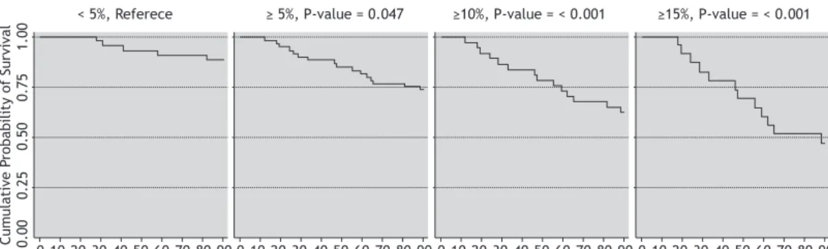
Kaplan-Meier survival curves for the fi rst 3 months
after diagnosis are shown in Figure 1. We observed a direct linear trend between the proportional WL and
mortality. Event-free survival at 3 months was 88% for the patients with WL < 5%, compared with 72% for those with WL ≥ 5% (p = 0.04 7), 61% for those with WL ≥ 10% (p < 0.001), and 45% for those with WL ≥ 15% (p < 0.001).
The outcomes of the Cox proportional hazards regression model are summarized in Table 3. The univariate and multivariate (adjusted) analyses both demonstrated that the risk of death during the 3-month follow-up period was higher when the higher WL cut-off points were applied. The adjusted multivariate analysis showed that the risk of death increases exponentially as the cut-off points of WL increase, the hazard ratios
for the 5%, 10%, and 15% cut-off points being 4.51 (95% CI: 1.32-15.39), 6.34 (95% CI: 2.31-17.40), and 14.17 (95% CI: 5.06-39.65), respectively.
DISCUSSION
NSCLC, as well as whether that association differs among four WL cut-off points. We have shown that greater WL translates to shorter overall survival for patients with NSCLC. We found that, among patients with advanced NSCLC, the 3-month mortality rate was almost two
times higher for those with WL ≥ 15% than for those with WL < 5%. Our results suggest that pre-treatment
WL is an important clinical parameter with relevant prognostic value in patients with advanced NSCLC.
Our findings are consistent with those of previous studies that evaluated survival and WL in populations of lung cancer patients that were more heterogeneous, including patients in different stages of the disease. (1-3,10-12,20-22) The prognostic signifi cance
of WL in stage IV NSCLC could be attributed to the
potential link to cachexia. Defi ned as a multifactorial
syndrome of progressive loss of skeletal muscle mass that cannot be completely reversed, cachexia has a heterogeneous clinical presentation that varies according to tumor type, site, and stage.(13,21) Lung cancer is often
accompanied by malnutrition, sarcopenia, and cachexia.
Following the cancer-specifi c cachexia classifi cations,
van der Meij et al.(23) demonstrated that, at the time
of diagnosis, approximately 18% and 23% of stage
III NSCLC patients had cachexia or were in a state of pre-cachexia, respectively. However, the exact basis of these prognostic differences remains unknown.
Several hypotheses have been proposed to explain the association between cachexia and poorer prognosis. Some authors have suggested that the survival advantage associated with obesity is due to the relatively large energy stores.(21-23) Conversely,
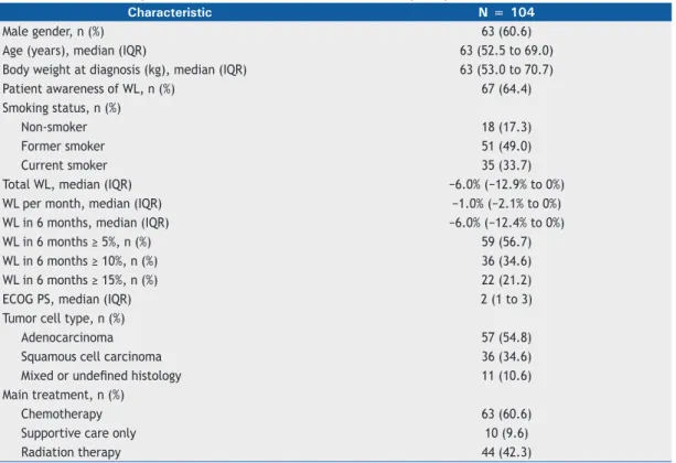
Table 1. Baseline anthropometric and clinical characteristics of the study sample.
Characteristic N = 104
Male gender, n (%) 63 (60.6)
Age (years), median (IQR) 63 (52.5 to 69.0)
Body weight at diagnosis (kg), median (IQR) 63 (53.0 to 70.7)
Patient awareness of WL, n (%) 67 (64.4)
Smoking status, n (%)
Non-smoker 18 (17.3)
Former smoker 51 (49.0)
Current smoker 35 (33.7)
Total WL, median (IQR) −6.0% (−12.9% to 0%)
WL per month, median (IQR) −1.0% (−2.1% to 0%)
WL in 6 months, median (IQR) −6.0% (−12.4% to 0%)
WL in 6 months ≥ 5%, n (%) 59 (56.7)
WL in 6 months ≥ 10%, n (%) 36 (34.6)
WL in 6 months ≥ 15%, n (%) 22 (21.2)
ECOG PS, median (IQR) 2 (1 to 3)
Tumor cell type, n (%)
Adenocarcinoma 57 (54.8)
Squamous cell carcinoma 36 (34.6)
Mixed or undefi ned histology 11 (10.6)
Main treatment, n (%)
Chemotherapy 63 (60.6)
Supportive care only 10 (9.6)
Radiation therapy 44 (42.3)
IQR: interquartile range; WL: weight loss; and ECOG-PS: Eastern Cooperative Oncology Group performance status.
Table 2. Kaplan-Meier survival analysis, by weight loss-related variable.
Variable Mean survival χ2 p*
Days (95% CI) WL, self-awareness
Yes 85 (80-89) 3.05 0.080
No 78 (73-84)
Proportional WL in 6 months
< 5% 85 (80-89) 3.94 0.047
≥ 5% 78 (72-84)
< 10% 85 (81-89) 11.58 < 0.001
≥ 10% 73 (64-82)
< 15% 85 (81-88) 23.78 < 0.001
≥ 15% 66 (53-78)
when the stores are depleted, the energy balance is negative.(21-23) Cachexia and a loss of skeletal muscle
mass are associated with poor prognosis in patients with advanced NSCLC who are receiving chemotherapy.(10,24)
Another hypothesis is that individuals with sarcopenia are susceptible to infections during hospitalization and residence in nursing homes, where such infections and premature termination of treatment are both possible contributors to shortened survival.(25) Accordingly, the well-recognized poor prognosis in advanced lung cancer masks some heterogeneity that could be
partially explained by WL stratifi cation and other, as yet unconfi rmed, biological factors.
Cachexia is a multifactorial syndrome with a complex pathogenesis. Therefore, multimodal interventions should be used in order to prevent WL among patients with cachexia.(26) Frequent high-calorie meals and
rest before meals are recommended. Currently, there is little evidence that nutritional supplementation is
effi cacious. (27) Physical activity has an anti-infl ammatory
effect and is effective in reducing muscle catabolism, increasing protein synthesis, and reversing protein degradation. (28) Pharmacological treatment can also
be used to prevent cachexia. Corticosteroids and progesterone analogs have been shown to increase appetite, thus resulting in modest weight gain. However, such drugs do not improve survival or quality of life.(29)
Our study has some limitations, not the least of which is the small sample size. In addition, there are many
confounding variables that could infl uence the analysis
of 3-month survival of patients with advanced NSCLC. One such variable is the main treatment adopted, which, in our sample. was quite heterogeneous, therefore potentially affecting the prognosis. However, we believe that using a Cox proportional hazards regression model
was an effective strategy to reduce the infl uence of those
confounders, and the associations detected retained
their signifi cance even after the multivariate analyses.
Studies including larger patient samples are needed
in order to corroborate our fi ndings. Further studies
should also include biomarkers linked to aggressive behavior of lung cancer, such as circulating tumor cells and circulating cell-free nucleic acids, which could also affect survival rates, and patients who have such biomarkers might show different rates of WL.(30)
Although increased efforts have been directed toward
the identifi cation of biological markers as prognostic
indicators of lung cancer, there are some important clinical indices that should be also considered more thoroughly. In conclusion, our results indicate that proportional WL is an important prognostic factor for 3-month survival after diagnosis in patients with stage IV NSCLC.
Table 3. Crude and adjusted hazard ratios for weight-loss related prognostic factors.
Variable Crude HR (95% CI) p Adjusted HR* (95% CI) p
WL, self-awareness 2.54 (0.86-7.49) 0.089 3.59 (1.03-12.48) 0.044
WL in 6 months ≥ 5% 2.65 (0.97-7.21) 0.055 4.51 (1.32-15.39) 0.016
WL in 6 months ≥ 10% 4.45 (1.80-10.99) 0.001 6.34 (2.31-17.40) < 0.001
WL in 6 months ≥ 15% 6.53 (2.76-15.44) < 0.001 14.17 (5.06-39.65) < 0.001 HR: hazard ratio; and WL: weight loss. *Cox proportional hazards regression model analysis, adjusted for gender, age, Eastern Cooperative Oncology Group performance status, and tumor cell type.
0.00
0.25
0.50
0.75
1.00
10
0 20 30 40 50 60 70 80 9001020 30 40 50 60 70 80 9001020 30 40 50 60 70 80 9001020 30 40 50 60 70 80 90 Weight loss in 6 months
< 5%, Referece
Time, days
≥ 5%, P-value = 0.047 ≥10%, P-value = < 0.001 ≥15%, P-value = < 0.001
Cumulative P
robability of Survival
REFERENCES
1. Ross PJ, Ashley S, Norton A, Priest K, Waters JS, Eisen T, et al. Do
patients with weight loss have a worse outcome when undergoing chemotherapy for lung cancers? Br J Cancer. 2004;90(10):1905-11. https://doi.org/10.1038/sj.bjc.6601781
2. Jeremić B, Miličić B, Milisavljevic S. Clinical prognostic factors in
patients with locally advanced (stage III) non-small cell lung cancer treated with hyperfractionated radiation therapy with and without concurrent chemotherapy: single-Institution Experience in 600 Patients. Cancer. 2011;117(13):2995-3003. https://doi.org/10.1002/ cncr.25910
3. Martin L, Birdsell L, Macdonald N, Reiman T, Clandinin MT, McCargar LJ, et al. Cancer cachexia in the age of obesity: skeletal muscle depletion is a powerful prognostic factor, independent of body mass index. J Clin Oncol. 2013;31(12):1539-47. https://doi.org/10.1200/ JCO.2012.45.2722
4. Fredrix EW, Wouters EF, Soeters PB, van der Aalst AC, Kester
AD, von Meyenfeldt MF, et al. Resting energy expenditure in patients with non-small cell lung cancer. Cancer. 1991;68(7):1616-21. https://doi.org/10.1002/1097-0142(19911001)68:7<1616::AID-CNCR2820680725>3.0.CO;2-3
5. Hyltander A, Drott C, Körner U, Sandström R, Lundholm K. Elevated energy expenditure in cancer patients with solid tumours. Eur J Cancer. 1991;27(1):9-15. https://doi.org/10.1016/0277-5379(91)90050-N
6. Staal-van den Brekel A, Schols A, ten Velde G, Buurman WA, Wouters EF. Analysis of energy balance in lung cancer patients. Cancer Res. 1994;54(24):6430-3.
7. Staal-van den Brekel A, Dentener MA, Schols AM, Buurman WA, Wouters EF. Increased resting energy expenditure and weight loss are related to a systemic infl ammatory response in lung cancer patients. J Clin Oncol. 1995;13(10):2600-5. https://doi.org/10.1200/ JCO.1995.13.10.2600
8. Melville S, McNurlan MA, Calder AG, Garlick PJ. Increased protein turnover despite normal energy metabolism and responses to feeding in patients with lung cancer. Cancer Res. 1990;50(4):1125-31.
9. Jatoi A, Daly BD, Hughes V, Dallal GE, Roubenoff R. The prognostic effect of increased energy expenditure prior to treatment for lung cancer. Lung Cancer. 1999;23(2):153-8. https://doi.org/10.1016/ S0169-5002(99)00008-2
10. Buccheri G, Ferrigno D. Importance of weight loss defi nition in the prognostic evaluation of non-small-cell lung cancer. Lung Cancer. 2001;34(3):433-40. https://doi.org/10.1016/S0169-5002(01)00273-2
11. Tartari RF, Ulbrich-Kulczynski JM, Filho AF. Measurement of mid-arm muscle circumference and prognosis in stage IV non-small cell lung cancer patients. Oncol Lett. 2013;5(3):1063-1067. https://doi. org/10.3892/ol.2013.1128
12. Kimura M, Naito T, Kenmotsu H, Taira T, Wakuda K, Oyakawa T, et al. Prognostic impact of cancer cachexia in patients with advanced non-small cell lung cancer. Support Care Cancer. 2015;23(6):1699-708. https://doi.org/10.1007/s00520-014-2534-3
13. Fearon K, Strasser F, Anker SD, Bosaeus I, Bruera E, Fainsinger RL, et al. Defi nition and classifi cation of cancer cachexia: an international consensus. Lancet Oncol. 2011;12(5):489-95. https:// doi.org/10.1016/S1470-2045(10)70218-7
14. Aapro M, Arends J, Bozzetti F, Fearon K, Grunberg SM, Herrstedt J, et al. (2014) Early recognition of malnutrition and cachexia in the cancer patient: a position paper of a European School of Oncology Task Force. Ann Oncol. 2014;25(8):1492-9. https://doi.org/10.1093/ annonc/mdu085
15. Goldstraw P, Crowley J, Chansky K, Giroux DJ, Groome PA, Rami-Porta R, et al. The IASLC Lung Cancer Staging Project: proposals for the revision of the TNM stage groupings in the forthcoming (seventh) edition of the TNM Classifi cation of malignant tumours. J Thorac Oncol. 2007;2(8):706-14. Erratum in: J Thorac Oncol. 2007;2(10):985. https://doi.org/10.1097/JTO.0b013e31812f3c1a
16. Oken MM, Creech RH, Tormey DC, Horton J, Davis TE, McFadden
ET, et al. Toxicity and response criteria of the Eastern Cooperative Oncology Group. Am J Clin Oncol. 1982;5(6):649-55. https://doi. org/10.1097/00000421-198212000-00014
17. Centers for Disease Control and Prevention--CDC [homepage in the internet]. Atlanta (GA): CDC; [updated 2017 Jan; cited 2017 Jul 29]. National Health and Nutrition Examination Survey (NHANES)--Anthropometry Procedures Manual. [Adobe Acrobat document, 102p.]. Available from: http://www.cdc.gov/nchs/data/nhanes/ nhanes_07_08/manual_an.pdf
18. Altman DG. Practical statistics for medical research, 1st ed. London: Chapman & Hall; 1991.
19. Cox DR. Regression models and life-tables. J Royal Statistical Soc. Series B (Methodological) 1972;34:187-220.
20. Buccheri G, Ferrigno D, Tamburini M. Karnofsky and ECOG performance status scoring in lung cancer: a prospective, longitudinal study of 536 patients from a single institution. Eur J Cancer. 1996;32A(7):1135-41. https://doi.org/10.1016/0959-8049(95)00664-8
21. Paralkar VR, Li T, Langer CJ. Population characteristics and prognostic factors in metastatic non-small-cell lung cancer: a Fox Chase Cancer Center retrospective. Clin Lung Cancer. 2008;9(2):116-21. https://doi. org/10.3816/CLC.2008.n.018
22. Blanchon F, Grivaux M, Asselain B, Lebas FX, Orlando JP, Piquet J, et al. 4-year mortality in patients with non-small-cell lung cancer: development and validation of a prognostic index. Lancet Oncol. 2006;7(10):829-36. https://doi.org/10.1016/S1470-2045(06)70868-3
23. van der Meij BS, Schoonbeek CP, Smit EF, Muscaritoli M, van
Leeuwen PA, Langius JA. Pre-cachexia and cachexia at diagnosis of stage III non-small-cell lung carcinoma: an exploratory study comparing two consensus-based frameworks. Br J Nutr. 2013;109(12):2231-9. https://doi.org/10.1017/S0007114512004527
24. Goldstraw P, Ball D, Jett JR, Le Chevalier T, Lim E, Nicholson AG, et al. Non-small-cell lung cancer. Lancet. 2011;378(9804):1727-40. https://doi.org/10.1016/S0140-6736(10)62101-0
25. Kovarik M, Hronek M, Zadak Z. Clinically relevant determinants of body composition, function and nutritional status as mortality predictors in lung cancer patients. Lung Cancer. 2014;84(1):1-6. https://doi.org/10.1016/j.lungcan.2014.01.020
26. Aversa Z, Costelli P, Muscaritoli M. Cancer-induced muscle wasting:
latest fi ndings in prevention and treatment. Ther Adv Med Oncol. 2017;9(5):369-382. https://doi.org/10.1177/1758834017698643
27. Koretz RL, Lipman TO, Klein S; American Gastroenterological Association. AGA technical review on parenteral nutrition. Gastroenterology. 2001;121(4):970-1001. https://doi.org/10.1053/ gast.2001.28031
28. Gould DW, Lahart I, Carmichael AR, Koutedakis Y, Metsios GS. Cancer cachexia prevention via physical exercise: molecular mechanisms. J Cachexia Sarcopenia Muscle. 2013;4(2):111-24. https://doi.org/10.1007/s13539-012-0096-0
29. Ruiz Garcia V, López-Briz E, Carbonell Sanchis R, Gonzalvez Perales JL, Bort-Marti S. Megestrol acetate for treatment of anorexia-cachexia syndrome. Cochrane Database Syst Rev. 2013;(3):CD004310. https://doi.org/10.1002/14651858.CD004310.pub3