Prognostic Markers in Non-Small Cell Lung Cancer
Thomas John1,2*, Maud H. W. Starmans3,4, Yao-Tseng Chen5, Prudence A. Russell6, Stephen A. Barnett2, Shane C. White2, Paul L. Mitchell2, Marzena Walkiewicz1, Arun Azad1,2, Philippe Lambin4, Ming-Sound Tsao7, Siddhartha Deb1, Nasser Altorki5, Gavin Wright6, Simon Knight2, Paul C. Boutros3,8,
Jonathan S. Cebon1,2
1Ludwig Institute for Cancer Research, Austin Health, Melbourne, Australia,2Medical Oncology and Thoracic Surgery departments, Austin Health, Melbourne, Australia, 3Informatics and Biocomputing Platform, Ontario Institute for Cancer Research, Toronto, Canada,4Department of Radiation Oncology (Maastro), GROW-School for Oncology and Developmental Biology, Maastricht University Medical Center, Maastricht, The Netherlands,5Weill Cornell Medical College-New York Presbyterian Hospital, New York City, New York, United States of America,6St Vincent’s Hospital, Melbourne, Australia,7University Health Network, Ontario Cancer Institute, Princess Margaret Hospital, Toronto, Canada,8Department of Medical Biophysics, University of Toronto, Toronto, Canada
Abstract
Background:Cancer-Testis Antigens (CTAs) are immunogenic proteins that are poor prognostic markers in non-small cell lung cancer (NSCLC). We investigated expression of CTAs in NSCLC and their association with response to chemotherapy, genetic mutations and survival.
Methods:We studied 199 patients with pathological N2 NSCLC treated with neoadjuvant chemotherapy (NAC;n= 94), post-operative observation (n= 49), adjuvant chemotherapy (n= 47) or unknown (n= 9). Immunohistochemistry for NY-ESO-1, MAGE-A and MAGE-C1 was performed. Clinicopathological features, response to neoadjuvant treatment and overall survival were correlated. DNA mutations were characterized using the Sequenom Oncocarta panel v1.0. Affymetrix data from the JBR.10 adjuvant chemotherapy study were obtained from a public repository, normalised and mapped for CTAs.
Results:NY-ESO-1 was expressed in 50/199 (25%) samples. Expression of NY-ESO-1 in the NAC cohort was associated with significantly increased response rates (P= 0.03), but not overall survival. In the post-operative cohort, multivariate analyses identified NY-ESO-1 as an independent poor prognostic marker for those not treated with chemotherapy (HR 2.61, 95% CI 1.28–5.33;P= 0.008), whereas treatment with chemotherapy and expression of NY-ESO-1 was an independent predictor of improved survival (HR 0.267, 95% CI 0.07–0.980; P= 0.046). Similar findings for MAGE-A were seen, but did not meet statistical significance. Independent gene expression data from the JBR.10 dataset support these findings but were underpowered to demonstrate significant differences. There was no association between oncogenic mutations and CTA expression.
Conclusions: NY-ESO-1 was predictive of increased response to neoadjuvant chemotherapy and benefit from adjuvant chemotherapy. Further studies investigating the relationship between these findings and immune mechanisms are warranted.
Citation:John T, Starmans MHW, Chen Y-T, Russell PA, Barnett SA, et al. (2013) The Role of Cancer-Testis Antigens as Predictive and Prognostic Markers in Non-Small Cell Lung Cancer. PLoS ONE 8(7): e67876. doi:10.1371/journal.pone.0067876
Editor:William C. S. Cho, Queen Elizabeth Hospital, Hong Kong
ReceivedFebruary 28, 2013;AcceptedMay 23, 2013;PublishedJuly 23, 2013
Copyright:ß2013 John et al. This is an open-access article distributed under the terms of the Creative Commons Attribution License, which permits unrestricted use, distribution, and reproduction in any medium, provided the original author and source are credited.
Funding:This work was supported by the Victorian Cancer Agency, Clinical Research Fellowship (CRF09-04), a Pfizer Cancer Research grant, the Center for Translational Molecular Medicine (www.ctmm.nl) (AIRFORCE Project Ref. 030-103), National Health & Medical Research Council (NHMRC) of Australia Practitioner Fellowship (487905) and Project grants (542510 & 1007381) as well as the Ontario Institute for Cancer Research through funding provided by the Government of Ontario to PB. Supported in part by the Operational Infrastructure Support Program Funding of the Victorian State Government. The funders had no role in study design, data collection and analysis, decision to publish, or preparation of the manuscript.
Competing Interests:The Pfizer Cancer Research grant is a peer reviewed, competitive grant. This does not alter the authors’ adherence to all the PLOS ONE policies on sharing data and materials.
* E-mail: tom.john@ludwig.edu.au
Background
Although the overall incidence of non-small cell lung cancer (NSCLC) appears to be declining in North America, mortality has only recently plateaued [1]. More deaths are attributable to NSCLC than any other cancer [1]. As symptoms occur late, the majority of patients are diagnosed with locally advanced or metastatic disease. Management of late-stage disease has evolved
observation, and it remains unclear whether tyrosine kinase inhibitors are beneficial for early stage NSCLC [2,3].
There is currently no consensus for the management of locally advanced (Stage IIIA) NSCLC. This remains controversial because of variations in staging techniques, conflicting trial results and idiosyncratic practices among different institutions [4]. Options include surgery followed by adjuvant chemotherapy (ACT), neoadjuvant chemotherapy (NAC) followed by surgery and definitive chemo-radiation alone or followed by surgery. To date, the only modality shown to significantly improve survival is surgical resection followed by ACT, as shown in a number of large studies [5]. Although initial studies of NAC appeared promising, enthusiasm diminished as more data from large adjuvant studies became available, leading to poor recruitment and underpowered analyses [6]. Only one study compared NAC to ACT and demonstrated equivalency in patients with early stage disease [7], although there were caveats [4].
One particular rationale for using NAC as opposed to post-operative adjuvant therapy, is that it provides valuable, potentially predictive information, based on drug sensitivityin vivo. In contrast
current predictive markers, cannot determine whether an individ-ual with NSCLC will respond to a given treatment [8]. This is important as response to treatment can improve resectability and may translate into improvement in survival [9]. Furthermore, the ability to predict a priori which patients will benefit makes it possible to optimize therapy and avoid unnecessary toxicity in those who are unlikely to respond.
Cancer-Testis antigens (CTAs) are molecules characterized by expression restricted to normal testis tissue but aberrant expression in a variety of cancer types, including 10–50% of NSCLC [10,11]. Their expression is often coordinated [12] and associated with poorer clinical outcome [13] and advanced disease [14,15]. There is some evidence that they are functional; short interfering RNA screening of a lung cancer cell line showed that several CTAs were associated with resistance to paclitaxel chemotherapy [16]. However, the CTAs identified in these screens have not been extensively studied and their presence in patient lung tumors has yet to be evaluated. The association with chemoresistance is analogous to observations in other tumor types where it has been speculated that CTAs mark more primitive, ‘‘stem-like’’ cells [10,17–19]. Indeed the current study was initially undertaken in order to assess the relationship between these antigens and chemoresistance. It therefore came as a surprise to discover that NY-ESO-1 and to a lesser extent MAGE-A3 were associated with chemosensitivity.
While the functional role of CTAs in cancer remains unclear, the immunogenicity of these molecules has been well documented [10]. Indeed CTAs were originally identified by cloning antigens that activated cytotoxic T lymphocytes and screening patient sera for immunoreactive antibodies [20]. To date over 100 distinct CTA families have been identified [21] with approximately 30 encoded by genes located on the X-chromosome (CT-X genes). The promoter regions for all studied CT-X genes contain CpG islands and are often methylated in somatic tissues and therefore not expressed. In cancer, global demethylation of the X-chromosome including the promoter regions of CT-X genes are believed to result in the coordinated activation of multiple CTAs [10], although it is unclear whether the CTAs are bystanders or functionally participate in tumorigenesis.
To evaluate the relationship between chemosensitivity and CTA expression in NSCLC, we performed immunohistochemical studies on tumors from patients with pathological N2 (pN2) nodal involvement (at least Stage IIIA). We investigated two clinical cohorts, a pre-operative and post-operative cohort. Patients had
either undergone pre-operative NAC, where we investigated the mediastinal nodal tissue prior to treatment or were treated surgically with or without adjuvant chemotherapy in the post-operative group; where we investigated the primary tumor tissue post-resection. Here we demonstrate that the CTA NY-ESO-1 is prognostic for poor clinical outcome, however also appears to be predictive for improved response and survival benefit from chemotherapy.
Materials and Methods
Patients and clinical specimens
Under a protocol approved by the ethics committees of Austin Health, St Vincent’s Hospital and Weill Cornell Medical Centre hospitals, patients who underwent pre-operative NAC or surgery with or without ACT for pN2 disease were identified and clinical information captured retrospectively. Consent for accessing tissues and clinical records was waived on the proviso that patient details were de-identified. NAC patients were treated with at least three cycles of platinum-based chemotherapy prior to surgery. ACT patients were treated with four cycles of platinum-based chemo-therapy post resection. Patients who did not receive ACT were observed without any further treatment. For the pre-operative cohort, samples obtained from mediastinal lymph node sampling prior to chemotherapy administration were used for CTA staining and DNA isolation. In the post-operative cohort, the primary surgical lung tumor specimen was used. Response to NAC was defined pathologically by down-staging of the initial lesion post-operatively. Patients were staged pre-operatively with PET scans or mediastinoscopy.
For further validation we investigated patients treated as part of the BR.10 study [22] whose tumors were subjected to molecular profiling using Affymetrix arrays. The original study randomized 482 patients to either receive adjuvant chemotherapy or observa-tion following surgical resecobserva-tion, however only 133 of these samples were available for gene expression arrays [23].
CT antigen staining
For CTA staining, 4-mm sections of formalin-fixed paraffin-embedded (FFPE) tissues were cut and stained as previously described [19]. NY-ESO-1 (E978) was obtained from the Ludwig Institute for Cancer Research and used at a concentration of 2.5mg/mL. MAGE-A antibody (6C1) was purchased from Abcam and used at 0.3mg/mL. MAGE-C1/CT7 antibody (CT7-33) was
obtained from Santa-Cruz (cat no sc-20034) and used at 0.5mg/
mL. The detection of nuclear and/or cytoplasmic staining in any percentage of tumor cells was considered positive. Complete absence of staining was considered negative for each CTA tested. Testis tissue was used as a positive control; normal lung tissue and absence of primary antibody were used as negative controls.
Mutational Profiling
Microarray Analysis
CTA mRNA abundances were evaluated in the BR.10 dataset [23]. These data are publically available at the National Centre for Biotechnology Information Gene Expression Omnibus (GSE14814) and from the Director’s Challenge Project [25]. These analyses were performed in R (v2.14.2). Data pre-processing was performed using RMA [26] (Affymetrix package version 1.20.0) and updated Entrez GeneID annotation [27] (hgu133ahsentrezgcdf package v14.1.0). CTA’s were matched to the gene expression microarray platform via their Entrez GeneIDs. Each gene was used to median dichotomize the patient cohort. Differences in survival between the two groups were assessed with Kaplan-Meier survival curves and Cox proportional hazard ratio modeling followed by the Wald test (survival package v2.36-12) in the whole cohort, patients treated with ACT and patients not treated with ACT.
Statistics
Differences in patient demographics for CTA positive and CTA negative patient groups were assessed with x2-tests. A p-value
,0.05 was considered significant. In addition, to evaluate prognostic and predictive value of CTA expression both univariate and multivariate Cox proportional hazard ratio modeling analyses were performed in R (v2.14.2, survival package v2.36-12).
Results
Patients
Clinicopathological data were available for 199 patients in two cohorts. The first, a pre-operative cohort, included 94 patients who underwent neoadjuvant chemotherapy followed by surgery. In the second, a post-operative cohort, 105 patients were included; 49 who had surgical resection alone, 47 who received additional adjuvant chemotherapy and 9 where adjuvant chemotherapy data was not recorded. The clinical information for each group is summarized in Table 1. Patients treated with NAC who proceeded to surgery represent a highly selected subgroup based mainly on resectability and good performance status post-chemotherapy. In the post-operative cohort, patients who did not receive chemotherapy were mainly treated prior to 2004 when adjuvant treatment was more broadly accepted.
CT Antigen expression
NY-ESO-1 stained similar proportions in each cohort, with 24/ 94 (26%) of pN2 nodes in the pre-operative cohort and 26/105 (25%) of resected lung tumors in the post-operative cohort (Figure 1). The median age of patients whose tumors stained positive for NY-ESO-1 was 62 years in the pre-operative and 66 years in the post-operative cohort, which were similar to patients with NY-ESO-1 negative tumors. More males were NY-ESO-1 positive in both cohorts with 14/24 (58%) positive in the pre-operative cohort (Px2= 0.728) and 16/26 (62%) in the
post-operative cohort (Px2= 0.605).
MAGE-A expression was observed in only 27/94 (29%) pre-operative patients but in 52/105 (50%) in the post-pre-operative cohort (Px2= 0.004). MAGE-C1 stained fewer samples, with 13/
94 (13%) in the NAC cohort but 32/105 (44%) in the post-operative cohort.
CTAs were expressed in both adenocarcinoma and squamous cell carcinomas (Figure 1E, 1F). However more squamous cell carcinomas were positive in both datasets, although this did not reach statistical significance in the pre-operative cohort. For NY-ESO-1 in the pre-operative cohort 8/22 (36%) squamous cell carcinomas were positive compared to 9/60 (15%)
adenocarcino-mas (Px2= 0.070). Similarly in the post-operative cohort 12/30
(30%) were positive compared to 8/59 (14%; Px2= 0.011) adenocarcinomas. These histological differences in CT antigen staining were similar for MAGE-A and MAGE-C1 and to that previously reported [13]. Furthermore expression of one CTA was often associated with expression of the others a phenomenon which has also been previously observed [13].
Genetic mutations and CT antigen expression
We next sought to determine if CTA expression was associated with specific genetic mutations. We screened 238 somatic mutations in 19 oncogenes using Sequenom’s OncoCarta panel v1.0. Lower rates of mutations in the pre-operative cohort to that expected in a Caucasian NSCLC population were observed, with only fourEGFR(4%) mutations and nineKRAS(9%) mutations, all occurring in adenocarcinomas as expected. Two additional cases harboredBRAF mutations, while three hadTP53mutations and one an NRAS mutation. The low rates of EGFR and KRAS
mutations may reflect a higher rate of false-negative results due to the low tumor cell percentage in proportion to non-neoplastic cells in these lymph nodes, although areas of high tumor cellularity were specifically selected for DNA isolation. In the post-operative cohort, rates of mutations were significantly higher, and closer to those expected for late-stage NSCLC, with 16EGFR mutations (15%) and 18 (17%)KRAS mutations. CTA expression was not associated with oncogenic mutations in adenocarcinomas (Figure 1E).
Response to neoadjuvant chemotherapy
Response to NAC was assessed based on pathological down staging. Of the 24 NY-ESO-1 positive tumors, two patients achieved a complete response (CR; i.e. no residual tumor was pathologically detectable), 14 a partial response (PR), six had stable disease (SD) and two progressed through chemotherapy (PD). In the 70 NY-ESO-1 negative patients, there was one CR, 23 PRs, 36 SD, nine PDs and one missing response (Figure 2). There was a significantly increased response rate (CR+PR) in NY-ESO-1 expressing tumors compared to NY-NY-ESO-1 negative ones (Px2= 0.034).
Despite improved responses to NAC, no survival differences were observed in patients expressing NY-ESO-1 or any other CTA compared to those tumors that did not. Pathological CR was associated with prolonged survival; however, for the majority of patients, the best response was a PR. It should be noted that patients that were known to have progressed through chemother-apy pre-operatively and therefore unable to proceed to surgery were not included in this study. Furthermore given the low rate of NY-ESO-1 expression, these analyses were underpowered to detect a survival difference.
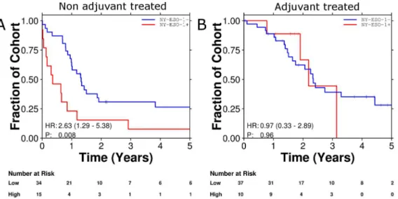
Survival following adjuvant chemotherapy
inde-pendent poor prognostic factor (HR 2.61, 95% CI 1.28–5.33; p = 0.008 Wald test) whereas treatment with chemotherapy and expression of NY-ESO-1 was an independent predictor of improved survival (HR 0.267, 95% CI 0.073–0.980; p = 0.046 Wald test. Table 2).
Survival in an independent cohort using gene expression data
We sought to validate our findings using microarray data previously published from the BR.10 adjuvant chemotherapy dataset [23]. This cohort contained Affymetrix microarray data for the mRNA abundances of 133 patients of which 71 received adjuvant chemotherapy. Unfortunately, although oligonucleotide probes representing MAGE-A1, MAGE-A4 and MAGE-C1 could be mapped to the array, NY-ESO-1 was not present after mapping to modern gene-annotations. Expression of MAGE-A1 and 4 antigens was associated with inferior survival in patients random-ized to observation. However MAGE-A1 and 4 positive patients
treated with chemotherapy had improved survival compared to similarly treated MAGE-A1 and 4 negative patients and survival curves similar to those seen in our post-operative cohort of patients were observed (Figure 4). However, these differences in survival did not reach the level of statistical significance. It is important to note that although survival in the overall BR.10 dataset demonstrated statistically significant differences for patients treated with adjuvant chemotherapy, in this subset of 133 patients, the trend toward improved survival remained, however was not significant. This may explain why in this underpowered cohort, similar trends were seen to those observed in our post-operative cohort although did not reach the level of statistical significance.
Discussion
These data provide further confirmation that CTA expression in NSCLC is associated with poor overall prognosis. In particular NY-ESO-1 was associated with significantly poorer outcome in patients not treated with adjuvant chemotherapy when compared
Table 1.Clinicopathological features associated with CTA expression in two cohorts of patients.
Pre-operative cohort (n = 94) Post-operative cohort (n = 105)
Age median (range) 63 (37–82) Age median (range) 66 (29–86)
Sex Sex
Male 50 Male 58
Female 44 Female 47
Stage Stage
IIB 3 IIIA 99
IIIA 88 IIIB 6
IIIB 3
Histology Histology
Adenocarcinoma 60 Adenocarcinoma 59
Squamous 22 Squamous 30
Other 11 Other 16
NA 1
Response Adjuvant chemo
Complete Response (CR) 3 No 49
Partial Response (PR) 37 Yes 47
Stable Disease (SD) 42 NA 9
Progressive Disease (PD) 11
NA 1
NY-ESO-1 NY-ESO-1
2 70 2 79
+ 24 + 26
MAGE-A MAGE-A
2 67 2 53
+ 27 + 52
MAGE-C1 MAGE-C1
2 81 2 73
+ 13 + 32
Mutation status (EGFR/KRAS/TP53) Mutation status (EGFR/KRAS/TP53)
2 62 2 50
+ 25 + 50
NA 7 NA 5
to NY-ESO-1 negative patients who were also not treated with adjuvant chemotherapy. Although similar trends were seen for MAGE-A and MAGE-C1, NY-ESO-1 remained the strongest predictor of response to chemotherapy in the neoadjuvant setting but also for benefit from adjuvant chemotherapy. These findings support NY-ESO-1 as a useful prognostic factor but also a potential predictive factor for significant benefit from adjuvant and neoadjuvant chemotherapy.
Despite the improved responses to chemotherapy in the neoadjuvant setting it was surprising that no translation from response into a survival benefit was observed for patients with NY-ESO-1+ tumors. However it should be emphasized that these analyses were underpowered to detect a survival difference. These results paralleled similar findings in patients treated with neoadjuvant chemotherapy in breast cancer. Breast tumors that were negative for estrogen receptor, progesterone receptor and HER2 gene amplification, otherwise known as ‘‘triple negative’’ breast cancers also strongly expressed CT-X antigens [28,29] compared to receptor-positive tumors. Furthermore, triple
nega-tive breast cancers have been shown to be more chemosensinega-tive to anthracycline and platinum based chemotherapy [30,31]. Inter-estingly, however, although response to chemotherapy was significantly improved in triple negative tumors, survival was not impacted in these studies, except in the minority of patients who achieved a complete response. This led to the hypothesis that the poorer phenotype associated with CTA expression and triple negativity resulted in increased relapse rates, despite good initial responses [31]. These findings may explain why patients in our study whose tumors expressed NY-ESO-1 had significantly improved responses to neoadjuvant chemotherapy and yet no survival benefit when compared to patients with NY-ESO-1 negative tumors.
One caveat to our study is that the tissue investigated for our two cohorts were different, with the pre-operative cohort consisting of lymph node tissue and the post-operative cohort the primary lung tumor. There were clear differences in the number of samples that were positive for CTAs as well as for mutations between these cohorts. However in patients undergoing
Figure 1. CTA expression by IHC in an individual patient tumor sample.A: Testis (positive control), B: NY-ESO-1, C: MAGE-A, D: MAGE-C1, E: Heat map detailing overlap in expression of CTAs and other clinicopathological features in the neoadjuvant cohort.
doi:10.1371/journal.pone.0067876.g001
Figure 2. Expression of N-ESO-1 and response to neoadjuvant chemotherapy (A). CR = Complete Response, PR = Partial Response, SD = Stable Disease, PD = Progressive Disease, NA = Not Assessable. Forest plot detailing factors associated with survival in patients who were treated in the post-operative cohort. Sq = Squamous Cell, ADC = Adenocarcinoma, ACT = Adjuvant Chemotherapy (B).
neoadjuvant chemotherapy, it is not possible to obtain a large portion of the primary tissue and using tissue post-chemotherapy may also be inaccurate. In our study we also tested the primary lung tumor sample after NAC for NY-ESO-1. In samples with viable tumor, NY-ESO-1 expression was not seen in the primary resected tumor if the pre-treatment lymph node was negative; however in 13 cases, despite NY-ESO-1+ lymph nodal tissue, following chemotherapy NY-ESO-1 expression was not detected in the primary resected tumor (data not shown).
As the functional role of CTAs is poorly understood, it is difficult to define a mechanism by which their expression could influence chemosensitivity. Whilst some data exist supporting
KRAS mutations as a marker of chemoresistance and EGFR
mutations as a marker of chemosensitivity [8], our data do not demonstrate an association between these mutations and NY-ESO-1 expression. Certainly our findings are in contradistinction to recent studies demonstrating that in cell lines, other CTAs including acrosin-binding protein (ACRBP) were associated with
resistanceto taxane chemotherapy [16]. However, unlike ACRBP
which is a non-X-CTA with a specific function associated with the mitotic spindle [32], CT-X antigens evaluated in his study have no well-defined function to date. This difference likely underlies their different influences on chemosensitivity. Other studies have demonstrated that the MAGE proteins form complexes with
Kap-1, a known co-repressor ofTP53and MAGE may thus act to prevent cells from undergoing apoptosis and promote tumorigen-esis. However, linking these pathways to explain the improved response to chemotherapy in breast and now lung cancer warrants further investigation.
As CTAs are known to be immunogenic, it is perhaps more plausible that the improved response and survival for patients whose tumors express NY-ESO-1 were not the result of increased cytotoxicity, but rather were mediated through immunological mechanisms. NY-ESO-1 is a potent stimulator of T-Cells and has been used as a tumor vaccine in a variety of tumor types [21,33] Chemotherapy induced tumor cell lysis could enable antigens previously ignorant to T-Cells to be processed and presented via antigen presenting cells with resultant T-Cell stimulation. This may explain in part the increased down-staging of tumors in the short term, although one would expect a T-Cell response to result in more durable remissions. Evidence for this is supported by recent studies investigating immune checkpoint inhibitors where the impact of immune activation can be delayed such that initial disease progression may be observed followed by more durable remissions [34]. Interestingly, NY-ESO-1 specific T cell responses have been reported to increase in frequency and functionality during anti-CTLA-4 treatment, an immune checkpoint inhibitor, with resultant durable disease remissions, therefore highlighting the importance of this antigen [35]. Previous studies have not shown a correlation between the development of antibodies to tumor antigens and spontaneous remissions. However in the context of chemotherapy, immunological recognition and re-sponse in addition to chemotherapy induced depletion of regulatory T-Cells could explain the improved response rates we have observed and the longer-term survival advantage in the adjuvant setting. An alternative to these hypotheses is that the survival advantage seen in the adjuvant setting may not represent improved tumor responses but poorer survival in the absence of chemotherapy. Further studies are warranted to better explain our findings.
Recent clinical trials investigating immune checkpoint inhibitors in NSCLC have demonstrated significant promise particularly in squamous cell carcinomas [36,37]. A study investigating a monoclonal antibody directed against CTLA-4, a molecule that inhibits T-Cell activation, demonstrated an overall benefit in
Figure 3. Survival curves demonstrating NY-ESO-1 to be a poor prognostic factor (A) and a predictive marker for benefit from adjuvant chemotherapy (B).
doi:10.1371/journal.pone.0067876.g003
Table 2.Multivariate analysis of factors associated with survival in patients treated with surgery initially.
HR 95% CI P (Wald test)
Stage (IIIB vs. IIIA) 1.245 0.4697–3.299 0.660
Histology (SQ vs. ADC) 1.542 0.8445–2.816 0.159
Histology (other vs. ADC) 1.331 0.5561–3.185 0.521
ACT 0.7427 0.3977–1.387 0.351
NY-ESO-1 2.609 1.278–5.329 0.008
NY-ESO-1:ACT 0.2674 0.07301–0.9795 0.046
HR = hazard ratio, ACT = adjuvant chemotherapy, SQ = squamous cell, ADC = adenocarcinoma histology.
prolonging immune related progression free survival. Although tumors of all different histological types appeared to benefit, a trend towards improved benefit was seen in patients with squamous cell histology, a subgroup that we have also observed to highly express CTAs [36]. These data have resulted in the launch of a randomized phase III study comparing the addition of ipilimumab to chemotherapy in patients with squamous cell histology. Similarly an inhibitor of programmed death-1 (PD-1), a molecule also involved in T-cell inhibition, has recently demon-strated efficacy in a Phase I study [37]. Significantly more responses were observed in patients with squamous cell carcinoma when compared to non-squamous tumor types. Although we are currently unable to demonstrate that CTAs play a role in these early studies, the strong association of these immunogenic molecules with the same histology to benefit from immunoactiva-tion warrants further investigaimmunoactiva-tion.
These data demonstrate that expression of the CTA NY-ESO-1 is associated with increased downstaging with chemotherapy given in the neoadjuvant setting and significant survival benefit with
chemotherapy given in the adjuvant setting. Our data therefore support NY-ESO-1 as both a (poor) prognostic marker and also a predictive marker. As the assay is straightforward and easy to interpret, NY-ESO-1 could easily be studied prospectively as a marker. Perhaps more importantly, the immunogenicity of this molecule may be a rationale for future studies using NY-ESO-1 as a therapeutic target, thereby better personalizing treatment towards those most likely to derive benefit.
Acknowledgments
The authors thank Dr. Syed Haider for technical support.
Author Contributions
Conceived and designed the experiments: TJ YC AA SD JC. Performed the experiments: MS PR MW YC SD PB. Analyzed the data: MW PB PL. Contributed reagents/materials/analysis tools: TJ MS PR SB MT NA YC JC SK. Wrote the paper: TJ MS PB JC. Patient clinical data: SB PM SW GW NA.
References
1. Siegel R, Naishadham D, Jemal A (2012) Cancer statistics, 2012. CA Cancer J Clin 62: 10–29. doi:10.3322/caac.20138.
2. Kelly K, Chansky K, Gaspar LE, Albain KS, Jett J, et al. (2008) Phase III trial of maintenance gefitinib or placebo after concurrent chemoradiotherapy and docetaxel consolidation in inoperable stage III non-small-cell lung cancer: SWOG S0023. J Clin Oncol 26: 2450–2456. doi:10.1200/JCO.2007.14.4824. 3. Goss GD, Lorimer I, Tsao M-S, O’Callaghan CJ, Ding K, et al. (2010) A phase III randomized, double-blind, placebo-controlled trial of the epidermal growth
factor receptor inhibitor gefitinb in completely resected stage IB-IIIA non-small cell lung cancer (NSCLC): NCIC CTG BR.19. ASCO Meeting Abstracts 28: LBA7005.
4. Strauss GM (2012) Induction Chemotherapy and Surgery for Early-Stage Non-Small-Cell Lung Cancer: What Have We Learned From Randomized Trials? Journal of Clinical Oncology 30: 128–131. doi:10.1200/JCO.2011.39.7570. 5. Pignon J-P, Tribodet H, Scagliotti GV, Douillard J-Y, Shepherd FA, et al. (2008)
Lung adjuvant cisplatin evaluation: a pooled analysis by the LACE
Collaborative Group. J Clin Oncol 26: 3552–3559. doi:10.1200/ JCO.2007.13.9030.
6. Song W-A, Zhou N-K, Wang W, Chu X-Y, Liang C-Y, et al. (2010) Survival benefit of neoadjuvant chemotherapy in non-small cell lung cancer: an updated meta-analysis of 13 randomized control trials. J Thorac Oncol 5: 510–516. doi:10.1097/JTO.0b013e3181cd3345.
7. Felip E, Rosell R, Maestre JA, Rodriguez-Paniagua JM, Moran T, et al. (2010) Preoperative Chemotherapy Plus Surgery Versus Surgery Plus Adjuvant Chemotherapy Versus Surgery Alone in Early-Stage Non-Small-Cell Lung Cancer. Journal of Clinical Oncology 28: 3138–3145. doi:10.1200/ JCO.2009.27.6204.
8. Coate LE, John T, Tsao M-S, Shepherd FA (2009) Molecular predictive and prognostic markers in non-small-cell lung cancer. Lancet Oncology 10: 1001– 1010. doi:10.1016/S1470-2045(09)70155-X.
9. Shanafelt TD, Loprinzi C, Marks R, Novotny P, Sloan J (2004) Are chemotherapy response rates related to treatment-induced survival prolonga-tions in patients with advanced cancer? J Clin Oncol 22: 1966–1974. 10. Simpson AJG, Caballero OL, Jungbluth A, Chen Y-T, Old LJ (2005) Cancer/
testis antigens, gametogenesis and cancer. Nat Rev Cancer 5: 615–625. doi:10.1038/nrc1669.
11. Scanlan MJ, Simpson AJG, Old LJ (2004) The cancer/testis genes: review, standardization, and commentary. Cancer Immun 4: 1.
12. Tajima K, Obata Y, Tamaki H, Yoshida M, Chen Y-T, et al. (2003) Expression of cancer/testis (CT) antigens in lung cancer. Lung Cancer 42: 23–33. 13. Scanlan MJ, Altorki NK, Gu¨re AO, Williamson B, Jungbluth A, et al. (2000)
Expression of cancer-testis antigens in lung cancer: definition of bromodomain testis-specific gene (BRDT) as a new CT gene, CT9. Cancer Lett 150: 155–164. 14. Shigematsu Y, Hanagiri T, Shiota H, Kuroda K, Baba T, et al. (2010) Clinical significance of cancer/testis antigens expression in patients with non-small cell lung cancer. Lung Cancer 68: 105–110. doi:10.1016/j.lungcan.2009.05.010. 15. Yoshida N, Abe H, Ohkuri T, Wakita D, Sato M, et al. (2006) Expression of the
MAGE-A4 and NY-ESO-1 cancer-testis antigens and T cell infiltration in non-small cell lung carcinoma and their prognostic significance. Int J Oncol 28: 1089–1098.
16. Whitehurst AW, Bodemann BO, Cardenas J, Ferguson D, Girard L, et al. (2007) Synthetic lethal screen identification of chemosensitizer loci in cancer cells. Nature 446: 815–819. doi:10.1038/nature05697.
17. Suzuki T, Yoshida K, Wada Y, Hamai Y, Sentani K, et al. (2007) Melanoma-associated antigen-A1 expression predicts resistance to docetaxel and paclitaxel in advanced and recurrent gastric cancer. Oncol Rep 18: 329–336. 18. Gedye C, Quirk J, Browning J, Svobodova´ S, John T, et al. (2009) Cancer/testis
antigens can be immunological targets in clonogenic CD133+melanoma cells. Cancer Immunol Immunother 58: 1635–1646. doi:10.1007/s00262-009-0672-0.
19. John T, Caballero OL, Svobodova´ SJ, Kong A, Chua R, et al. (2008) ECSA/ DPPA2 is an embryo-cancer antigen that is coexpressed with cancer-testis antigens in non-small cell lung cancer. Clin Cancer Res 14: 3291–3298. doi:10.1158/1078-0432.CCR-07-1322.
20. van der Bruggen P, Traversari C, Chomez P, Lurquin C, De Plaen E, et al. (1991) A gene encoding an antigen recognized by cytolytic T lymphocytes on a human melanoma. Science 254: 1643–1647.
21. Caballero OL, Chen Y-T (2009) Cancer/testis (CT) antigens: potential targets for immunotherapy. Cancer Science 100: 2014–2021. doi:10.1111/j.1349-7006.2009.01303.x.
22. Winton T, Livingston R, Johnson D, Rigas J, Johnston M, et al. (2005) Vinorelbine plus cisplatin vs. observation in resected non-small-cell lung cancer. N Engl J Med 352: 2589–2597. doi:10.1056/NEJMoa043623.
23. Zhu CQ, Ding K, Strumpf D, Weir BA, Meyerson M, et al. (2010) Prognostic and Predictive Gene Signature for Adjuvant Chemotherapy in Resected Non-Small-Cell Lung Cancer. Journal of Clinical Oncology 28: 4417–4424. doi:10.1200/JCO.2009.26.4325.
24. John T, Kohler D, Pintilie M, Yanagawa N, Pham N-A, et al. (2011) The ability to form primary tumor xenografts is predictive of increased risk of disease recurrence in early-stage non-small cell lung cancer. Clin Cancer Res 17: 134– 141. doi:10.1158/1078-0432.CCR-10-2224.
25. Director’s Challenge Consortium for the Molecular Classification of Lung Adenocarcinoma, Shedden K, Taylor JMG, Enkemann SA, Tsao M-S, et al. (2008) Gene expression-based survival prediction in lung adenocarcinoma: a multi-site, blinded validation study. Nat Med 14: 822–827. doi:10.1038/ nm.1790.
26. Irizarry RA, Hobbs B, Collin F, Beazer-Barclay YD, Antonellis KJ, et al. (2003) Exploration, normalization, and summaries of high density oligonucleotide array probe level data. Biostatistics 4: 249–264. doi:10.1093/biostatistics/4.2.249. 27. Dai M, Wang P, Boyd AD, Kostov G, Athey B, et al. (2005) Evolving gene/
transcript definitions significantly alter the interpretation of GeneChip data. Nucleic Acids Research 33: e175. doi:10.1093/nar/gni179.
28. Grigoriadis A, Caballero OL, Hoek KS, da Silva L, Chen Y-T, et al. (2009) CT-X antigen expression in human breast cancer. Proc Natl Acad Sci USA 106: 13493–13498. doi:10.1073/pnas.0906840106.
29. Curigliano G, Viale G, Ghioni M, Jungbluth AA, Bagnardi V, et al. (2011) Cancer-testis antigen expression in triple-negative breast cancer. Annals of Oncology 22: 98–103. doi:10.1093/annonc/mdq325.
30. Carey LA, Dees EC, Sawyer L, Gatti L, Moore DT, et al. (2007) The Triple Negative Paradox: Primary Tumor Chemosensitivity of Breast Cancer Subtypes. Clinical Cancer Research 13: 2329–2334. doi:10.1158/1078-0432.CCR-06-1109.
31. Silver DP, Richardson AL, Eklund AC, Wang ZC, Szallasi Z, et al. (2010) Efficacy of neoadjuvant Cisplatin in triple-negative breast cancer. J Clin Oncol 28: 1145–1153. doi:10.1200/JCO.2009.22.4725.
32. Whitehurst AW, Xie Y, Purinton SC, Cappell KM, Swanik JT, et al. (2010) Tumor Antigen Acrosin Binding Protein Normalizes Mitotic Spindle Function to Promote Cancer Cell Proliferation. Cancer Res 70: 7652–7661. doi:10.1158/ 0008-5472.CAN-10-0840.
33. Nicholaou T, Ebert L, Davis ID, Robson N, Klein O, et al. (2006) Directions in the immune targeting of cancer: lessons learned from the cancer-testis Ag NYE S O 1 . I m m u no l C e l l B i o l 8 4 : 3 0 3 – 3 1 7 . d o i : 1 0 . 1 1 1 1 / j . 1 4 4 0 -1711.2006.01446.x.
34. Wolchok JD, Hoos A, O’Day S, Weber JS, Hamid O, et al. (2009) Guidelines for the evaluation of immune therapy activity in solid tumors: immune-related response criteria. Clin Cancer Res 15: 7412–7420. doi:10.1158/1078-0432.CCR-09-1624.
35. Yuan J, Gnjatic S, Li H, Powel S, Gallardo HF, et al. (2008) CTLA-4 blockade enhances polyfunctional NY-ESO-1 specific T cell responses in metastatic melanoma patients with clinical benefit. Proceedings of the National Academy of Sciences 105: 20410–20415. doi:10.1073/pnas.0810114105.
36. Lynch TJ, Bondarenko I, Luft A, Serwatowski P, Barlesi F, et al. (2012) Ipilimumab in Combination With Paclitaxel and Carboplatin As First-Line Treatment in Stage IIIB/IV Non-Small-Cell Lung Cancer: Results From a Randomized, Double-Blind, Multicenter Phase II Study. Journal of Clinical Oncology 30: 2046–2054. doi:10.1200/JCO.2011.38.4032.