ISSN: 1553-619X
©2014 Science Publication
doi:10.3844/ajisp.2014.116.130 Published Online 10 (3) 2014 (http://www.thescipub.com/aji.toc)
Corresponding Author: Raghda A. Hafiz,Department of Medical Microbiology and Immunology, Faculty of Medicine, Zagazig University, Egypt
THE IMMUNOMODULATORY EFFECTS OF PROBIOTIC
BACTERIA ON PERIPHERAL BLOOD MONONUCLEAR CELLS
(PBMCS) OF ALLERGIC PATIENTS
1
Somaya M. El Sheikh,
1Mona A.M. Shalaby,
1Raghda A. Hafez,
1
Wafaa S.A. Metwally and
2Yassin M. El-Ayoty
1Department of Medical Microbiology and Immunology, Faculty of Medicine, Zagazig University,Egypt 2
Department of Botany, Faculty of Science, Zagazig University, Egypt
Received 2013-12-03; Revised 2014-05-31; Accepted 2014-08-08
ABSTRACT
Allergic diseases represent major health burden. An allergic reaction is characterized by a disrupted T-helper 1⁄T-T-helper 2 balance toward a preferential allergen specifically induced TH2 cytokine profile, causing allergic inflammation Probiotic bacteria have various benificial effects in many pathologic situation. Studies have shown that the bacteria present in the intestinal micro flora play a role in the TH1/TH2 balance and its modulation can promote the control of infectious and immune processes.Testing the effects of probiotic bacteria on TH1/TH2 cytokine production by peripheral blood mononuclear cells of allergic patients and control subjects. This study included 24 patients allergic to date pollen and 16 healthy control subjects. PBMC of both groups were separated and cultured for 72 h with date pollen allergen (home-made) in the presence or absence of Lactobacillus rhamnosus ATCC 7469 (Living and dead) and C- phycocyanin (extracted from Spirulina platensis). The cell culture supernatants were collected to measure Interlukin 4 and Interferon gamma by quantitative ELISA. Incubation of PBMCs of allergic patients with living Lactobacillus rhamnosus ATCC 7469 showed marked reduction in IL4 production (median IL4 concentarion = 3.9 pg.) compared to PBMCs callenged with pollen alone (mediam IL4 conentration = 52.6 pg). When PBMC were incubated with living Lactobacillus rhamnosus in absence of allergen significant increase in and IFNγ (median concentration = 42.75 pg.) was obtained, compared to PBMC challenged with allergen alone (median = 22.8 pg). When PBMCs incubated with heat killed Lactobacillus rhamnosus either in presence or absence of the offending allergen, marked reduction in IL4 production was obtained (median = 10.6, 3.6 pg respectively) compared to PBMC incubated with allergen alone (median = 52.6 pg). When PBMCs incubated with dead Lactobacillus rhamnosus, marked increase in IFNγ production was obtained (median = 49 pg) when compared to IFNγ production by PBMC challenged with allergen (median 22.8 pg). PBMCs challenged with PC in the presence or absence of allergen showed marked decrease of IL4 production (median = 19.8, 17 pg respectively) when compared to PBMC incubated with the offending allergen alone (median = 52.6 pg). PBMCs incubated with PC showed significant increase of and IFNγ production (median= 319.6 pg) when compared to PBMC incubated with the offending allergen alone (median = 22.8 pg). ConclusionLactobacillus rhamnosus ATCC 7469 and C-phycocyanin (extracted from Spirulina platensis) inversed the TH1: TH2 polarization in allergic patients and could be a promissing line of treatmen.
Keywords: TH: T Helper Cells, PC: Phycocyanin, IL 4: Interlukine 4, IFNγ: Interferon Gamma, PBMCs:
1. INTRODUCTION
Allergy is a reaction characterized by a disrupted TH1/TH2 balance with predominance of TH2 cytokines (IL-4, IL-5, IL-9 and IL-13). These cytokines induce IgE antibody formation, promote eosinophil development and recruitment and increase the production of mucus in the gut and airways (Vissers et al., 2011).
Studies have shown that the bacteria present in the intestinal micro flora play a role in the TH1/TH2 balance and its modulation can promote the control of infectious and immune processes (Huang et al., 2014).
Intestinal flora differs in both atopic and healthy subjects. Atopic children were found to to have higher levels of Clostridia and lower levels of Bifidobacterium. (Ozdemir et al., 2010).
Administration of probiotics, defined as living microorganisms that, when administered in adequate amounts, confer a health benefit on the host (Rijkers et al., 2010), has shown to be able to reduce the incidence of many atopic disorders (Niers et al., 2009; Kalliomäki et al., 2010). The most popular probiotic strains are represented by the following genera: Lactobacillus, Streptococcus and
Bifidobacterium. Enterococci and yeasts have also been included (Chow, 2002; Shah, 2007).
Spirulina platensis is symbiotic, multicellular and filamentous blue-green bacteria with various health benifits (Ali and Saleh, 2012). C Phycocyanin (C-PC) is the major photosynthetic pigment of Spirulina platemsis. C-PC has shown potential therapeutic benefits for immunostimulation. It was proved to enhance proliferation and differentiation of bone marrow hematopoietic cells (Hayashi et al., 2006). Taurine isa free amino acid with many probiotic actions, (Abdel-Rahman, 2014) found
that
co- administration of taurine protects hepatic and cardiac tissues against histopathological changes and apoptosis induced by hypercholesterolemia.This study tried to investigate the effects of
Lactobacillus rhamnosus ATCC 7469 (living and dead) and phycocyanin extracted from Spirulina platensis on TH1/TH2 paradigm. The peripheral blood mononuclear cells system was used to evaluate theses effects.
2. MATERIALS AND METHODS
This study included 24 allergic patients and 16 control subjects. Patient group: They were 10 males (41.6%) and 14 females (58.4%). Their ages ranged from 20-32 years with a mean age (26.5±0.7) years. 20 of them
had positive family history (83%). They were referred to Allergy and Immunotherapy unit, Faculty of medicine, Zagazig University. All selected patients suffered from allergic rhinitis and diagnosed by an ENT specialist and were monosensitized to date pollen allergen.
Control group: They were 6 males (37.5%) and 10 females (62.5%). Their ages ranged from 20-31 years with a mean age (25.7±0.9) years. They had never suffered from allergy symptoms (asthma, sneezing, itching). They showed negative skin test aginst the allergen pannel used in the unit.
2.1. Allergen Extraction
2.1.1. Method (Home Made, According to the
Protocol
Used
in
the
Unit
of
Immunotherapy, Zagazig University)
Addition of crude pollen (50 gm) to the Coca’s solution (500 mL) at a concentration of 1:10 in a flask with shaking in a shaker for 48 h at room temprature.
The extract was filtered through Whatman No1 filter paper. Then, through a stirilized Seitez filter using membrane filter pore size 0.45 um. Lastly filtration through syringe filter pore size: 0.22 um.
The sterility of the extract was checked by cultivation on nutrient and blood agar both aerobically and anaerobically to exclude bacterial contamination.
2.2. Preparation of Bacteria
The Lactobacillus rhamnosus ATCC 7469 were provided in MRS broth (de Man, Rogosa, Sharpe (MRS broth with tween 80); biolife, Italy. Selective medium for lactobacilli) with 30% glycrol. First, they were grown anaerobically (Oxoid anaerobic gas generating system) in MRS agar medium at 37°C for 48 h. Subculture was successfully achieved in Candle jar.
NB: All figures and tables owned to the auther research and are not taker from other articles or papers.
The growth was examined by Gram staining, catalase test, oxidase test and triple sugar iron test and it confirmed to be Gram positive bacill, Catalase and oxidase negative, ferment glucose, sucrose and lactose with produaction of acid only.
anaerobically incubated at 37°C for 24-48 h. Bacteria were then washed three times with Phosphate Buffered Saline (PBS) and adjusted to final concentrations of 3×10⁸ CFU/mL. An aliquot of the bacterial samples were also stored at 20°C in MRS broth containing 20% glycerol for subsequent experiments. Each aliquit contains 3×10⁸ CFU/mL.
2.3. Extraction of C-Phycocyanin from Spirulina
Platensis
C-PC was extracted from S. platensis according to (Boussiba and Richmond 1979; Silva et al., 2009) as follow:
• 20 gm of cultured S. platensis was suspended in 200 ml of 0.1 sodium phosphate buffer pH 7.2, containing 100 ug mL−1 lysozome and 10 mL EDTA • The enzymatic disintegration of the cell wall occurred by placing the algae in shaking water path at 30°C for 24 h
• The resultant slurry centrifuged for 1 h at 10.000 rpm to remove cell debris, yielding a clear supernatant of crude C-PC
• Ammonium sulfate was gradually added to the crude extract to achieve 25 and 50% saturation with continuous stirring. The resulting solution was kept for 2 h and centrifuged at 12.000 g for 30 min • The obtained precipitate was dissolved in
Na-phosphate buffer and dialyzed over night at 4°C against the same
According to (Bennett and Bogorad, 1973), the C-Phycocyanin Concentration (PC) was defined as:
615 652
[ 0.474 ]
5.34
OD OD
PC= − ×
where, PC is the C-phycocyanin concentration (mg mL-1), OD615 is the optical density of the sample at
615 nm and OD652 is the optical density of the sample
at 652 nm. OD was measures by spectrophotometer at 615 nm and at 652 nm 0.73-(0.47×0.47)/5.34 = 0.09 mg mL−1 = 90 ug mL−1.
Filtration of phycocyanin extract via syringe filter pore size: 0.22 um to ensure sterility.
2.4. Skin Testing and Selection of Patients
Involved in the Study
2.4.1. Skin Tests
Disinfection of forearm skin by 70% ethyle alcohol.
Intradermal injection of 100 microliter of home made extracts of: House dust, smoke, wool, cotton, mixed fungi, date pollen and hey dust, in addition to normal saline as negative control. The reaction is observed withen 20 min.
2.4.2. Patient Selection
Allergic subjects sensitive date palm pollen (Phoenix dactylifera; Pho d) allergen. The allergic patients presented with a history of allergic rhinitis and referred diagnosed by a physician and showed positive skin prick test responses. Consent was taken from each individual after explaining the nature of investigation and the purpose of the study in accordance with the ethical standards of the responsible regional committee.
2.5. Blood Sampling
About 5 mL of peripheral blood were obtained from the study participants by venous puncture and collected into preservative-free heparin containing tubes at 10 unit mL−1 final concentration.
2.6. Separation
of
Peripheral
Blood
Mononuclear Cells
Heparinized blood was diluted 1:1 with normal saline in 15 mL conical centrifuge tubes (Falcon tubes) and mixed gently by inversion. Diluted blood (5 mL) was completely layered on an identical volume of the density gradient which contained 5.6% Ficoll and 9.6% diactrizoate with a density of 1.077 g mL−1 and an osmolarity of 300 mOsm. The tubes were kept at a 45° angle and the diluted blood was allowed to run down side of tubes without allowing the two solutions to mix. The tubes were transferred to the centrifuge without disturbing the interface. Samples were centrifuged for 30 min at 400× g at room temperature without applying a brake.
The PBMCs interface (Buffy coat) was carefully removed by Pasteur pipettes and was washed twice ; the first wash with normal saline supplemented with 2% heat-inactivated fetal calf serum and the second wash with RPMI 1640 complete medium (RPMI 1640 medium that contained 10% heat-inactivated fetal bovine serum, penicillin/streptomycin 1%) by centrifugation for 10 min at 400× g.
2.7. Activation of Mononuclear Cells
PBMCs (2×10⁶ mL) from each participant were cultured in 300 mL/well in flat-bottomed 96-well microtiter (Pochard et al., 2002).
bovine serum penicillin/streptomycin (1%) were added to wells from A-F and other components were added as the following protocol:
A: +20 uL of complete medium as negative control B: +20 uL of bacterial susspension (Thawed, washed
andresusspened in complete medium)
C: +20 uL of bacterial susspension (Thawed, washed and resusspened in complete medium) +20 uL of date pollen allergen (1:1000)
D: +20 uL of phychocyianin 90 mg mL−1
E: +20 uL of phychocyianin 90 mg mL−1 + 20 uL of date pollen allergen (1: 1000)
F: +20 uL of date pollen allergen (1:1000)
G: +20 uL of killed bacterial susspension (1 h incubation at 70°C)
H: +20 uL of killed bacterial susspension + 20 uL of date pollen allergen (1: 1000)
The microtitre plate then covered by its lid and incubated in CO2 incubator providing 5% CO2 for 72 h. Cell-free supernatants were harvested and centrifuged. The supernatants were stored at -20°C until analysis of cytokines.
2.8. Cytokine Assays
IL-4 and IFN-γ were quantified in the supernatants by means of specific ELISA (e Bioscience) according to the manufacturer’s recommendations.
2.9. Statistical Analysis
Because of a non-normal distribution of most of the data the nonparametric Wilcoxon signed-rank test was used. This test allowed to compare data from cultures in the absence and presence of lactobacillus rhamnosus and to compare data from cultures of phycocyanin. When p≤0.05, the difference was considered to be statistically significant The statistical analysis was performed using SPSS software (version 15.0; SPSS Inc., Chicago).
3. RESULTS
3.1. Standaredization Experments
As multiple factors were involved in the study, standardization experments were done on 3 cases to select the optimum conditions:
• Concentration of the allergen: 1:10, 1:100 and 1:1000 cencentration s were examined, the concentration 1:1000 were the optimum one in terms cytokine induction
• Time of incubation: 48, 72, 96 h of incubation were examined. Incubation for 72 h were the optimum regarding cytokine production and cell viability
Phycocyanin concentration: We examined 90 and 45 ug per mL and found that 90 ug per mL were the optimum in terms of cytokine production.
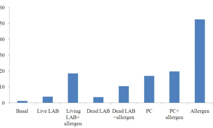
3.1.1. Effects Date Pollen Allergen on Cytokine
Production by PBMCs of Allergic Patients.
(Table 1, 2. Fig 1, 2)
Significant increase in IL4 level occurred when PBMCs incubated with the allergen compared to the basal level. Significant reduction in IFN gamma occurred when PBMCs incubated with the allergen compared to the basal level.
3.1.2. Effects of Living Lactobacillus Rhamnosus
on Cytokine Production by PBMCs of
Allergic Patients (Table 3, 4. Fig 1, 2)
Significant reduction in IL4 production obtained when PBMCs of allergic patients were incubated with living lactobacilli rhamnosus in the presence or absence of the offending allergen. Interferon gamma significantly increased when PBMCs incubated with living lactobacilli alone, However, PBMCs incubated with living lactobacilli in the presence of the allergen showed insignificant difference in interferon gamma production.
3.1.3. Effects of Dead Lactobacillus Rhamnosus
on Cytokine Production by PBMCs of
Allergic Patients (Table 5, 6 and Fig. 1, 2)
Significant reduction in IL4 production occured when PBMCs of allergic patients were incubated with dead lactobacilli in the presence or absence of the offending allergen.
Interferon gamma significantly increased when PBMCs incubated with dead lactobacilli in the presence or absence of the allergen.
3.1.4. Effects of PC on Cytokine Production by
PBMCs of Allergic Patients. (Table 7, 8
and Fig. 1, 2)
Significant reduction in IL4 production happened when PBMCs of allergic patients were incubated with PC in the presence or absence of the offending allergen.
Table 1. Effect of date pollen allergen on IL4 production by allergic patients PBMCS
Condition 1 Condition 2 W for N = 24 Critival value at p≤0.05 Significance Basal IL4 PBMCs+ allergen
(median: 1.25) (median: 52.6) 0 81 Sig.
Table 2. Effect of date pollen allergen on IFN gamma production by allergic patients PBMCS
Condition 1 Condition 2 W for N = 24 Critival value at p≤0.05 Significance
Basal IFNγ (median: 22.8) 34 81 Sig.
(median:39.5) PBMCS+allergen
Table 3. Effect of Living Lactobacillus rhamnosus on IL4 production by allergic patients PBMCS
Condition 1 Condition 2 W for N = 24 Critival value at p≤0.05 Significance
PBMCS+allergen PBMCS+ Living LAB 11 81 Sig.
(median:52.6) (median: 3.9)
PBMCS+allergen PBMCS+ Living LAB+ allergen 50 81 Sig.
(median:52.6 (median: 18.5)
Table 4. Effect of living Lactobacillus rhamnosus on IFN gamma production by allergic patients PBMCS
Condition 1 Condition 2 W for N = 24 Critival value at p≤0.05 Significance
PBMCS+allergen PBMCS+ Living LAB 27 81 Sig.
(median:22.8) (median: 42.75)
PBMCS+allergen PBMCS+ Living LAB+ allergen 90 81 Non Sig.
(median:22.8) (median: 28.7)
Table 5. Effect of dead Lactobacillus rhamnosus on IL4 production by allergic patients PBMCS
Condition 1 Condition 2 W for N = 24 Critival value at p≤0.05 Significance
PBMCS+allergen PBMCS+ Dead LAB 3 81 Sig.
(median:52.6 (median: 3.6)
PBMCS+allergen PBMCS+ Dead LAB+ allergen 26 81 Sig.
(median:52.6 (median: 10.6)
Table 6. Effect of dead Lactobacillus rhamnosus on IFN gamma production by allergic patients PBMCSs
Condition 1 Condition 2 W for N = 24 Critival value at p≤0.05 Significance
PBMCS+allergen PBMCS+ Dead LAB 7 81 Sig.
(median:22.8) (median: 49)
PBMCS+allergen PBMCS+ Dead LAB+ allergen 72 81 Sig.
(median:22.8) (median: 36.4)
Table 7. Effect of PC on IL4 production by allergic patients PBMCSs
Condition 1 Condition 2 W for N = 24 Critival value at p≤0.05 Significance
PBMCS+allergen PBMCS+ PC 20 81 Sig.
(median:52.6 (median: 17)
PBMCS+allergen PBMCS+PC+allergen 37 81 Sig.
(median:52.6 (median: 19.8)
Table 8. Effect of PC on IFN gamma production by allergic patients PBMCSs
Condition 1 Condition 2 W for N = 24 Critival value at p≤0.05 Significance
PBMCS+allergen PBMCS+ PC 0 81 Sig.
(median:22.8) (median: 319.6)
PBMCS+allergen PBMCS+PC+allergen 73 81 Sig.
Fig. 1. Median IL 4 concentration (in picograms) in supernatant after 72 h in allergic patients
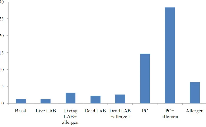
Fig. 3. Median IL 4 concentration (in picograms) in supernatant after 72 h in control subjects
3.1.5. Effects of living Lactobacillus Rhamnosus
on Cytokine Production by PBMCs of
Control Subjects. (Table 9, 10 and Fig. 3, 4)
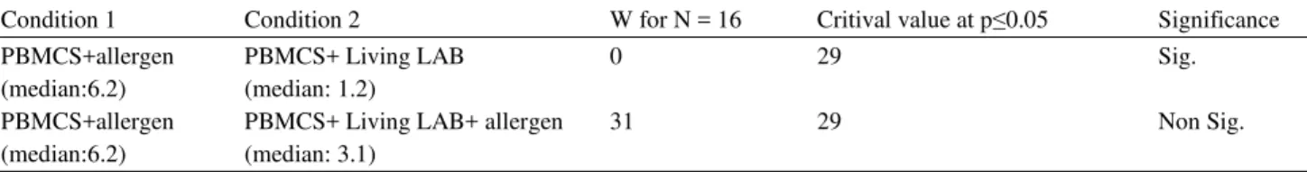
IL4 was significantly decreased when PBMCs incubated with living lactobacilli alone, however, PBMCs incubated with living lactobacilli in the presence of the allergen showed insignificant difference in IL 4.
Interferon gamma significantly increased when PBMCs incubated with living lactobacilli alone. Incubation of PBMCs with living lactobacilli and the allergen showed significant IFN gamma reduction.
3.1.6. Effects of dead Lactobacillus Rhamnosus on
Cytokine Production by PBMCs of Control
Subjects. (Table 11, 12 and Fig. 3, 4)
IL 4 was significantly decreased when PBMCs incubated with dead lactobacilli in the presence or absence of the allergen.
Interferon gamma significantly increased when PBMCS incubated with dead lactobacilli alone. Simultaneous incubation of PBMCs with dead lactobacilli and the allergen revealed insignificant difference in IFN gamma production.
3.1.7. Effects of PC on Cytokine Production by
PBMCs of Control Subjects. (Table 13, 14
and Fig. 5)
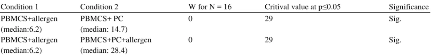
PC induced significant IL4 increase in the presence or absence of pollen allergen. PBMCs incubated with PC produced significatly increased IFN γ while PBMCs stimulated with both PC and pollen allergen showed significant decrease of IFN γ. However, the overall IFNγ/IL4 (Fig. 5) ratio showed significant increase (median 28.8) when PBMCs incubated with PC compared to PBMCs challenged with allergen (median 10)(W = 0, p≤0.05) Fig. 5.
Table 9. Effect of living lactobacilli on IL4 production by control subject PBMCS
Condition 1 Condition 2 W for N = 16 Critival value at p≤0.05 Significance
PBMCS+allergen PBMCS+ Living LAB 0 29 Sig.
(median:6.2) (median: 1.2)
PBMCS+allergen PBMCS+ Living LAB+ allergen 31 29 Non Sig.
(median:6.2) (median: 3.1)
Table 10. Effect of living lactobacilli on IFN gamma production by control subject PBMCS
Condition 1 Condition 2 W for N = 16 Critival value at p≤0.05 Significance
PBMCS+allergen PBMCS+ Living LAB 0 29 Sig.
(median:58.5) (median: 251.2)
PBMCS+allergen PBMCS+ Living LAB+ allergen 0 29 Sig.
(median:58.5) (median: 30.5)
Basal PBMCS+ Living LAB 0 29 Sig.
(median: 397.4) (median: 251.2)
Table 11. Effect of dead lactobacilli on IL4 production by control subject PBMCS
Condition 1 Condition 2 W for N = 16 Critival value at p≤0.05 Significance
PBMCS+allergen PBMCS+ Dead LAB 0 29 Sig.
(median:6.2) (median: 2.17)
PBMCS+allergen PBMCS+ Dead LAB+ allergen 1 29 Sig.
(median:6.2) (median: 2.6)
Table 12. Effect of dead lactobacilli on IFN gamma production by control subject PBMCS
Condition 1 Condition 2 W for N = 16 Critival value at p≤0.05 Significance
PBMCS+allergen PBMCS+ Dead LAB 0 29 Sig.
(median:58.5) (median:345.2)
PBMCS+allergen PBMCS+ Dead LAB+ allergen 31 29 NonSig.
Table 13. Effect of PC on IL4 production by control subject PBMCS
Condition 1 Condition 2 W for N = 16 Critival value at p≤0.05 Significance
PBMCS+allergen PBMCS+ PC 0 29 Sig.
(median:6.2) (median: 14.7)
PBMCS+allergen PBMCS+PC+allergen 0 29 Sig.
(median:6.2) (median: 28.4)
Table 14. Testing of significant differences IFN gamma production by control subjects PBMCS at different conditions
Condition 1 Condition 2 W for N = 16 Critival value at p≤ 0.05 Significance
PBMCS+allergen PBMCS+ PC 0 29 Sig.
(median:58.5) (median: 414)
PBMCS+allergen PBMCS+PC+allergen 0 29 Sig.
(median:58.5) (median: 28.2)
Fig. 5. IFN γ/IL4 ratio. after 72 h incubation (PC, control subjects)
4. DISCUSSION
This study tried to investigate the effects of
Lactobacillus rhamnosus ATCC 7469 (living and dead) and phycocyanin extracted from Spirulina platensis on TH1/TH2 paradigm. The peripheral blood mononuclear cells system was used to evaluate theses effects.
The current study revealed that simulation of PBMCs of allergic patients with the offending allergen (date pollen) induced significant increase of IL4 level and significant reduction of IFNγ level when compared to the basal levels (unstimulated PBMCs), this could be explained by polarization of immune system of allergic patients toward TH2, more IL4 and less IFNγ (Table 1 and 2). This observation was previously documented
by Imada et al. (1995), they found that IL-4 production was more significantly increased among atopic individuals, than normal subjects when their PBMCs stimulated by pollen allergen.
The resultant TH2 shifted response of allergic patients indicates the role of impaired TH1/TH2 balance in allergic diseases and was proved by (Pochard et al., 2002; Ghadimi et al., 2008), their experiments included challenging of PBMCs of patients allergic to house dust mite (Dpt allergen). IFN-γ mRNA expression and production was reported to be reduced in atopic subjects (Parronchi et al., 1991) during in vitro
stimulation experiments.
cultures of PBMC from allergic but not from healthy children. In contrast, IL-10 and IFN-γ were induced in cultures from both allergic and nonallergic children.
There was significant reduction of IL4 production when PBMCs of allergic patients incubated with living
Lactobacillus rhamnosus in the presence or absence of the allergen. The reduction was more significant when PBMC incubated with lactobacillus rhamnosus in absence of the offending allergen (Table 3).
Interferon gamma significantly increased when PBMCs incubated with living lactobacilli alone, However, PBMCS incubated with living lactobacilli in the presence of the allergen showed insignificant difference in interferon gamma production (Table 4). This may be due to the TH2 shift of the allergic patients when their PBMC challenged with this probiotic bacterial strain in the presence of the offending allergen, meaning, the presence of the specific allergen prevents
Lactobacillus rhamnosus from exerting their effect on IFN gamma, the predominant cytokine (IL4) downregulates IFNγ production. This was in accordance with Pohjavuori et al. (2004), they observed that that peripheral blood mononuclear cells in patients with atopic disease have a reduced TH1 cytokine IFNγ secretion capacity.
Heat-killed Lactobacillus rhamnosus was examined to evaluate the immunomodulatory effects of the dead strain, the results obtained were more or less similar to those of living bacteria. Significant reduction in IL4 production when PBMCs of allergic patients were incubated with dead lactobacilli in the presence or absence of the offending allergen (Table 5). Interferon gamma significantly increased when PBMCS incubated with dead lactobacilli in the presence or absence of the allergen (Table 6).
These results are quite similar to those obtained by Pochard et al. (2002), they found that the level of IL4 inhibition was not affected by the physiologic state of the bacteria because live bacteria induced the same effect as heat-killed or paraformaldehyde-treated LAB. This observation may point to the ability of lactobacilli to modulate the immune response via their cell wall composition, namely by their recognition by PRR. Toll- Like Receptors (TLR) are important PRRs that recognize a range of MAMPS such as Lipoteichoic acid (TLR2) and Lipopolysaccharide (TLR4) on Gram-positive and Gram-negative bacteria, respectively. Taylor et al. (2006) found that TLR2 ligands had the capacity to inhibit TH2 cytokine production by mononuclear cells stimulated with mite allergen. The inhibition of TH2
cytokine production was observed with a variety of TLR2 ligands, including high and low concentrations of heat-killed S aureus, LTA and Pam3CSK4. Pinto et al. (2009) reported that L. rhamnosus GG and L. plantarum BFE 1685, enhanced TLR2 at both the mRNA and the protein level in human intestinal cells.
The ability of L. lactis subsp. Lactis G50 killed by heat to induce cytokine production of macrophages in culture, was previously proved by Kimoto et al. (2004), it was observed that this strain continued to induce cytokine production, suggesting that such activity is associated with elements on the bacterial wall.
Ghadimi et al. (2008) proved that Lactobacillus rhamnosus GG, Lactobacillus gasseri (PA16/8),
Bifidobacterium bifidum (MP20/5) and Bifidobacterium
longum (SP07/3), as well as their genomic DNA, dose-dependently modulated the TH1/TH2 response to allergens. DNA seemed to contribute to about 50% of the immunomodulatory effects exerted by live bacteria.
Lactobacilli can also affect cytokine production by PBMCs of control subjects, when PBMCs of the controls challenged only by living lactobacilli IL4 was significantly decreased compared to its level after stimulation with the tested allergen. This finding was in accordance with Rutten et al. (2011), they reported that IL4 was significantly reduced when healthy PBMCs stimulated by different lactobacillus strain compared to cultures stimulated with Phytoheamagglutinin (PHA).
Simultaneous stimulation of control PBMCs with living lactobacilli and the tested allergen (pollen allergen) resulted in insignificant IL4 difference.
Interferon gamma significantly increased when PBMCs incubated with living lactobacilli alone. Incubation of PBMCs with living lactobacilli and the allergen showed significant IFN gamma reduction. These findings were similar to those obtained by Rasche et al. (2007), they co-stimulated peripheral mononuclear cells of individuals allergic to grass pollen and those non-allergic with inactive Lactobacillus acidophilus and the non-pathogenic Nissle strain of Escherichia, they reported that stimulation with lactobacilli plus allergen resulted in a TH2-like response in allergic and non-allergic individuals.
Other studies, Rutten et al. (2011), found that probiotic mixtures were able to induce significant amounts of IFN-γ compared to PBMCs which were cultured in medium only (for the four mixtures together compared to unstimulated medium).
affected by the PBMC: Bacteria ratio and the type of used antigen during stimulation experiments.
IL 4 was significantly decreased when PBMCs incubated with dead lactobacilli in the presence or absence of the allergen (Table 11). Interferon gamma significantly increased when PBMCs incubated with dead lactobacilli alone. Simultaneous incubation of PBMCS with dead lactobacilli and the allergen revealed insignificant difference in IFN gamma production (Table 12).
To date, the real pathways of probiotic immunomodulatory effects are not fully understood and some types of immune cells that are primed by probiotics might be the connection between in vivo
and in vitro stimulation.
The overall effect of Lactobacillus rhamnosus was skewing of the cytokine production toward TH1. More than one mechanism could be involved, PBMCs have been used widely for screening of the ability of probiotics to induce cytokine production. In vitro studies using PBMCs from allergic patients have shown reduced expression of TH2-associated cytokines (IL-4 and IL-5) on stimulation with total extract of Dermatophagoides pteronyssinus (house dust mite) and prior treatment with lactic acid bacteria strains such as Lactobacillus plantarum (Pochard et al., 2002). Both Lactococcus lactis and Lactobacillus plantarum, induce high levels of IL-12 and IFNγ, suppressing Th2 differentiation (Ghadimi et al., 2008). In contrast, suppression of contact dermatitis in mice has been shown to be mediated by a Lactobacillus acidophilus L92-dependent generation of Tregs (Shah et al., 2012). This effect is thought to result from strain-dependent tolerisation of DCs, increasing suppressor activity of natural Tregs as well as inducing Foxp3+conversion through high expression of IL-10, TGFβ, COX-2 and indoleamine 2,3-dioxygenase (Kwon et al., 2010).
Several studies, focussing on probiotic administration in type-1 hypersensitivity responses, revealed inhibitory characteristics for both Lactobacillus and
Bifidobacterium strains. Mouse models have
demonstrated the ability of L. casei and B. longum to inhibit IgE production (Schiffer et al., 2009). Inhibition of antibody production prevents binding to Fc receptors on mast cells, thus inhibiting the secretion of vasoactive amines, such as histamines and other inflammatory mediators, such as TNF-α. TNF-α production was found to be inhibited by L. reuteri
(Thomas et al., 2012).
The inhibition of IgE production is thought to be a consequence of direct action by probiotics on TH2 cells or APCs, which prime B-cell activation and class-switching. A large body of evidence demonstrates a role for Lactobacillus and Bifidobacterium strains in decreasing the levels of secreted IL-4 and IL-5. Both cytokines are TH2-derived, with IL-4 acting on B-cells to induce class-switching and on mast B-cells to induce degranulation and further cytokine production and IL-5 inducing eosinophil degranulation. Specific strains found to inhibit IL-4 and IL-5 production include L. casei (Schiffer et al., 2009), L. rhamnosus
(Ghadimi et al., 2008), B. longum (Takahashi et al., 2006) and B. infantis (Dev et al., 2008).
L. casei treatment in mice inhibited IgE production by inhibition of Syk/Lyn and MAPK signalling (Schiffer et al., 2009).
Evrard et al. (2011) found that the probiotic L.
rhamnosus Lcr35 induces a dose-dependent
immunomodulation of human DCs leading, at high bacterial doses, to the semi-maturation of the cells and a strong synthesis of pro-TH1/TH17 cytokines.
The current present study also evaluated the effect of Spirulina platensis on cytokine production of PBMCs of allergic patients and control subjects. Previous studies on immunomodulatory effects of spirulina proved that the photosynthetic pigment phycocyanin has a part to play in modulating the immune system.
Allergic PBMCs challenged with PC in the presence or absence of the offending allergen resulted in significant IL4 reduction and IFN γ increase, denoting TH1 polarizing effect of this compound (Table 7 and 8). Mao et al. (2005) had shown that allergic patients consuming 2,000 mg of Spirulina
daily can reduce the production of IL-4 from PHA stimulated PBMCs by 32%. Another double-blind, placebo-controlled study from Turkey evaluating the effectiveness and tolerability of Spirulina for treating patients with allergic rhinitis, Spirulina consumption significantly improved the symptoms and physical findings compared with placebo (p<0.001), including nasal discharge, sneezing, nasal congestion and itching (Cingi et al.,2008).
(median 28.8) when PBMC incubated with PC compared to PBMC challenged with allergen (Fig. 5).
Mao et al. (2000) reported that in vitro culture of resting and PHA-stimulated PBMCs of healthy individuals with Spirulina platensis extract significantly increased the levels of IL-4. Although
Spirulina platensis stimulates several cytokines, it is clearly more effective in the generation of a Thl-type response. Basha et al. (2009), also proved that PC had induced significant elevation of IFN γ level from PBMC of healthy subjects and chronic HCV patients.
Subhashini et al. (2004) found that PC had an inhibitory effect on the release of histamine from mast cells during an allergic inflammatory response.
Lactobacilli and bifidobacteria are generally considered to be safe (FAO/WHO, 2002). They have been used in various types of foods for a long time and they rarely cause infections in humans. The numbers of reported lactobacilli-induced bacteremia have not increased, despite the rapid increase of probiotic use the last decade (Salminen et al., 2002).
In a Swedish study, the incidence of lactobacilli-induced bacteremia and the presence in blood cultures of three commercially available probiotic strains were followed for five years. The incidence of bacteremia caused by lactobacilli constituted <1% of the total number of bacteremia cases with no increase during this five year-period. Lactobacilli-induced bacteremia was not caused by any of the three commercially available strains in any of the reported cases (Sullivan and Nord, 2006).
It has been suggested that probiotics should be used with caution in patients who are immunocompromised, have cardiac valvular disease, short bowel, jejunostomy or a central venous catheter (Besselink et al., 2008). No severe adverse events have been reported in any of the intervention studies performed in full term neonates and healthy infants (Boyle et al., 2008).
Although the effective dosage range of Phycocyanin in various animal models of inflammation was from 25 to 300 mg kg−1 p.o, the safety of the phycobiliprotein is good. The measured LD50 values were estimated to be greater than 3 g kg−1 for rats and mice (Romay et al., 1998).
5. CONCLUSION
In this study we showed that Lactobacillus rhamnosus ATCC 7469 strain can modulate the
Th1/Th2 balance by reducing TH2 cytokine (IL4) release and enhancing TH1 cytokine production (IFN-γ). The same effect was also obtained by the phycocyanin, pigmented extract from the cyanobacterium Spirulina platensis. The mechanism by which Lactobacillus rhamnosus exerted these effect seemed to be their cell wall composition, as the results obtained by dead bacteria were similar to those obtained by the living ones.
As the best anti-allergic effects of these bacteria were obtained when acting in absence of the allergen, it is better to use this bacteria in the prophylaxis rather than in treatment of allergic attacks. As the bacteria gave quite similar results in patients and healthy subjects, we assume that it could be a permissive line of prevention of allergy.
The safety of lactobacilli is very good, it could be the future treatment for allergy.
PC is good IFN-γ inducer in both allergic and control subjects. As Spirulina and its content C-phycocyanin are generally safe, in vivo expirment to evaluate their effect on allergic patients may be a new promising anti allergic therapy.
6. RECOMMENDATIONS
Further studies are needed to determine the exact source of IFN γ, using ELISpot or flowcytometry.
Both living and dead Lactobacillus rhamnosus ATCC 7469 should be considered as new line of allergy prevention.
C- phycocyanin gave promising results in allergic patients and could be a new line of treatment of allergy.
Further studies on animals and human subjects are needed.
7. ACNOWLEDGEMENT
Deep thanks to Dr. Shaimaa Hatab, lecturer of food sience, faculty of agriculture, Suize canal university, for supplying us with the lactobacillus strain used in this study.
8. REFERENCES
Abdel-Rahman, G.H., 2014. Taurine attenuates hepatic and cardiac damage and apoptosis in rabbits fed a high fat diet. OnLine J. Biol. Sci., 14: 12-20. DOI: 10.3844/ojbsci.2014.12.20
Basha, O.M., S.G. Zaghloul, R.A. Sadeq, R.A. Hafez and and A. El-Mohsen et al., 2009. C-phycocyanin effect on IFN-γ production in patients with chronic hepatitis c infection: In vitro study. Egypt. J. Med. Microbiol., 18: 81-90.
Bennett, A. and L. Bogorad, 1973. Comparative chromatic adaptation in a filamentous blue-green alga. J. Cell. Biol., 58: 419-35. DOI: 10.1083/jcb.58.2.419
Besselink, M.G., H.C. Van Santvoort, E. Buskens, M.A. Boermeester and H Van Goor et al., 2008. Probiotic prophylaxis in predicted severe acute pancreatitis: A randomised, doubleblind, placebo-controlled trial. Lancet, 371: 651-9. DOI: 10.1016/S0140-6736(08)60207-X
Boussiba, S. and A.E. Richmond, 1979. Isolation and characterization of phycocyanin from the blue green algae spirulina platensis. Arch. MicroBiol., 120: 155-159. DOI: 10.1007/BF00409102
Boyle, R.J., F.J. Bath-Hextall, J. Leonardi-Bee, D.F. Murrell and M.L. Tang, 2008. Probiotics for treating eczema. Cochrane Database Syst Rev. PMID: 18843705
Bullens, D.M., A. De Swerdt, E. Dilissen, A. Kasran and R.A. Kroczek et al., 2005. House dust mite-specific T cells in healthy non-atopic children. Clin. Exp. Allergy, 35:1535-41.PMID: 16393318
Chow, J., 2002. Probiotics and prebiotics: A brief overview, J. Ren.Nutr., 12: 76-86. PMID: 11953920 Cingi, C., M. Conk-Dalay, H. Cakli and C. Bal, 2008.
The effects of spirulina on allergic rhinitis. Eur. Arch Otorhinolaryngol., 265: 1219-23. PMID: 18343939
Dev, S., H. Mizuguchi, A.K. Das, C. Matsushita and K. Maeyama et al., 2008. Suppression of histamine signalling by probiotic Lac-B: A possible mechanism of its anti-allergic effect. J. Pharmacol. Sci., 107: 159-166. DOI: 10.1254/jphs.08028FP Evrard, B., S. Coudeyras, A. Dosgilbert, N. Charbonnel
and J. Alame et al., 2011. Dose-dependent immunomodulation of human dendritic cells by the probiotic Lactobacillus rhamnosus Lcr35. PLoS
One, 6: e18735-e18735. DOI:
10.1371/journal.pone.0018735
FAO/WHO, 2002. Guidelines for the evaluation of probiotics in food. Report of a joint FAO/WHO working group on drafting guidelines for the evaluation of probiotics in food. World Health Organization, London Ontario, Canada.
Ghadimi, D., R. Folster-Holst, M. De Vrese, P. Winkler and K.J. Heller et al., 2008. Effects of probiotic bacteria and their genomic DNA on TH1/TH2-cytokine production by Peripheral Blood Mononuclear Cells (PBMCs) of healthy and allergic subjects. Immunobiology, 213: 677-692. DOI: 10.1016/j.imbio.2008.02.001
Hayashi, O., S. Ono, K. Ishii, Y. Shi and T. Hirahashi
et al., 2006. Enhancement of proliferation and differentiation in bone marrow hematopoietic cells by Spirulina (Arthrospira) platensis in mice. J. Applied Phycol., 18: 47-56. DOI: 10.1007/s10811-005-9014-6
Huang, X., Y. Chen, F. Zhang, Q. Yang, and G. Zhang, 2014. Peripheral Th17/Treg cell-mediated immunity imbalance in allergic rhinitis patients. Braz. J. Otorhinolaryngol., 80: 152-5. PMID: /24830974
Imada, M., F. Estelle, R. Simon, T. Jay and K.T. Hayglass, 1995. Allergen-stimulated interleukin-4 and interferon-gamma production in primary culture: Responses of subjects with allergic rhinitis and normal controls. Immunology, 85: 373-380. PMID: 7558124
Kalliomäki, M., J.M. Antoine, U. Herz, G.T. Rijkers and J.M. Wells et al., 2010. Guidance for substantiating the evidence for beneficial effects of probiotics: Prevention and management of allergic diseases by probiotics. J. Nutr., 140: 713S-721S. DOI: 10.3945/jn.109.113761
Kimoto, H., K. Mizumachi, T. Okamoto and J. Kurisaki, 2004. New Lactococcus strain with immunomodulatory activity: Enhancement of Th1-type immune response. Microbial Immunol.,
48: 75-82. DOI:
10.1111/j.1348-0421.2004.tb03490.x
Kwon, H.K., C.G. Lee, J.S. So, C.S. Chae and J.S. Hwang et al., 2010. Generation of regulatory dendritic cells and CD4+Foxp3+ T cells by probiotics administration suppresses immune disorders. Proc. Nat. Acad. Sci. USA., 107: 2159-2164. DOI: 10.1073/pnas.0904055107
Mao, T.K., M.E. Gershwin and V.D. Water, 2000. Effect of Spirulina on the secretion of cytokines from peripheral blood mononuclear cells. J. Med. Food, 3: 135-40. DOI: 10.1089/jmf.2000.3.135 Niers, L., R. Martin, G. Rijkers, F. Sengers and H.
Timmerman et al., 2009. The effects of selected probiotic strains on the development of eczema (the PandA study). Allergy, 64: 1349-1358. DOI: 10.1111/j.1398-9995.2009.02021.x
Ozdemir, C., M. Akdis and C.A. Akdis, 2010. T-cell response to allergens. Chem. Immunol. Allergy, 95: 22-44. DOI: 10.1159/000315936
Parronchi, P., D. Macchia, M.P. Piccinni, P. Biswas and C. Simonelli et al., 1991. Allergen- and bacterial antigen-specific T-cell clones established from atopic donors show a different profile of cytokine production. Proc. Natl Acad
Sci. USA., 88: 4538-42. DOI:
10.1073/pnas.88.10.4538
Pinto, M.G.V., M.R. Gómez, S. Seifert, B. Watzl and W.H. Holzapfel et al., 2009. Lactobacilli stimulate the innate immune response and modulate the TLR expression of HT29 intestinal epithelial cells in vitro. Int. J. Food Microbiol.,
133: 86-93. DOI:
10.1016/j.ijfoodmicro.2009.05.013
Pochard, P., P. Gosset, C. Grangette, C. Andre and A.B. Tonnel et al., 2002. Lactic acid bacteria inhibit TH2 cytokine production by mononuclear cells fromallergic patients. J. Allergy Clin.
Immunol., 110: 617-623. DOI:
10.1067/mai.2002.128528
Pohjavuori, E., M. Viljanen, R. Korpela, M. Kuitunen and M. Tiittanen et al., 2004. Lactobacillus GG effect in increasing IFN-g production in infants with cow’s milk allergy. J. Allergy Clin.
Immunol., 114: 13. DOI:
10.1016/j.jaci.2004.03.036
Rasche, C., C. Wolfram, M. Wahls and M. Worm, 2007. Differential immunomodulating effects of inactivated probiotic bacteria on the allergic immune response. Acta Derm Venereol., 87: 305-11. DOI: 10.2340/00015555-0232
Rijkers, G.T., S. Bengmark, P. Enck, D. Haller and U. Herz et al., 2010. Guidance for substantiating the evidence for beneficial effects of probiotics: Current status and recommendations for future research. J. Nutr., 140: 671S-676S. DOI: 10.3945/jn.109.113779
Romay, C., J. Armesto, D. Remirez, R. González and N. Ledon et al., 1998. Antioxidant and anti-inflammatory properties of C-phycocyanin from blue-green algae. Inflamm Res., 47: 36-41. Rutten, N.B.M., I. Besseling-Van der Vaart, M. Klein,
S. De Roock Vlieger and A.M. Vlieger et al., 2011. In vitro assessment of the immunomodulatory effects of multispecies probiotic formulations for management of allergic diseases. Beneficial Microbes, 2: 183-192. PMID: 21986357
Salminen, M.K., S. Tynkkynen, H. Rautelin, M. Saxelin and M. Vaara et al., 2002. Lactobacillus bacteremia during a rapid increase in probiotic use of Lactobacillus rhamnosus GG in Finland. Clin. Infect. Dis., 35: 1155-60. DOI: 10.1086/342912
Schiffer, C., A.I. Lalanne, L. Cassard, D.A. Mancardi and O. Malbec et al., 2009. A strain of
Lactobacillus casei inhibits the effector phase of immune inflammation. J. Immunol., 187: 1-10. DOI: 10.4049/jimmunol.1002415, PMID: 21810608
Shah, N.P., 2007. Functional cultures and health benefits, Int. Dairy J., 17: 1262-1277. DOI: 10.1016/j.idairyj.2007.01.014
Shah, M.M., M. Saio, H. Yamashita, H. Tanaka and T. Takami et al., 2012. Lactobacillus acidophilus strain L-92 induces CD4+ CD25+ Foxp3+ regulatory T cells andsuppresses allergic contact dermatitis. Biol. Pharm. Bull., 35: 612-616. DOI: 10.1248/bpb.35.612
Silva, L.A., K.R. Kuhn, C.C. Moraes, C.A.V. Burkert and S.J. Kalil, 2009. Experimental design as a tool for optimization of C-phycocyanin purification by precipitation from Spirulina platensis. J. Braz. Chem. Soc., 20: 5-12. DOI: 10.1590/S0103-50532009000100003
Subhashini, J., S.V. Mahipal, M.C. Reddy, M. Mallikarjuna Reddy and A. Rachamallu et al., 2004. Molecular mechanisms in C-Phycocyanin induced apoptosis in human chronic myeloid leukemia cell line-K562. Biochem. Pharmacol., 68: 453-462. DOI: 10.1016/j.bcp.2004.02.025 Sullivan, A. and C.E. Nord, 2006. Probiotic
Takahashi, N., H. Kitazawa, N. Iwabuchi, J.Z. Xiao and K. Miyaji et al., 2006. Immunostimulatory oligodeoxynucleotide from Bifidobacterium
longum suppresses Th2 immune responses in a murine model. Clin. Exp. Immunol., 145: 130-138. DOI: 10.1111/j.1365-2249.2006.03111.x Taylor, R.C., P. Richmond and J.W. Upham, 2006.
Toll-like receptor 2 ligands inhibit TH2 responses to mite allergen. J. Allergy Clin. Immunol., 17: 1148-1154. DOI: 10.1016/j.jaci.2006.02.014
Thomas, C.M., T. Hong, J.P. van Pijkeren, P. Hemarajata and D.V. Trinh et al., 2012. Histamine derived from probiotic Lactobacillus reuteri suppresses TNF via modulation of PKA and ERK signalling. PLoS One 7, e31951. DOI: 10.1371/journal.pone.0031951 Vissers, K.C.P., K. Besse, M. Wagemans,W. Zuurmond