Rev Odontol UNESP. 2013 July-Aug; 42(4): 237-242 © 2013 - ISSN 1807-2577 ORIGINAL ARTICLE
Surface properties and color stability of an acrylic resin
combined with an antimicrobial polymer
Propriedades de superfície e estabilidade de cor de uma resina acrílica
combinada com um polímero antimicrobiano
Ana Carolina PERO
a, Jaqueline IGNÁCIO
a, Gabriela GIRO
a, Danny Omar MENDOZA-MARIN
a,
André Gustavo PALEARI
a, Marco Antonio COMPAGNONI
aaFaculdade de Odontologia, UNESP – Univ Estadual Paulista, Araraquara, SP, Brasil
Resumo
Introdução: A ocorrência de estomatite protética é relativamente comum uma vez que as superfícies das próteses bucais representam um ambiente propício para adesão de microrganismos orais e formação de biofilme.
Objetivo: Avaliar as propriedades de superfície (molhabilidade/rugosidade) e estabilidade de cor de uma resina acrílica combinada com o polímero antimicrobiano poli (2-tert-butilaminoetil) metacrilato (PTBAEMA).
Material e método: Trinta espécimes em formato de disco de uma resina acrílica (Lucitone 550) foram divididos em três grupos: 0% (controle), 5% e 10% PTBAEMA. Os valores de rugosidade (Ra) foram medidos utilizando um rugosímetro, e a molhabilidade foi determinada através de medições de ângulo de contato utilizando um goniômetro e água deionizada como líquido teste. Dados de cores foram medidos com um espectrofotômetro. Os testes de Kruskal-Wallis e Dunn foram utilizados para comparar os valores de rugosidade. Os dados de molhabilidade foram analisados utilizando-se ANOVA e teste de Tukey. Os dados de cor foram comparados com o teste t de Student e valores ΔE foram classificados de acordo com o National Bureau of Standards (NBS). Todas as análises estatísticas foram realizadas considerando α = 0,05. Resultado: Diferenças significativas (p<0,05) foram detectadas entre os grupos para rugosidade, molhabilidade, e estabilidade de cor. De acordo com o NBS, as alterações de cor obtidas nos grupos 5% e 10% PTBAEMA foram “marcante” e “extremamente marcante”, respectivamente. Conclusão: Pode-se concluir que a incorporação de PTBAEMA em uma resina acrílica aumentou a rugosidade e molhabilidade das superfícies e produziu alterações de cor com relevância clínica.
Descritores: Resinas acrílicas; produtos com ação antimicrobiana; molhabilidade; cor, propriedades físicas.
Abstract
Introduction: The occurrence of stomatitis is common since the surface characteristics of the dentures may act as reservoirs for microorganisms and have the potential to support biofilm formation. Purpose: To assess the surface properties (wettability/roughness) and color stability of an acrylic resin combined with the antimicrobial polymer poly (2-tert-butylaminoethyl) methacrylate (PTBAEMA). Material and method: Thirty disc-shaped specimens of an acrylic resin (Lucitone 550) were divided into three groups: 0% (control); 5% and 10% PTBAEMA. Surface roughness values (Ra) were measured using a profilometer and wettability was determined through contact angle measurements using a goniometer and deionized water as a test liquid. Color data were measured with a spectrophotometer. Kruskal-Wallis and Dunn’s test were used to compare roughness values. Wettability data were analyzed using ANOVA and Tukey’s test. Color data were compared using the Student’s t-test and ∆E values were classified according to the National Bureau of Standards (NBS). All statistical analyses were performed considering α=.05. Result: Significant differences (p<.05) were detected among the groups for roughness, wettability and color stability. According to the NBS, the color changes obtained in the 5% and 10% PTBAEMA groups were “appreciable” and “much appreciable”, respectively. Conclusion: It could be concluded that PTBAEMA incorporation in an acrylic resin increased the roughness and wettability of surfaces and produced color changes with clinical relevance.
INTRODUCTION
Microbial growth on the denture base surface is caused by the adherence of microbial cells that are promoted by surface roughness and hydrophobic interactions between Candida species and oral bacteria. his mainly occurs with Candida spp and oral streptococci1,2, species commonly associated with the use of dentures.
Since the surface characteristics of the substratum are important to a microorganisms’ adherence3,several approaches have been proposed to induce a chemical modiication of the denture base surface and to prevent denture stomatitis. Among these reports, there have been investigations related to the chemical modiication of surface charge of denture resin4, the incorporation of antimicrobial agents5-7, the application of coatings8,9 and plasma treatment10.
Poly (2-tert-butylaminoethyl) methacrylate (PTBAEMA), which acts as a very eicient contact biocide, is a polycationic polymer functionalized with pendant amino groups11. According to Seyfriedsberger et al.11, (2006) polycationic polymers are macromolecular substances that can act as antimicrobial agents which substitute low-molecular-weight biocides. hey are advantageous due to their reduced toxicity and the fact that they do not cause bacterial resistance. In a previous study11, PTBAEMA was successfully incorporated into polyethylene surfaces and highly antimicrobial properties were achieved for Escherichia coli and Staphylococcus aureus.
he incorporation of PTBAEMA into dental materials was irst reported by Marra et al.6 (2012). hese authors demonstrated the high antimicrobial activity of an acrylic resin combined with PTBAEMA for Staphylococcus aureus and Streptococcus mutans bioilm. However, no signiicant efect on Candida albicans bioilm formation was recorded.
It could be hypothesized that PTBAEMA incorporation into acrylic resins modiies the denture surface, thereby preventing these surfaces from acting as bioilm reservoirs. Consequently, it is reasonable to assume that PTBAEMA incorporation could afect the physical properties of the acrylic resin surfaces, such as its wettability and roughness. In addition, considering that aesthetics is an important factor in terms of treatment acceptance by the patient, the color of dental materials should remain stable over a long period12.
he aim of this in vitro study was to assess the physical properties of an acrylic resin combined with the antimicrobial polymer poly (2-tert-butylaminoethyl) methacrylate (PTBAEMA). he null hypothesis tested was that the incorporation of PTBAEMA would have no efect on the roughness, wettability and color stability of an acrylic resin.
MATERIAL AND METHOD
1.
Specimen Fabrication
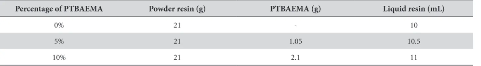
hirty discs (15 mm in diameter and 3 mm thick) of a heat-polymerized acrylic resin (Lucitone 550 – Dentsply International Inc., York, PA, USA) were produced and divided into three groups (n=10), according to the PTBAEMA incorporation: 0% (control); 5% and 10% (Table 1).
A metal mold was used to obtain disc-shaped silicone patterns (Zetaplus/Indurent-Zhermack, Badia Polesine, Rovigo, Italy), which were placed in a lask, sandwiched between two glass slides and supported by dental stone (Herodent, Vigodent S/A Ind. Com., Rio de Janeiro, RJ, Brazil). Ater the material had set, the silicone patterns were removed and the acrylic resin was mixed and packed into the mold. A pneumatic press (Delta, Delta Máquinas Especiais, Vinhedo, SP, Brazil) was used for trial packing of the acrylic resin at 1500 psi initially and later at 3500 psi, maintained for 30 minutes. Specimens were polymerized using an automatic polymerization tank (Solab Equipamentos para Laboratórios Ltda, Piracicaba, SP, Brazil) for 90 minutes at 73 °C followed by 30 minutes at 100 °C. Ater polymerization, the lasks were allowed to bench cool at room temperature. he specimens were delasked and excess lash was removed with a bur (Max-Cut, Malleifer AS, Ballaigues, Switzerland). Before the roughness, contact angle and color measurements were assessed, the samples were cleaned ultrasonically for 5 minutes in water.
2.
Surface Roughness Measurements
Surface roughness was measured using a proilometer (Mitutoyo SJ-400, Mitutoyo Corporation, Tokyo, Japan) with a resolution of 0.01µm, at a stylus speed of 0.5 mm/s, a cut-of length of 2.4 mm and a diamond stylus tip radius of 5 µm. hree measurements were made at diferent sites by the same operator for each specimen and a mean value was obtained and expressed as Ra (µm). he Ra value describes the overall roughness of a surface and is deined as the arithmetic mean value of all absolute distances of the roughness proiles from the center line within the measuring length13.
3.
Contact Angle Measurements
he contact angle measurement was used to characterize the surface wettability. he liquid drop was placed onto the substrate using a microsyringe. Droplets of deionised water (volume of ~ 1.0 µl) were used to measure the contact angle. An automated goniometer (Ramé-hart 200, Ramé-hart instrument
Table 1. Experimental groups, according to the percentage of PTBAEMA
Percentage of PTBAEMA Powder resin (g) PTBAEMA (g) Liquid resin (mL)
0% 21 - 10
5% 21 1.05 10.5
co., Netcong, New Jersey, USA), connected to a computer, was used to measure the contact angles produced by the droplets on the specimens. A CCD camera was used to record the image of the droplets on the surface and the contact angles were measured using DROPimage Standard sotware (Ramé-hart instrument co., Netcong, New Jersey, USA).
he measurements were performed optically with an accuracy of ±1°. hree drops were deposited at diferent random locations on each sample and then a mean value was obtained. his procedure allows one to take into account a possible non-uniformity of the surface probed by the contact angle. he experiments were carried out by the same operator in a controlled temperature (25±1°C).
4.
Color Stability
Color measurements were performed using a spectrophotometer Color Guide 45/0 (BYK-Gardner, Santo André, SP, Brazil) according to the CIE (Commission Internationale de l’Eclairage) L*a*b* system14. CIE L*a*b* is an approximate uniform color space with coordinates for lightness: white-black (L*); redness-greenness (a*) and yellowness-blueness (b*). he measurements were made by the same operator, using a standard illuminant D6515. he specimens were placed on a sample sighting device, which had a circular hole of 15 mm in diameter, provided by the spectrophotometer manufacturer.
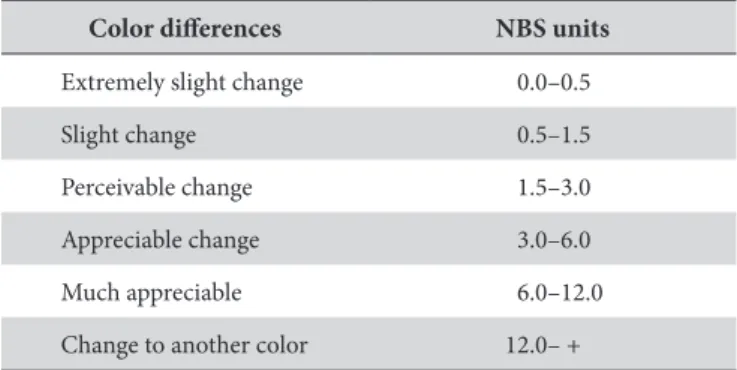
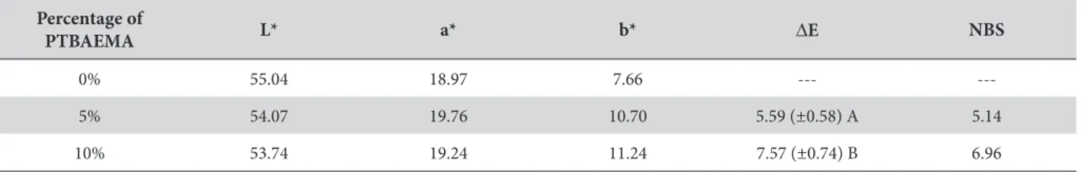
L*, a* and b* values were obtained for the three groups: 0% (control); 5% and 10% PTBAEMA. Color diferences were calculated for the two groups with 5% and 10% PTBAEMA, in comparison to the control group, using the formula ∆E* = [(∆L*)2 + (∆a*)2 + (∆b*)2]1/2, where ∆L*, ∆a* and ∆b* represent diferences in L*, a* and b* values. ∆E* values obtained for the groups with 5% and 10% PTBAEMA were converted to NBS (National Bureau of Standards) units, using the formula NBS units = ∆E* x 0.92 to denote the color diferences in a clinical perspective16,17. Table 2 displays the ratings of color diferences, according to NBS units.
RESULT
he efect of PTBAEMA incorporation into acrylic resin specimens on the roughness of surfaces is shown in Figure 1. Data were analyzed using the Kruskal-Wallis non-parametric test, followed by Dunn’s test, with a level of signiicance of 5%. here were signiicant diferences among the groups (p<.001),
which indicates that roughness increased with PTBAEMA incorporation.
Contact angle measurements are used to characterize surface wettability. he mean contact angle values of each group were compared by one-way analysis of variance (ANOVA), followed by Tukey’s post hoc tests with a level of signiicance of 5%. he statistical analysis demonstrated that the incorporation of PTBAEMA made the acrylic resin surfaces more wettable. Figure 2 displays the contact angle measurements obtained for the groups with 0%, 5% and 10% PTBAEMA. Signiicant diferences (p<.05) were detected among the groups (Group 0%= 57.3°±2.2A, Group 5%= 28.7°±3.2B, Group 10%= 44.3°±3.9C).
Table 3 displays the color data in CIE L*a*b* color space, the mean and standard deviation values of ∆E, as well as the NBS classiication for the 5% and 10% groups. he mean ∆E values for the 5% and 10% groups were compared using the student’s t-test with α=.05. Signiicant diferences were detected between the groups (p<.05). According to NBS, the incorporation of PTBAEMA at 5% and 10% produced “appreciable” and “much appreciable” color changes, respectively.
DISCUSSION
he study of the incorporation of antimicrobial agents into acrylic resins may represent a viable alternative to control the development of oral infections and improve the oral health of denture wearers. he null hypothesis of the present study was
Table 2. NBS units for expressing color diferences
Color diferences NBS units
Extremely slight change 0.0–0.5
Slight change 0.5–1.5
Perceivable change 1.5–3.0
Appreciable change 3.0–6.0
Much appreciable 6.0–12.0
Change to another color 12.0– +
Figure 1. Efect of PTBAEMA incorporation into acrylic resin specimens on the roughness of surfaces, according to the group. Diferent capital letters indicate signiicant diferences among the groups (Kruskal-Wallis test and Dunn’s test, p<.001).
rejected since the physical properties assessed were afected ater the incorporation of the polymer PTBAEMA.
he results of this study showed that the incorporation of PTBAEMA into a heat-polymerized acrylic resin increased the surface roughness. Surface properties such as roughness and surface free energy can interfere with microbial colonization and the maturation of bioilm18-20. he initial adhesion of microorganisms is directly inluenced by the surface roughness21. Previous studies have suggested that a surface roughness greater than 0.2µm enhances the adhesion of microorganisms, which is considered a critical value of roughness for acrylic resins13. he retention of microorganisms occurs faster on rough surfaces due to their larger contact area and also to obstruct the action of mechanical cleaning22. However, previous studies have also shown that the adhesion of C. albicans was not inluenced by the roughness of the acrylic resin10,23.
In the present study, the water contact angle was used to provide information about the surface wettability. he contact angle on the acrylic resin surface was reduced ater the incorporation of the polymer (2-tert-butylaminoethyl) (PTBAEMA) and, consequently, it determines a surface more hydrophilic. his behavior was observed even with the increase of roughness. It is well established that the contact angle on a solid surface depends on several factors, such as roughness, surface energy (hydrophilic surfaces are characterized by high surface energy) and surface cleanliness24. herefore, it could be hypothesized that the incorporation of PTBAEMA modiied the surface energy of acrylic resin and this contributes to the increase of surface wetting.
According to some authors, hydrophilic surfaces are less susceptible to adhesion of Candida albincans9,19. However, Minagi et al.20 (1985)observed that a decrease in the contact angle resulted in an increased adherence of Candida albicans on acrylic resin materials and a decrease in the adherence of Candida tropicalis. hese contrasting indings demonstrate that the exact mechanism by which the adhesion of microorganisms occurs is dependent on other factors related to the substrate such as surface free energy, surface tension and electrostatic interactions4,10,19,25.
Furthermore, in a clinical situation simulated in in vivo studies, the oral environment is inluenced by many dynamic factors4. he presence of saliva, the inluence of pH, and interaction with other microorganisms may also moderate the adhesion of C. albincans23,26.
Despite the controversy regarding the inluence of such factors on microorganisms’ adhesion, a previous in vitro study6 has demonstrated that the incorporation of PTBAEMA into an
acrylic resin produced antimicrobial activity for Staphylococcus aureus and Streptococcus mutans, but had no signiicant efect on Candida albicans bioilm formation. hus, it seems that the incorporation of PTBAEMA into dental materials may be promising in terms of an attempt to reduce the adherence of microorganisms such as the Streptococcus species, which are considered essential in the initial formation of oral bioilms27, and Staphylococcal infections, particularly those caused by Staphylococcus aureus, which lead to substantial morbidity and mortality in hemodialysis patients28.
he incorporation of 5% and 10% PTBAEMA into a denture base resin produced noticeable color changes. According to previous studies12,29, color diferences with corresponding ∆E values lower than 1.0 are not visually detectable by the human eye, and 3.3 NBS units are acceptable in clinical dentistry. In the present study, the color diferences were considered “appreciable” and “much appreciable” for the 5% and 10% PTBAEMA groups, respectively. his could be explained by the chemical ainity of the polycationic polymer PTBAEMA with acrylic resin, since copolymerization between PTBAEMA and a denture base resin have been observed in a previous study30. Considering that the clinical relevance of color diferences is subjective, future in vivo studies could be performed to assess the impact of the color stability of acrylic resins modiied by PTBAEMA on the satisfaction of denture wearers.
he present study focused on the physical properties of a denture base resin ater the incorporation of the polymer PTBAEMA. his study has limitations since surface topography and surface free energy were not assessed. hese characteristics are extremely important in the microbial adhesion process25. Further studies should be conducted to investigate other properties of acrylic resins ater the incorporation of the polymer PTBAEMA, thereby contributing to the prevention of oral infections.
CONCLUSION
Within the limitations of this study, it was concluded that the incorporation of the antimicrobial polymer (2-tert-butylaminoethyl - PTBAEMA) into an acrylic resin increased the roughness of surfaces and the wettability, as well as producing color changes with clinical relevance.
ACKNOWLEDGEMENTS
he authors would like to thank FAPESP – São Paulo Research Foundation (Grant 2010/18967-7) for inancial support.
Table 3. Color data in CIE L*a*b* color space, means and standard deviations of ΔE, and NBS classiication for the groups
Percentage of
PTBAEMA L* a* b* ∆E NBS
0% 55.04 18.97 7.66 ---
---5% 54.07 19.76 10.70 5.59 (±0.58) A 5.14
10% 53.74 19.24 11.24 7.57 (±0.74) B 6.96
REFERENCES
1. Nair RG, Samaranayake LP. he efect of oral commensal bacteria on candidal adhesion to denture acrylic surfaces. An in vitro study. APMIS. 1996;104:339-49. PMid:8703439. http://dx.doi.org/10.1111/j.1699-0463.1996.tb00725.x
2. Radford DR, Challacombe SJ, Walter JD. Denture plaque and adherence of Candida albicans to denture-base materials in vivo and in vitro. Crit Rev Oral Biol Med. 1999;10:99-116. PMid:10759429. http://dx.doi.org/10.1177/10454411990100010501
3. Yildirim MS, Hasanreisoglu U, Hasirci N, Sultan N. Adherence of Candida albicans to glow-discharge modiied acrylic denture base polymers. J Oral Rehabil. 2005; 32:518-25. PMid:15975132. http://dx.doi.org/10.1111/j.1365-2842.2005.01454.x
4. Park SE, Blissett R, Susarla SM, Weber HP. Candida albicans adherence to surface-modiied denture resin surfaces. J Prosthodont. 2008; 17:365-9. PMid:18266657. http://dx.doi.org/10.1111/j.1532-849X.2007.00292.x
5. Casemiro LA, Gomes Martins CH, Pires-de-Souza FC, Panzeri H. Antimicrobial and mechanical properties of acrylic resins with incorporated silver-zinc zeolite - part I. Gerodontology. 2008;25:187-94. PMid:18194331. http://dx.doi.org/10.1111/j.1741-2358.2007.00198.x
6. Marra J, Paleari AG, Rodriguez LS, Leite AR, Pero AC, Compagnoni MA. Efect of an acrylic resin combined with an antimicrobial polymer on bioilm formation. J Appl Oral Sci. 2012;20:643-8. PMid:23329246. http://dx.doi.org/10.1590/S1678-77572012000600009 7. Pesci-Bardon C, Fosse T, Serre D, Madinier I. In vitro antiseptic properties of an ammonium compound combined with denture base
acrylic resin. Gerodontology. 2006;23:111-6. PMid:16677185. http://dx.doi.org/10.1111/j.1741-2358.2006.00088.x
8. Redding S, Bhatt B, Rawls HR, Siegel G, Scott K, Lopez-Ribot J. Inhibition of Candida albicans bioilm formation on denture material. Oral Surg Oral Med Oral Pathol Oral Radiol Endod. 2009;107:669-72. PMid:19426921. http://dx.doi.org/10.1016/j.tripleo.2009.01.021 9. Yoshijima Y, Murakami K, Kayama S, Liu D, Hirota K, Ichikawa T, et al. Efect of substrate surface hydrophobicity on the adherence of
yeast and hyphal Candida. Mycoses. 2010;53:221-6. PMid:19671080. http://dx.doi.org/10.1111/j.1439-0507.2009.01694.x
10. Zamperini CA, Machado AL, Vergani CE, Pavarina AC, Giampaolo ET, da Cruz NC. Adherence in vitro of Candida albicans to plasma treated acrylic resin. Efect of plasma parameters, surface roughness and salivary pellicle. Arch Oral Biol. 2010; 55:763-70. PMid:20667522. http://dx.doi.org/10.1016/j.archoralbio.2010.06.015
11. Seyfriedsberger G, Rametsteiner K, Kern W. Polyethylene compounds with antimicrobial surface properties. Eur Polym J. 2006; 42:3383-9. http://dx.doi.org/10.1016/j.eurpolymj.2006.07.026
12. Silva PM, Acosta EJ, Jacobina M, Pinto LR, Porto VC. Efect of repeated immersion solution cycles on the color stability of denture tooth acrylic resins. J Appl Oral Sci. 2011; 19: 623-7. PMid:22230997.
13. Zissis AJ, Polyzois GL, Yannikakis SA, Harrison A. Roughness of denture materials: a comparative study. Int J Prosthodont. 2000;13:136-40. PMid:11203622.
14. International Commission on Illumination. Colorimetry: oicial recommendations of the International Commission on Illumination. 2nd ed. Vienna: Bureau Central de la CIE; 1986.
15. Polyzois GL, Yannikakis SA, Zissis AJ, Demetriou PP. Color changes of denture base materials ater disinfection and sterilization immersion. Int J Prosthodont. 1997;10:83-9. PMid:9484075.
16. Dozic A, Voit NFA, Zwartser R, Khashayar G, Aartman I. Color coverage of a newly developed system for color determination and reproduction in dentistry. J Dent. 2010;38(Suppl. 2):e50–6. PMid:20638437. http://dx.doi.org/10.1016/j.jdent.2010.07.004
17. Nimerof I. Colorimetery National Bureau of Standards Monograph 104; 1968:47.
18. Cunha TR, Regis RR, Bonatti MR, Souza RF. Inluence of incorporation of luoroalkyl methacrylates on roughness and lexural strength of a denture base acrylic resin. J Appl Oral Sci. 2009;17:103-7. PMid:19274394. http://dx.doi.org/10.1590/S1678-77572009000200006 19. Klotz SA, Drutz DJ, Zajic JE. Factors governing adherence of Candida species to plastic surfaces. Infec Immun. 1985; 50: 97-101.
PMid:3899942 PMCid:262141.
20. Minagi S, Miyake Y, Inagaki K, Tsuru H, Suginaka H. Hydrophobic interaction in Candida albincans and Candida tropicalis adherence to various denture base resin materials. Infect Immun. 1985; 47:11-4. PMid:3880719 PMCid:261449.
21. Kolenbrander PE, Andersen RN, Blehert DS, Egland PG, Foster JS, Palmer RJ Jr. Communication among oral bacteria. Microbiol Mol Biol Rev. 2002;66:486-505. PMid:12209001 PMCid:120797. http://dx.doi.org/10.1128/MMBR.66.3.486-505.2002
22. Radford DR, Sweet SP, Challacombe SJ, Walter JD. Adherence of Candida albicans to denture-base materials with diferent surface inishes. J Dent. 1998;26:577-83. http://dx.doi.org/10.1016/S0300-5712(97)00034-1
23. Moura JS, Silva WJ, Pereira T, Del Bel Cury AA, Rodrigues Garcia RC. Inluence of acrylic resin polymerization methods and saliva on the adherence of four Candida species. J Prosthet Dent. 2006;96:205–11. PMid:16990072. http://dx.doi.org/10.1016/j.prosdent.2006.07.004 24. Compagnoni MA, Pero AC, Ramos SM, Marra J, Paleari AG, Rodriguez LS. Antimicrobial activity and surface properties of an acrylic
resin containing a biocide polymer. Gerodontology. 2012; Dec 20. [Epub ahead of print]. http://dx.doi.org/10.1111/ger.12031
25. Kang SH, Lee HJ, Hong SH, Kim KH, Kwon TY. Inluence of surface characteristics on the adhesion of Candida albicans to various denture lining materials. Acta Odontol Scand. 2013;71:241-8. PMid:22428860. http://dx.doi.org/10.3109/00016357.2012.671360 26. Pereira-Cenci T, Cury AADB, Cenci MS, Rodrigues-Garcia RC. In vitro Candida colonization on acrylic resins and denture liners:
inluence of surface free energy, roughness, saliva, and adhering bacteria. Int J Prosthodont. 2007;20: 308-10. PMid:17580465.
28. Balaban N, Gov Y, Bitler A, Boelaert JR. Prevention of Staphylococcus aureus bioilm on dialysis catheters and adherence to human cells. Kidney Int. 2003;63:340-5. PMid:12472801. http://dx.doi.org/10.1046/j.1523-1755.2003.00733.x
29. Rosentritt M, Esch J, Behr M, Leibrock A, Handel G. In vivo color stability of resin composite veneers and acrylic resin teeth in removable partial dentures. Quintessence Int. 1998;29:517-22. PMid:9807133.
30. Rodriguez LS, Paleari AG, Oliveira Junior NM, Giro G, Pero AC, Compagnoni MA. Chemical characterization and lexural strength of a poly (methyl methacrylate) resin with monomer 2-tert-butylaminoethyl methacrylate. J Prosthodont. 2012 Oct 25. [Epub ahead of print]. PMid:23106690.
CONFLICTS OF INTERESTS
he authors declare no conlicts of interest.
CORRESPONDING AUTHOR
Ana Carolina Pero
Departmento de Materiais Odontológicos e Prótese, Faculdade de Odontologia, UNESP – Univ Estadual Paulista, Rua Humaitá, 1680, 14801-903 Araraquara - SP, Brasil
e-mail: [email protected]