*e-mail: [email protected]
Thermal and Microestructural Characterization of
Epoxy-Infiltrated Hydroxyapatite Composite
Pedro Barrionuevo Roesea*, Sandro Campos Amicob, Wilson Kindlein Júniora
a
LDSM – Universidade Federal do Rio Grande do Sul
90035-190 Porto Alegre - RS, Brazil
b
LAPOL/PPGEM - Universidade Federal do Rio Grande do Sul,
91501-970 Porto Alegre - RS, Brazil
Received: December 18, 2008; Revised: February 17, 2009
In this work, hydroxyapatite (HAp) obtained from the deorganification of bovine bones using hot NaOH solution was used to synthesize a HAp/epoxy infiltrated composite. Infiltration was carried out by vacuum assisted immersion in hot epoxy resin. The resulting composite was characterized regarding polymer content, morphological aspects and flexural strength. The infiltration method used resulted in thorough infiltration of the HAp but some residual porosity remained. Although the epoxy resin showed good interaction with the HAp, high polymer content was achieved and the flexural strength of the composite was higher than that of the original resin or the HAp, composite strength was lower than that of the human cortical bone.
Keywords: composite, microstructure, mechanical properties, glass transition
1. Introduction
Hydroxyapatite (HAp) is a ceramic material with superior bio-compatibility and osteoconductivity properties because of its
simi-larity to the mineral phase of bones1-3. Besides its biocompatibility,
HAp also shows the ability to promote bone growth and to integrate with surrounding bone tissues playing the role of support and filler
of bones3, making it a very promising source of biomaterials.
Hydroxyapatite for the synthesis of new biomaterials may come from different sources although synthetic routes are commonly used. However, it is also possible to use HAp from bone, which is obtained
by processing bovine or pig bones4 resulting in a porous and
brit-tle material. The characteristics of HAp from processed bone and
synthetic HAp were studied2 and it has been reported that synthetic
HAp, despite its chemical similarity to the bone mineral, shows significant differences in crystal morphology compared to bone and HAp obtained from processed bone.
The limitations of using HAp for the synthesis of biomaterials reside in its mechanical properties. For surgery and orthopedic ap-plications, good mechanical properties are mandatory and HAp has
very low toughness5-7. The most common approach to toughen HAp is
to use polymers as a second phase to obtain a HAp-based composite material with mechanical properties comparable to those of bone.
There are two main approaches for obtaining HAp-polymer composites. The most common is to use HAp in particulate form
dispersed in a polymer matrix8-10. The usual methods to obtain such
composites are melt blending and slurry (solution), each of them yielding a different set of mechanical properties.
Typical polymers for slurry and melt blending are PCL (poly
ε-caprolactone), PLA (poly L-lactic acid) and UHMWPE (ultra
high molecular weight polyethylene). For melt blended HAp-PCL,
a tensile strength of 18 MPa was reported11, whereas melt blended
HAp UHMWPE, reached 28 MPa10. On the other hand, 130 MPa
flexural strength was reported for hot pressed PLA-HAp slurry8.
A disadvantage of the slurry method is the need to use an organic solvent for the solubilization of the polymer. Superior mechanical properties obtained with the slurry method come from the improved contact of the polymer matrix with the particles, which is possible
due to the low viscosity of the solution in comparison with the polymer melt.
An alternative method to obtain composites with good reinforce-ment-matrix interaction is the infiltration of a monomer into HAp
with interconnected porosity followed by monomer polymerization5,6.
This method takes advantage of the low viscosity of the monomer and yields improved mechanical properties. These composites are referred to as infiltration composites and a PCL infiltrated HAp achieved
toughness comparable to that of human cortical bone12.
The porous HAp for infiltration composites may be obtained from
two routes: press sintering of HAp particles or foaming3. The latter
yelds macroporosity whereas the former results in microporosity. In the case of HAp from processed bovine bone, natural porosity results from the deorganification process, but there is a current lack of reported research work addressing infiltration composites with HAp. Thus, the aim of the present work was to obtain and characterize an epoxy infiltrated HAp composite taking advantage of the natural porosity of the HAp, in an attempt to approach the flexural strength
of a human bone, which ranges from 60 to 130 MPa8.
2. Materials and Methods
Hydroxyapatite was obtained from pieces of bovine cortical bone
(101 x 22 x 4.5 mm). Deorganification process was carried out based
on the procedure described elsewhere13. The bones were boiled in a
NaOH solution (4 mol/L) for 24 hours. Then, a new batch of NaOH solution was used and boiling was carried out for other 24 hours. The HAp was then washed with distilled water until neutral pH and dried at 100 °C for 24 hours.
A monoglycidyl ether-based epoxy resin (LY1316) and Epodur 231 curing agent from Huntsman, with a 100:27.5 ratio (wt:wt), were used. Viscosity measurements indicated 60 °C to be the optimum processing temperature, minimizing viscosity without compromising gel time.
The mould was then placed inside a vacuum chamber and vacuum was applied until the evolution of bubbles ceased. Lastly, the pieces were removed, wiped with towel paper to remove excess resin and left to cure overnight. This method is similar to that used to produce
ceramic-polymer composites14. A disadvantage of such method is
that the volume fraction of each component is unknown and needs to be determinated. For comparison, casted samples of pure epoxy resin were molded on the same dimensions of the HAp pieces and following the same heating and vacuum conditions.
Cross-sections of natural bone and HAp were examined with
optical (BX51M Olympus®) and scanning electron (JSM 6060 – Jeol)
microscopes. Samples for optical microscopy were ground with sand-paper up to grit #2000 and then polished using 1-µm diamond paste. For scanning electron microscopy, fractured samples were mounted on aluminum stubs and gold sputtered prior to observation. Chemical etching was not necessary.
The specific surface area of the HAp was determined by gas adsorption in a Quantachrome Autosorb Automated Gas Sorption System and calculated according to BET (Brunauer-Emmett-Teller) theory. Density and apparent porosity of the samples were determined based on the Archimedes principle. Thermogravimetric analysis was carried out in a Mettler Toledo TGA/SDTA 851e Thermoanalyser at 20 °C/min under oxidizing atmosphere.
Dynamic mechanical analysis of the samples (18.3 x 16 x 2.3 mm)
was performed in a TA instruments DMA 2980 with the single
can-tilever clamp, at 5 °C/min heating rate, 1 Hz frequency and 10 μm
amplitude. Four-point flexural testing was carried out in an 1105C Applied Test Systems universal testing machine. The upper and lower spans used were 25 and 50 mm, respectively, and five samples
(dimensions: 101 x 22 x 4.5 mm) were tested at 5 mm/min.
3. Results and Discussion
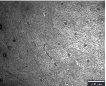
To verify the removal of organic matter, optical micrographs (Figure 1) and TGA (Figure 2) of the HAp were compared to those of the original bone. In the micrographs, it is possible to see a typical bone morphology (Figure 1a) characterized by the Haversian canal and a circular pattern around it caused by the orientation of the
col-lagen, which constitutes most of the organic matter of bone15. Figure
1a also shows the presence of two resorption cavities, found in most micrographs. In the HAp (Figure 1b), this pattern disappears, leaving
only the Haversian canals. Thus, it may be concluded that most of the collagen was removed.
In the TGA curve of the natural bone, there are three weight loss events: from 30-160 °C, related to the evaporation of water as-sociated with organic matter, between 200-500 °C, attributed to the
oxidation of organic matter, and 700-900 °C, attributed to the CO3–2
decomposition13. On the other hand, HAp shows only two major
events, attributed to the loss of water adsorbed on the HAp surface13
and the CO3–2 decomposition. Therefore, the obtained HAp, lacking
organic matter, may be considered a suitable ceramic reinforcement for the composite synthesis.
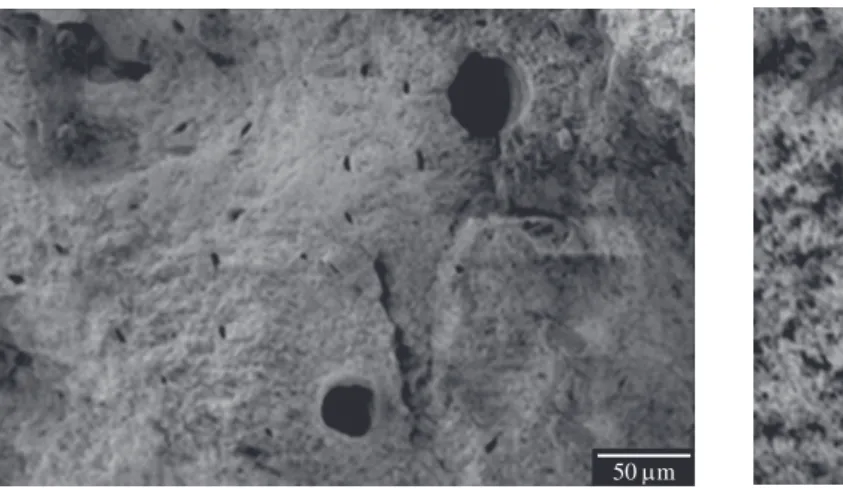
With the scanning electron microscopy (Figure 3), it is possible to verify that the HAp presents the pores, which were part of the Haversian system of the original bone, and also an intricate
micro-porosity, which explains its high specific surface area (91 m2.g–1).
In Figure 3b, it is also possible to see micro-cracks in the HAp that appeared as a consequence of the deorganification process.
The experimentally determined density of the composite
(1.90± 0.03 g.cm–3) was intermediate to that of the epoxy resin
(1.16 ± 0.03 g.cm–3) and of the HAp (3.16 g.cm–3)14, resulting in an
apparent porosity of (3.3% ± 1.4%), which is much lower than that of
200 m
(a)
200 m
(b)
Figure 1. Optical micrographs: a) Bone and b) HAp.
100
95
90
85
80
75
70
65
60
55
50
0 100 200 300 400 500 600 700 800 900 1000
Temperature (°C)
0.45
0.40
0.35
0.30
0.25
0.20
0.15
0.10
0.05
0.00 Bone HAp
wt. (%)
deri
v.
W (%/°C)
the original HAp, (55.4% ±1.4%). Thus, the infiltration process used was able to yield an epoxy infiltrated HAp composite structure.
From the optical micrographs of Figure 4, one can see a radial pattern around the Haversian canals, indicating that those structures were infiltrated first and then the infiltration proceeded radially toward inner regions. Besides, the presence of darker and clearer regions suggests that complete infiltration was not achieved. The presence of residual porosity corroborates the apparent porosity results.
The possible reasons for the incomplete impregnation may be that the viscosity of the resin was too high or the use of vacuum was not fully successful, retaining air bubbles inside the Haversian canals, which, in turn, led to partial filling. Figure 5 shows SEM images of different regions of the composite. In Figure 5a, one can see a region of incomplete infiltration of the Haversian system at the bottom right. Figure 5b shows that, despite displaying incomplete infiltration in some regions, the epoxy resin was capable of filling the HAp porous structure.
Figure 6 shows TGA curves of the composite samples. Compar-ing these curves with the HAp curve in Figure 2, a particularly large weight loss event may be seen between 180-540 °C, attributed to the
degradation of the epoxy resin. This weight loss was used to estimate the polymer content in the composite, which was found to be in the 33.9-36.1% (weight basis) range, or 29.2-31.12% in volume, i.e. the infiltration method used was able to incorporate a considerable amount of resin into the HAp porous structure. Combining TGA with the density results, the theoretical density of the composite was
calculated as 2.05 g.cm–3. Considering that the composite density
was 1.90 g.cm–3 and that the apparent porosity was 3.3%, 92% of the
theoretical density was reached and approximately 4.7% of closed porosity was present.
The DMA analysis (Figure 7) showed the composite to be much stiffer than the epoxy resin. Besides, for the composite, the glass transition spreads over a wider temperature range, with a higher
glass transition temperature (Tg). This indicates a good
matrix-rein-forcement interaction in the composite16, reducing interface segment
mobility and increasing Tg, contributing for the observed mechanical
properties.
Regarding flexural testing, the HAp fractured with just the pre-tensioning load of 2.5 N due to its high brittleness, which may be partially attributed to micro-cracks in the HAp pieces. The flexural
(a)
50 m
(b)
1 m
Figure 3. Scanning electron micrographs of HAp detailing the a) Haversian system and b) micro-pores.
(a)
200 m
(b)
200 m
strength of the composite reached 26.2 ± 2.7 MPa, which is higher than that of the pure resin, 17.6 MPa, but still below the 60 MPa lower limit of human cortical bone. The literature lacks similar research works for comparison, but PLA-HAp composite obtained by solution
techniques reached 100 to 130 MPa8. The lower strength observed in
the composite produced may be attributed to a few factors: the resin chosen - epoxy, which is not as tough as PCL, the presence of residual porosity in the composite, and the presence of cracks in the HAp.
4. Conclusions
Hydroxyapatite was obtained from bones via deorganification in hot NaOH solution. Most of their organic matter was removed, but the process originated micro-cracks in the pieces, yielding a very brittle material with high porosity, which was used to obtain an infiltration composite with epoxy resin.
Morphological analysis of the composite suggested that the infil-tration began at the Haversian canal, proceeding then radially toward inner regions. The polymer content in the composite reached 36% in weight, 92% of the theoretical density. Good interaction between resin and hydroxyapatite was found, increasing the glass transition temperature of the epoxy.
The composite presented flexural strength superior to the origi-nal materials, but still low in comparison to natural bone and other
hydroxyapatite infiltration composites. This was attributed to the limited toughness of the epoxy resin chosen as matrix, the residual porosity due to incomplete infiltration and the presence of micro-cracks in the HAp pieces.
References
1. Kalita SJ, Bhardwaj A, Bhatt HA. Nanocrystalline calcium phosphate ceramics in biomedical engineering. Materials Science and Engineering-C.
2007; 27(3):441-449.
2. Rosena VB, Hobbsa LW, Spectorb M. The ultrastructure of anorganic bovine bone and selected synthetic hyroxyapatites used as bone graft substitute materials Biomaterials. Biomaterials. 2002, 23(3):921-928.
3. Huang X, Miao XG. Novel porous hydroxyapatite prepared by combining H2O2 foaming with PU sponge and modified with PLGA and bioactive
glass. Journal Biomaterials Applications. 2007; 21(4):351-374.
4. Chlopek J, Rosol P, Morawska-Chochol A. Durability of polymer-ceramics composite implants determined in creep tests. Composites Science and Technology. 2006; 66(11-12):1615-1622.
5. Nakahira A, Tamai M, Miki S, Pezzotti G. Fracture behavior and biocompatibility evaluation of nylon-infiltrated porous hydroxyapatite.
Journal of Materials Science. 2002; 37(20):4425-4430.
50 m (a)
1 m (b)
Figure 5. Residual porosity in a) a Haversian canal and b) in its surroundings.
36.1% 35.1% 33.9%
Composite 1 Composite 2 Composite 3
Temperature (°C) 0 100
100
95
90
85
80
75
70
65
60
55
50
deri
v.
W (%/°C)
wt. (%)
200 300 400 500 600 700 800 900 1000 0.25
0.20
0.15
0.10
0.05
0.00
Figure 6. Thermogravimetric curves for three composite samples.
1000
800
600
400
200
0
Epoxy resin Composite
50
40
30
20
10
0
Loss modulus (MP
a)
Temperature (°C)
20 40 60 80 100 120 140 160
Storage modulus (MP
a)
6. Lien SM, Chien CH, Huang TJ. A novel osteochondral scaffold of ceramic–gelatin assembly for articular cartilage repair. Materials Science and Engineering C. 2009; 29(1):315-321.
7. Zhang P, Hong Z, Yu T, Chen X, Jing X. In vivo mineralization and osteogenesis of nanocomposite scaffold of poly(lactide-co-glycolide) and hydroxyapatite surface-grafted with poly(L-lactide). Biomaterials.
2009; 30(1):58-70.
8. Russias J, Saiz E, Nalla RK, Gryn K, Ritchie RO, Tomsia AP. Fabrication and mechanical properties of PLA/HA composites: a study of in vitro degradation. Materials Science and Engineering C. 2006; 26(8):1289-1295.
9. Jun-Liang Z, Tao F, Yong H, Ke-Wei X. Reinforcing hydroxyapatite/ thermosetting epoxy composite with 3-D carbon fiber fabric through RTM processing. Materials Letters. 2003; 58(1-2):163-168.
10. Liming F, Yang L, Ping G. Processing and mechanical properties of HA/ UHMWPE nanocomposites. Biomaterials. 2006; 27(20):3701-3707.
11. Biqiong C, Kang S. Mechanical and dynamic viscoelastic properties of hydroxyapatite reinforced poly (ε-caprolactone). Polymer Testing. 2005; 24(8):978-982.
12. Pezzoti G, Asmus SMF, Ferroni LP, Miki S. In situpolimerization into porous ceramics: a novel route to tough biomimetic materials. Journal of Materials Science: Materials in Medicine. 2002; 13(8):783-787. 13. Haberko K, Bucko MM, Brzezinska-Miecznik J, Haberko M, Mozgawa
W, Panz T, Pyda A, Zarebski J. Natural hydroxyapatite: its behaviour during heat treatment. Journal of European Ceramic Society. 2006; 26(4-5):537-542.
14. Konopka K, Boczkowska A, Batorski K, Szafran M, Kurzydlowski KJ. Microstructure and properties of novel ceramic–polymer composites.
Materials Letters. 2004; 58(30):3857-3862.
15. Nyman JS, Reyes M, Wang XD. Effect of ultrastructural changes on the toughness of bone. Micron. 2005; 36(7-8):566-582.