A Phytase-Based Reporter System for
Identification of Functional Secretion Signals
in Bifidobacteria
Annika Osswald1, Christina Westermann1, Zhongke Sun1,2*, Christian U. Riedel1*
1Institute of Microbiology and Biotechnology, University of Ulm, 89068, Ulm, Germany,2College of Life Sciences and Agriculture, Zhoukou Normal University, Chuanhui District, 466001, Zhoukou City, P.R. China
*[email protected](CUR);[email protected](ZS)
Abstract
Health-promoting effects have been attributed to a number ofBifidobacteriumsp. strains. These effects as well as the ability to colonise the host depend on secreted proteins. More-over, rational design of protein secretion systems bears the potential for the generation of novel probiotic bifidobacteria with improved health-promoting or therapeutic properties. To date, there is only very limited data on secretion signals of bifidobacteria available. Usingin silicoanalysis, we demonstrate that all bifidobacteria encode the major components of Sec-dependent secretion machineries but onlyB.longumstrains harbour Tat protein transloca-tion systems. A reporter plasmid for secretransloca-tion signals in bifidobacteria was established by fusing the coding sequence of the signal peptide of a sialidase ofBifidobacterium bifidum
S17 to the phytase geneappAofE.coli. The recombinant strain showed increased phytase activity in spent culture supernatants and reduced phytase levels in crude extracts com-pared to the control indicating efficient phytase secretion. The reporter plasmid was used to screen seven predicted signal peptides inB.bifidumS17 andB.longumE18. The tested signal peptides differed substantially in their efficacy to mediate protein secretion in different host strains. An efficient signal peptide was used for expression and secretion of a thera-peutically relevant protein inB.bifidumS17. Expression of a secreted cytosine deaminase led to a 100-fold reduced sensitivity ofB.bifidumS17 to 5-fluorocytosine compared to the non-secreted cytosine deaminase suggesting efficient conversion of 5-fluorocytosine to the cytotoxic cancer drug 5-fluorouracil by cytosine deaminase occurred outside the bacterial cell. Selection of appropriate signal peptides for defined protein secretion might improve therapeutic efficacy as well as probiotic properties of bifidobacteria.
Introduction
Bifidobacteria are an important component of the normal human gastrointestinal microbiota and, besides lactobacilli, the most frequently used microbial supplements in functional foods and probiotic formulations [1]. The concept of functional foods containing live microbial
OPEN ACCESS
Citation:Osswald A, Westermann C, Sun Z, Riedel CU (2015) A Phytase-Based Reporter System for Identification of Functional Secretion Signals in Bifidobacteria. PLoS ONE 10(6): e0128802. doi:10.1371/journal.pone.0128802
Academic Editor:Roman G. Gerlach, Robert Koch-Institute, GERMANY
Received:March 8, 2015
Accepted:April 30, 2015
Published:June 18, 2015
Copyright:© 2015 Osswald et al. This is an open access article distributed under the terms of the Creative Commons Attribution License, which permits unrestricted use, distribution, and reproduction in any medium, provided the original author and source are credited.
Data Availability Statement:All relevant data are within the paper and its Supporting Information files.
supplements, i.e. probiotics, is based on the observation that some of the commensal bacteria of the human microbiota have beneficial effects in differentin vitrosettings, small animal mod-els or clinical trials [1,2]. These beneficial effects are, in most cases, strain-specific and include maintenance of remission in paediatric ulcerative colitis (UC), prevention ofClostridium diffi-cile- and antibiotic-associated diarrhoea, and a reduced mortality in necrotizing enterocolitis (NEC) [2].
Some of the health-promoting effects of probiotics and bifidobacteria seem to be mediated by secreted factors. Several anti-inflammatory factors of bifidobacteria potentially involved in their effects in UC and NEC are secreted proteins. One example is a eukaryotic-type serine pro-tease inhibitor (serpin). Serpin was initially identified in the genome sequence ofB.longum
NCC2705 [3] and shown to inhibit pancreatic and neutrophil elastases [4]. Later, serpin-like proteases were also identified in other bifidobacteria [5,6]. Serpin is hypothesized to confer protection against proteolysis by pancreatic elastase in the gastrointestinal environment as well as to inhibit proteolytic damage by neutrophil elastase during intestinal inflammation [4]. A secreted protein ofB.animalissubsp.lactiswas shown to inhibit inflammatory chemokine secretion by TNF-αchallenged cultured intestinal epithelial cells. Similarly, a released peptide factor ofB.infantiswas able to prevent loss of epithelial integrity in response to TNF-αor
IFN-γchallenge in anin vitrosetting [7].
The ability to exert a health-promoting effect via secreted proteins depends on an active metabolism and, thus, on acquisition of nutrients. Various strains and species of bifidobacteria were shown to ferment different high molecular weight substrates derived from both the host and its diet including mucus [8], human milk oligosaccharides [9,10], starch [11], and other plant-derived polysaccharides [12]. All these substrates require degradation by extracellular enzymes prior to uptake and further breakdown for energy conservation. The ability to utilize these substrates is thought to provide a selective advantage over other bacteria, aiding the colo-nization of breast-fed infants and persistence during later stages of life [13–16] and hence sup-port their health-promoting effects.
In addition to their reported health-promoting effects, some strains of bifidobacteria were shown colonise solid tumours in various mouse models [17–20] and thus have gained increas-ing interest as vectors for delivery of therapeutic genes in cancer therapy [21–23]. The most widely used approach in bacterial tumour targeting is expression of enzymes that convert non-toxic prodrugs to therapeutically active compounds [24]. One example of a well-studied pro-drug-converting enzyme (PCE) is cytosine deaminase (CD). This enzyme converts the non-toxic prodrug 5-fluorocytosine to 5-fluorouracil, which interferes with DNA synthesis and thus inhibits proliferation of tumour cells. In order to improve the efficacy of prodrug conver-sion, while at the same time avoiding inhibitory effects on the proliferation of the delivery vector, PCEs are mostly used as secreted proteins.
Altogether, these findings indicate that secreted proteins might be important for establish-ment and maintenance of stable bifidobacterial populations in the gastrointestinal tract. More-over, efficient protein secretion is crucial for functionality of bifidobacteria as probiotics and gene delivery vectors for tumour targeting approaches. In bacteria, the majority of extracellular proteins is secreted by either the Sec or the Twin arginine translocation (Tat) pathway [25,26]. The Sec pathway exports proteins across the cytoplasma membrane in an unfolded state, whereas the Tat-pathway transports folded proteins [27,28]. Both pathways depend on secre-tion signals usually located in the N-terminus of the substrate that are distinct yet share struc-tural similarities [27].
In bifidobacteria, protein secretion has not been analysed in great detail and there is only one study employing a nuclease reporter to identify bifidobacterial signal sequences [29]. In the present study, we aim at providing a more systematic analysis of protein secretion and
no role in study design, data collection and analysis, decision to publish, or preparation of the manuscript.
associated signal peptides of bifidobacteria, developing a system to analyse these SPs, and devising a tool for efficient expression of extracellular proteins in bifidobacteria.
Materials and Methods
Bacterial strains, plasmids and growth conditions
All strains and plasmids used in this study are listed inS1 Table.E.coliDH10B was used as cloning host and for propagation of plasmids and grown in Luria broth (LB) at 37°C. Bifido-bacteria were grown anaerobically at 37°C in Reinforced Clostridial Medium (RCM, BD Difco, Germany) or Lactobacilli MRS (BD Difco, Germany) broth supplemented with 0.5 g/L L-cyste-ine hydrochloride-monohydrate (MRSc). For cultivation ofE.coliandBifidobacterium sp. strains harbouring plasmids, 100μg/ml spectinomycin were added to culture media. All media were prepared with ultrapure water.
Cloning procedures
Genomic DNA ofE.coliDH10B was used as template for amplification ofappA. Coding sequences of predicted SPs were amplified from genomic DNA ofB.bifidumS17 orB.longum
E18. PCRs were performed using Phusion DNA Polymerase (Thermo Scientific, Germany). All primers used in this study (S2 Table) were purchased from Eurofins Genomics GmbH (Ger-many). Thermo cycling was performed on a FlexCycler (Analytik Jena, Germany) with anneal-ing temperature optimized for each primer pair. TheappAgene ofE.coliK-12 encoding a phytase was amplified without its native signal peptide sequence using primers PhytF and PhytR. The obtained PCR product was digested with restriction enzymesXhoI andHindIII and ligated to the 4,423 bp fragment of similarly digested pMDY23-Pgap[30], i.e. the vector
backbone including Pgapbut lacking thegusAgene. This yielded pMgapP, which harbours the appAgene fused directly to Pgapwithout any signal sequence.
Coding sequences of signal peptides were fused to theappAgene by splicing-by-overlap-extension (SOEing) PCR [31]. The coding sequences for different SPs were amplified using a forward primer and a SOEing reverse primer. The PCR was designed to include two additional amino acid residues after the predicted cleavage site to preserve the recognition sequence for cleavage. In parallel, a SOEing forward primer with complementary sequence to the SP reverse primer was used together with primer PhytR for amplification ofappA. To fuse the SP coding sequence toappA, a second round of PCR was performed using the two PCR products of the first round as template, the SP forward primer, and primer PhytR. To increase specificity, DMSO was added to the PCR reaction to a final concentration of 5% (v/v) and annealing tem-perature was set to 70°C. The obtained PCR products were digested with restriction enzymes
XhoI andHindIII and ligated to the 4,423 bp fragment ofXhoI/HindIII cut pMDY23-Pgap
yielding plasmids with exact translational fusions of the different SPs to AppA.
In order to generate a vector for expression of a secreted cytosine deaminase (CD), the sig-nal peptide of thebbif_1734gene encoding a sialidase was amplified fromB.bifidumS17 chro-mosomal DNA by PCR using primersSP_fw_SalI andSP_rev_HindIII. ThecodAgene was amplified fromE.coliK-12 chromosomal DNA with the primerscodA_fw_HindIII and
codA_rev_SacII by PCR. Both PCR products were digested withHindIII and subsequently joined by a ligase reaction. The fusion was then amplified by PCR using primersSP_fw_SalI
codA_rev_SacII and the product was digested withSalI andSacII and ligated to the 4,423 bp fragment ofSalI/SacII cut pMGS-Pgap-bopAHis6[32] containing Pgapto yield pAO-S0-CD.
Following transformation intoE.coli, plasmids of spectinomycin resistant colonies were checked for correct inserts by PCR. Plasmids of positive clones were verified by restriction analysis and Sanger sequencing and constructs with correct sequences were transformed into eitherB.bifidumS17 orB.longumE18 as described elsewhere [33].
Phytase assay
For phytase samples, special attention is needed to deplete potential free phosphate contamina-tion. All related reagents and medium were prepared in ultrapure ddH2O (18 MOcm;
Milli-pore, USA).
RecombinantB.bifidumS17 orB.longumE18 strains were grown in 50 ml RCM containing 100μg/ml spectinomycin under anaerobic conditions. At the indicated time points, 5 ml of the cultures were harvested and centrifuged (5 000 × g, 5 min, 4°C). Supernatants were filter-steril-ized and used for determination of extracellular phytase activity. Bacterial pellets were washed twice in 1 ml of 0.2 M sodium citrate buffer, pH 5.5, and resuspended in 500μl of the same buffer. Bacterial suspensions were transferred to cryotubes containing 250μg glass beads and disrupted during 2 cycles of 35 s at 6500 rpm in a Precellys 24 homogenisator (PEQLAB Bio-technologie GmbH, Germany). Lysates were centrifuged (13 000 × g, 5 min, 4°C) and the supernatant was retained as crude extracts for determination of intracellular phytase activity. Total protein in crude extracts was quantified using the Pierce BCA protein assay kit (Thermo Scientific, Germany).
Phytase activity in supernatants and crude extracts was quantified using an assay described elsewhere [34] with minor modifications. Briefly, 100μl sample (supernatant or crude extract) were pre-incubated for 5 min at 37°C and then mixed with 100μl 10.8 mM sodium phytate (50% (w/w) phytic acid diluted in 0.2 M sodium citrate buffer). The reaction was carried out at 37°C for 15 min, and then stopped by adding 200μl 15% tricholoroacetic acid (TCA). After centrifugation (14 000 × g, 2 min), an aliquot of 20μl was mixed with 480μl ultrapure ddH2O
and 500μl color reagent (mix of 1M sulfuric acid, 2.5% (w/v) ammonium molybdate in ddH2O, and 10% (w/v) ascorbic acid in ddH2O at a ratio of 3:1:1). The mixture was incubated
at 50°C for 15 min, and 100μl were transferred to a transparent 96-well microtiter plate (Thermo Scientific, Germany). Absorbance at 820 nm was measured in triplicate using an Infi-nite M200 multimode microplate reader (Tecan, Switzerland). Phytase activity equivalents were calculated using to a standard curve of two-fold serial dilutions of a 9 mM potassium dihydrogen phosphate in water. Phytase activity equivalents were defined as the amount of enzyme that catalyses the release of 1μmol of inorganic phosphate per minute from 5.4 mM sodium phytate (i.e. 100μl sample + 100μl 10.8 mM sodium phytate solution, see above) under the conditions of the assay. Activities were expressed as relative phytase units (RPU) per ml in supernatant and RPU/mg in crude extracts.
Phytate degradation assays
To test for phytate degradation an agar plate assay developed for yeasts [35] was adapted for bifidobacteria using RCM agar (RCA) containing calcium phytate (Ca-phytate) as substrate. RCM phytate agar was prepared by adding 3 g/l calcium carbonate (Sigma, Germany) and 1.5 ml of a 50% (w/w) phytic acid solution in H2O (Sigma) to standard RCM agar prior to
Growth inhibition by 5-FC
The effect of the conversion of 5-FC to 5-FU by cytosine deaminase was determined forB. bifi-dumS17 wildtype and its isogenic derivatives carrying plasmids pAO-CD or pAO-S0_CD. OD600of overnight cultures grown in MRSc was adjusted to 0.1 in fresh medium containing
5-FC at 5, 1, 0.5, 0.1, 0.05, 0.01, 0.005 or 0.001 mg/ml (final concentration). 200μl aliquots were pipetted into wells of a 96-well microtiter plate in four technical replicates per strain and concentration. After 24 h of anaerobic incubation at 37°C, OD600was measured using an
Infi-nite M200 multimode reader.
Bioinformatic analysis
The sequenced and annotated genomes ofB.longumE18 (GenBank accession: CM002287),B.
bifidumS17 (CP002220),B.breveS27 (CP006716),B.animalissubsp.lactisATCC27673 (CP003941),B.adolescentisATCC15703 (AP009256), andB.dentiumBd1 (CP001750) were searched for genes encoding Sec proteins. Homologies of the deduced amino acid sequences of bifidobacterial Sec homologues to the respectiveE.coliK12-W3110 proteins were calculated using the multiple alignment function of the Basic Local Alignment Search Tool (BLAST, ww. ncbi.nlm.nih.gov/BLAST/).
B.bifidumS17 proteins with predicted extracellular localization were extracted from the precomputed genome results on the cPSORTdb database (version 3) [36]. The sequences of all proteins were analysed for potential SPs using SignalP Version 4.1 [37], TatP [38], and TAT-FIND [39].
Results
Analysis of protein secretion pathways in bifidobacteria
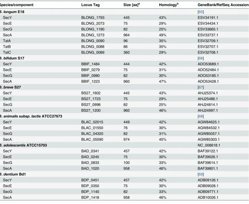
As a basis to establish a protein secretion reporter in bifidobacteria, the genome sequences of a number of representativeBifidobacterium sp. strains were analysed. As expected, all analysed genomes harboured genes for SecY, SecE, and SecG, i.e. the major components of the Sec trans-locon, and the associated ATPase SecA with reasonable homology to the respective proteins of
E.coliK12-W3110 (Table 1). By contrast, genes for Tat-dependent protein secretion were only found in the genomes ofB.longumE18 (Table 1) and other strains of this species (data not shown).
Construction of a secretion reporter for bifidobacteria
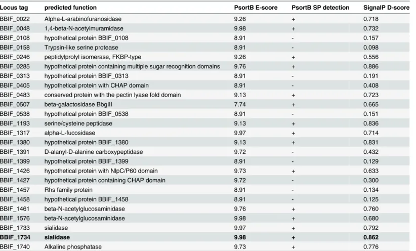
We next extracted a list of all proteins ofB.bifidumS17 predicted to be localized to the extra-cellular compartment. The N-terminal 60 residues of all proteins retrieved were analysedin silicofor potential SPs (Table 2). The SP of a sialidase (BBIF_1734) had the highest PSORTb E-score for extracellular localization and the second highest SignalP D-E-score for SP prediction. Moreover, the sialidase ofMicromonospora viridifaciens, a closely related member of the phy-lum Actinobacteria, has been experimentally confirmed to be secreted into the extracellular environment [40,41]. Thus, the SP of BBIF_1734 was named S0 and selected to develop a secre-tion reporter using the phytase geneappAofE.coliDH10B.
The S0 sequence was fused to theappAgene by SOEing PCR and cloned into pMDY23-Pgap
under the control of Pgap, replacing the glucuronidase reporter genegusA(Fig 1A) to yield
pMgapS0P. As a control vector, pMgapP was constructed, which harbours an identicalappA
construct fused directly to Pgapwithout a signal sequence.
measured at various time points during growth. Both strains displayed almost identical growth (data not shown) ruling out any effect of plasmids on growth or phytase activity. Phytase activ-ity markedly increased over time in supernatants ofB.bifidumS17/pMgapS0P (Fig 1B). By contrast, phytase activity in supernatants of the control strainB.bifidumS17/pMgapP were barely above background until later time points during growth, i.e. stationary growth phase (Fig 1B). On the other hand, phytase activities were higher in crude extracts ofB.bifidumS17/ pMgapP than in the strain harbouring pMgapS0P, i.e. the construct with a SP, throughout the experiment (Fig 1C).
Table 1. Components of the major protein secretion machineries encoded on the genomes of representativeBifidobacterium sp.
Species/component Locus Tag Size [aa]a Homologyb GeneBank/RefSeq Accession
B.longumE18 [65]
SecY BLONG_1793 445 43% ESV34191.1
SecE BLONG_2073 75 29% ESV34434.1
SecG BLONG_1190 82 25% ESV33665.1
SecA BLONG_1273 964 49% ESV33737.1
TatA BLONG_0090 96 35% ESV32709.1
TatB BLONG_0088 86 35% ESV32707.1
TatC BLONG_0089 360 29% ESV32708.1
B.bifidumS17 [66]
SecY BBIF_1484 444 42% ADO53689.1
SecE BBIF_0279 75 31% ADO52484.1
SecG BBIF_0980 82 30% ADO53185.1
SecA BBIF_1223 960 47% ADO53428.1
B.breveS27 [67]
SecY BS27_1602 445 43% AHJ25374.1
SecE BS27_1723 75 29% AHJ25486.1
SecG BS27_0998 82 25% AHJ24814.1
SecA BS27_1200 960 46% AHJ24997.1
B.animalis subsp.lactisATCC27673 [68]
SecY BLAC_02015 449 42% AGW84625.1
SecE BLAC_01550 76 30% AGW84532.1
SecG BLAC_04320 82 31% AGW85057.1
SecA BLAC_05590 974 45% AGW85303.1
B.adolescentisATCC15703 NC_008618.1
SecY BAD_0341 457 42% BAF39122.1
SecE BAD_0245 75 30% BAF39026.1
SecG BAD_0833 100 33% BAF39614.1
SecA BAD_1020 958 46% BAF39801.1
B.dentiumBd1 [69]
SecY BDP_0451 457 42% ADB09126.1
SecE BDP_0350 75 30% ADB09028.1
SecG BDP_1140 82 33% ADB09771.1
SecA BDP_1418 958 46% ADB10026.1
a
protein size in amino acid residues (aa).
bpercent identity on amino acid sequence level to the respective homologue ofE.coliK12-W3110.
Comparative analysis of various SPs in different
Bifidobacterium sp
.
hosts
Following the successful establishment ofappAas a secretion reporter inB.bifidumS17, this system was used to test various other bifidobacterial SPs. For this purpose, a total of six SPs with high D-scores according to the SignalP prediction were selected (Table 3). Of these SPs, two belong to proteins fromB.bifidumS17 (BBIF_1681 and BBIF_1761) and two other to pro-teins fromB.longumE18 (BLONG_1728 and BLONG_0476). Moreover, two SPs of (putative) Tat-secreted proteins ofB.longumE18 (BLONG_0223 and BLONG_1620) were included. To test their functionality, all SPs were fused to theappAreporter by SOEing PCR, cloned into pMDY23-Pgapreplacing thegusAreporter gene, and the obtained plasmids were introduced
intoB.bifidumS17 orB.longumE18 by electroporation. Monitoring of growth in RCM broth indicated that all recombinant strains show the same growth pattern (data not shown).
All recombinant strains were analysed for phytase secretion using a phenotypic assay based on the degradation of insoluble Ca-phytate in solid medium (Fig 2). Clear zones of Ca-phytate degradation were observed forB.bifidumS17 strains harbouring pMgapS0P, pMgapS1P, pMgapS3P, pMgapS4P, and pMgapS6. By contrast, strains harbouring plasmids pMgapS2P and pMgapS5P did not display Ca-phytate degradation above background levels (pMgapP). A similar pattern of Ca-phytate degradation was observed forB.longumE18 strains, however at somewhat lower levels.
Table 2. List ofB.bifidumS17 proteins with predicted extracellular localization and information on the signal peptides identified in their amino acid sequences.
Locus tag predicted function PsortB E-score PsortB SP detection SignalP D-score
BBIF_0022 Alpha-L-arabinofuranosidase 9.26 + 0.718
BBIF_0048 1,4-beta-N-acetylmuramidase 9.98 + 0.732
BBIF_0108 hypothetical protein BBIF_0108 8.91 - 0.157
BBIF_0158 Trypsin-like serine protease 8.91 - 0.098
BBIF_0246 peptidylprolyl isomerase, FKBP-type 9.26 + 0.556
BBIF_0285 hypothetical protein containing multiple sugar recognition domains 9.76 + 0.886
BBIF_0313 hypothetical protein BBIF_0313 8.91 - 0.191
BBIF_0405 hypothetical protein with CHAP domain 8.91 - 0.408
BBIF_0483 conserved protein with the pectin lyase fold domain 9.13 + 0.723
BBIF_0507 beta-galactosidase BbgIII 7.74 + 0.665
BBIF_0538 hypothetical protein BBIF_0538 8.91 - 0.151
BBIF_1193 serine/cysteine peptidase 9.13 + 0.836
BBIF_1317 alpha-L-fucosidase 9.97 + 0.714
BBIF_1380 hypothetical protein BBIF_1380 9.13 + 0.831
BBIF_1391 D-alanyl-D-alanine carboxypeptidase 9.72 - 0.432
BBIF_1399 hypothetical protein BBIF_1399 8.91 - 0.129
BBIF_1426 hypothetical protein with NlpC/P60 domain 9.73 + 0.633
BBIF_1427 hypothetical protein containing CHAP domain 9.72 - 0.300
BBIF_1457 Rhs family protein 8.91 - 0.134
BBIF_1458 hypothetical protein BBIF_1458 8.91 - 0.125
BBIF_1461 beta-N-acetylglucosaminidase 9.76 + 0.760
BBIF_1576 beta-N-acetylglucosaminidase 9.98 + 0.680
BBIF_1733 sialidase 9.97 + 0.792
BBIF_1734 sialidase 9.98 + 0.862
BBIF_1740 Alkaline phosphatase 9.73 + 0.776
In order to get a more quantitative comparison of protein secretion effected by the different SPs, phytase activity was measured in the supernatants of all strains generated. Using S0, high-est levels of phytase activity in supernatants ofB.bifidumS17 were observed in stationary growth phase (Fig 1). The sequence upstream of the SP-appAconstructs including the pro-moter (Pgap) and all elements relevant for translation, i.e. ribosome binding site, start codon,
and the sequence and distance between them, were identical in all strains. Thus, equal levels of expression at least amongst derivatives of the same strain was assumed and samples were col-lected from cultures grown for 16 h, i.e. early stationary growth phase, for determination of phytase activity. ForB.bifidumS17 strains, highest levels of phytase activity were observed for S6 and somewhat lower yet still efficient secretion was observed when S0, S1 and S4 were used as SP (Fig 3A). Statistical analysis suggests that protein secretion mediated by S4 and S6 is sig-nificant different (higher) compared to all other SPs and no difference is observed between S0 and S1 (Fig 3A;S3 Table). InB.longumE18, S0 and S1 were the most efficient secretion signals (Fig 3B). Again no statistically significant difference between S0 and S1 was observed but both SPs differed from all other SPs (Fig 3B;S3 Table).
Fig 1. Phytase reporter system to monitor protein secretion by bifidobacteria.(A) Schematic representation of the secretion reporter plasmid pMgapS0P. The vector was constructed by fusing the SP of BBIF_1734 (S0) to the phytase geneappAofE.coliK12 and cloning of the construct under the control of the gappromoter (Pgap) ofB.bifidumS17 in the vector backbone of pMDY23-PgapusingXhoI andHindIII. Relevant other features are:rep(origin of replication forE.coli),repAB(origin of replication for bifidobacteria), spc(spectinomycin resistance gene). (B)+(C) Phytase activity in supernatants (B) or crude extracts (C) ofB. bifidumS17/pMgapP (S17 pMgapP) andB.bifidumS17/pMgapS0P (S17 pMgaS0P) during growth in RCM batch cultures. Values are relative phytase units (RPU) per ml supernatant (B) or mg protein in crude extracts (C) and are mean +/- standard deviation of three independent cultures measured in technical triplicates.
doi:10.1371/journal.pone.0128802.g001
Table 3. Signal peptides used to establish or tested in the phytase reporter assay and corresponding information on the predicted localization.
Signal peptide locus tag, predicted function PsortB
E-scorea
SignalP D-scoreb
predicted cleavage sitec
Sec-dependent signal peptides
S0 BBIF_1734, sialidase 9.98 0.862 aa 33–37: ASA*AS
S3 BLONG_1728, hypothetical secreted protein with NlpC/P60 domain
3.33 0.891 aa 27–31: ATA*AE
S4 BLONG_0476, conserved hypothetical secreted protein with CHAP domain
3.33 0.838 aa 32–36: AQA*DT
S5 BBIF_1681, subtilisin family peptidase (lactocepin) 9.98 0.798 aa 26–30: ALA*AP
S6 BBIF_1761, surface protein with Gram positive anchor and Cna protein B-type domains
0d 0.879 aa 29–33: ANA*AD
Tat-dependent signal peptides
S1 BLONG_0223, Tat-secreted glycosidase 4.31 0.865 aa 30–34: AQA*AD
S2 BLONG_1620, putative Tat-secreted pectin lyase-like protein 9.13 0.772 aa 28–32: AFA*QS
aE-score, extracellular score, calculated by cPSORTdb (version 3) bD-score, discrimination score, calculated by SignalP 4.1.
camino acid positions (aa), the sequence and the exact cleavage site (
*) of the predicted SPs are indicated
dThis SP belong to a protein which is predicted to be located in the cell wall. The respective C-score for cell wall localization calculated by PSORTdb was
9.26.
Secretion of cytosine deaminase
In order to demonstrate applicability of the identified SPs for secretion of a therapeutically rele-vant protein, we aimed at implementing a PCE approach using CD. For this purpose, we selected S0 based on the efficient secretion of phytase by bothB.bifidumS17 andB.longum
E18. The coding sequence of S0 was fused to thecodAgene ofE.coliK12. The fusion was cloned under control of Pgapresulting in pAO-S0_CD, which was transformed intoB.bifidum
S17. As a control,codAwas cloned under control of Pgapwithout a SP in the same vector
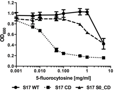
back-bone (pAO-CD). Growth of all strains was comparable in the absence of 5-FC (data not shown). Both recombinant strains as well as the wildtype were tested for growth inhibition by the prodrug 5-FC (Fig 4). This revealed thatB.bifidumS17 showed good resistance to the pro-drug and growth was only inhibited at the highest propro-drug concentration tested (5 mg/ml). Expression of CD without a SP markedly increased sensitivity of the recombinant strain (B.
bifidumS17/pAO-CD). Growth of this strain was already inhibited at 0.005 mg/ml 5-FC. By contrast, growth ofB.bifidumS17/pAO-S0_CD was comparable to that of the wild type in the presence of up to 0.1 mg/ml 5-FC. A slight inhibition of growth was observed only at 5-FC con-centrations of 0.5 mg/ml and above but final OD600were higher than that of the strain
Fig 2. Ca-phytate degradation of recombinant bifidobacteria expressing phytase with different signal peptides.Calcium phytate degradation by recombinant strains ofB.bifidumS17 (A) andB.longumE18 (B) harbouring pMgapP-derived plasmids containing different SPs (S0-S6). The control plasmid pMgapP contains no SP and serves as a background control for expression of a non-secreted phytase. Overnight cultures of all strains were spotted in triplicate on RCM agar supplemented with 0.15% calcium phytate and imaged after anaerobic incubation for 48 h at 37°C. One representative spot of three independent cultures is shown.
expressing the SP-less CD at all concentrations tested. Thus, the sensitivity to 5-FC was reduced by expressing CD as a secreted protein by two orders of magnitude suggesting efficient protein export.
Discussion
A number of traits of bacteria require extracellular proteins. These traits include acquisition of nutrients e.g. by secretion of glycosyl hydrolases for degradation of polysaccharides [42]. Extra-cellular proteins are not only required for mere survival, but are also involved in the interaction with the host. Thus, survival and interaction with the host of most bacteria crucially depends on secretion of functional proteins. Bifidobacteria as members of the normal human gut micro-biota are no exception to this rule. In fact, in the highly competitive environment of the
Fig 3. Phytase activity in supernatants recombinant bifidobacteria strains expressing phytase with different signal peptides.Phytase activity was measured in culture supernatants ofB.bifidumS17 (A) orB. longumE18 (B) harbouring pMgapP-derived plasmids containing different SPs (S0-S6) during growth in RCM batch cultures. The control plasmid pMgapP (-) contains no SP and serves as a background control for expression of a non-secreted phytase. Values are relative phytase units (RPU) per ml supernatant (B) and are mean +/- standard deviation of three independent cultures measured in technical triplicates. Statistical analysis was performed by one-way ANOVA with Bonferroni post-tests for multiple comparisons. Phytase activity in the supernatants of each strain was compared to all other strains. Letters indicate statistical significance of the difference (a:p<0.001 for all comparisons; b:p<0.001 for all comparisons except S0 vs. S1; differences for all other signal peptides were significant at p<0001 for less than 5 other signal peptides). For complete results of the analysis seeS3 Table.
gastrointestinal tract, secretion of proteins might be even more important than in other less densely populated habitats [42].
The vast majority of bacterial proteins are transported by the Sec pathway. Tat-dependent secretion is essential in only a few bacteria [25–27] and was shown to be important for viru-lence of a wide range of bacterial pathogens [43]. AllBifidobacterium sp. genomes analysed harboured genes for a Sec translocon. By contrast, only theB.longumgenomes contained genes for Tat protein export machineries. This is consistent with the conservation and distribu-tion of Sec and Tat systems amongst bacteria.
Various bifidobacterial signal sequences have been used for expression of secreted recombi-nant proteins. These include the SPs of the galacto-N-biose/lacto-N-biose I-binding protein [44] and exo-xylanase [45–47] ofB.longum, theβ-galactosidase ofB.bifidum[48], and Sec2 and ApuB ofB.breve[49,50]. However, in none of these cases the authors provide a rationale for selecting the respective SP and in most cases the sequences have not been analysed. Com-parative and systematic studies on secretion systems and signal peptides of bifidobacteria are largely missing. In one study, a nuclease reporter was used to screen a genomic library ofB.
breveUCC2003 for signal sequences inE.coliand subsequent confirmation of positive clones inL.lactis[29]. This identified three Sec-dependent SPs and three signal sequences of putative membrane proteins. Further analysis of these SPs confirmed that the three Sec-dependent SP are functional for secretion but quantitative analysis of nuclease activity in the supernatants showed no differences in efficiency of protein export.
We used a slightly different approach by selecting SPs of potentially secreted proteins of dif-ferent bifidobacterial species predictedin silico. Using the predicted SPs of the sialidase BBIF_1734, we were able to establish a reporter system for protein secretion in bifidobacteria employing the phytase AppA ofE.colilacking its native SP. Heterologous expression and secretion of a phytase has previously been demonstrated in lactic acid bacteria [51,52]. The
Fig 4. Growth inhibition of recombinant bifidobacteria by 5-fluorocytosine is reduced by secretion of cytosine deaminase.Effect of different concentrations of 5-FC on growth ofB.bifidumS17 wildtype (S17 WT) or isogenic derivatives carrying plasmids pAO-CD (S17 CD) or pAO-S0_CD (S17 S0_CD). Overnight cultures were adjusted to an final optical density at 600 nm (OD600) of 0.1 in fresh medium containing the indicated concentrations of 5-FC and incubated o/N anaerobically at 37°C and OD600was recorded. Values are mean +/- standard deviation of four technical replicates and results of one representative of three independent experiments are shown.
relative levels of extra- and intracellular phytase activity inB.bifidumS17/pMgapS0P com-pared toB.bifidumS17/pMgapP (the strain expressing AppA without SP) suggest that S0 mediates efficient secretion of phytase. Although it is not possible to quantitatively compare levels of intracellular and extracellular levels of phytase due to intrinsic limitations of the Phy-tex method (RPU/mg of protein in crude extracts vs. RPU/ml supernatant), our results demon-strate that phytase is a valuable reporter system for the identification and analysis of secretion signals in bifidobacteria. The enzyme is resistant against proteases, active in a wide range of pH values, and activity is optimal at pH 4–5, which is the normal pH in stationary phase batch fer-mentations of bifidobacteria [53].
Bifidobacteria do not encodeappAhomologues since BLAST searches revealed no signifi-cant hits in the genusBifidobacterium(data not shown). Nevertheless, phytase activity has been detected in differentBifidobacterium sp. [54,55]. Recently, two enzymes of bifidobacteria with phytase activity have been characterized. Based on sequence comparisons, the enzymes belong to a different phylogenetic cluster than theE.coliAppA enzyme and are more closely related to the phytases of plants, fungi and vertebrates [56]. Intrinsic activity of non-AppA phytases or other phosphatases might explain the slight background observed forB.bifidum
S17/pMgapP.
Using the phytase reporter, we were able to screen a number of other SPs of potentially secreted proteins ofB.bifidumS17 andB.longumE18. All strains were initially screened for secreted phytase by detection of Ca-phytate degradation in agar plates and measuring phytase activity in culture supernatants. Some of the SPs did not produce zones of phytate degradation on agar, but phytase activity well above background was measured in the supernatants. Thus, Ca-phytate degradation in agar plates is rather a first, but not definite, indicator for the func-tionality of highly efficient SPs and measuring phytase activity in supernatants allows a more quantitative analysis.
All SPs analysed yielded phytase activities above background in bothB.bifidumS17 andB.
longumE18, suggesting that bifidobacterial SPs might be functional in otherBifidobacterium
sp. besides their original hosts. Despite their annotation as (putative) Tat substrates, SPs S1 and S2 mediated phytase export inB.bifidumS17, which lacks a Tat system. Furtherin silico
analysis revealed that both SPs yielded no prediction for Tat recognition using TATFIND and no Tat motifs were found by TatP (data not shown). Moreover, S1 and S2 had D-scores of 0.865 and 0.772, respectively, in the SignalP analysis. Collectively, this suggests that the pre-dicted Tat-dependence of these SPs might be false and they might actually be Sec-dependent secretion signals.
Amongst the tested SPs, the SP of BBIF_1734 (S0) mediated efficient protein secretion in bothB.bifidumS17 andB.longumE18. Thus, we selected S0 to clone a vector for expression and secretion of a therapeutically relevant protein. A number of bacteria including bifidobac-teria are investigated as gene delivery vectors for cancer therapy. One of the approaches pur-sued, is the so-called Bacterial Directed Enzyme Prodrug Therapy (BDEPT), i.e. the use of recombinant bacteria expressing secreted enzymes for conversion of non-toxic prodrugs into their active, cytotoxic form [24]. An enzyme frequently used in BDEPT is the cytosine deami-nase [24]. This enzyme is present in a wide range of microorganisms but not in mammalian cells. Its physiological role is deamination of cytosine to uracil and, as a specific, non-physiologic side reaction, also catalyses conversion of the prodrug 5-FC to the tumour thera-peutic 5-FU.
In a proof-of-principle approach, a construct was generated for expression of a secreted form of the CodA cytosine deaminase ofE.coliin bifidobacteria using the Pgappromoter and
inhibited at concentration of 0.005 mg/ml and above. This demonstrates that a therapeutically relevant protein can be expressed and secreted in its active form using the SP of BBIF_1734. Moreover, the highest systemic concentration of 5-FC that is tolerated during therapy without adverse side effects is 0.1 mg/ml [57]. Thus, expression of CD as a secreted form byB.bifidum
S17 renders this strain tolerant to therapeutically relevant doses.
A number of genetically engineered bifidobacteria expressing various therapeutic genes were successfully used to inhibit tumour growth in mouse models [58–62]. CD has also been expressed in bifidobacteria [63,64] and one of these recombinant strains was successfully used in combination with 5-FC to inhibit growth of a subcutaneous tumours of a melanoma cell line in mice [64]. Expression CD and other proteins in secreted form by bifidobacteria using appro-priate secretion signals might improve their therapeutic efficacy.
In summary, our data shows that we have successfully developed a reporter system for iden-tification and analysis of secretion signals in bifidobacteria. We furthermore demonstrated that an SP identified using this report is functional in mediating secretion of a therapeutically rele-vant protein. Rationally designed systems for secreted proteins might improve the efficacy of recombinant bifidobacteria as probiotic supplements in functional foods or in therapeutic applications such as cancer therapy.
Supporting Information
S1 Data. Amino acid sequences of Sec and Tat homologues of representative Bifidobacter-ium sp.
(DOCX)
S1 Table. Bacterial strains.
(DOCX)
S2 Table. Oligonucleotides.
(DOCX)
S3 Table. Statistical analysis of phytase secretion using different signal peptides.
(DOCX)
Author Contributions
Conceived and designed the experiments: ZS CUR. Performed the experiments: AO CW ZS. Analyzed the data: AO CW ZS CUR. Wrote the paper: AO ZS CUR.
References
1. Kleerebezem M, Vaughan EE (2009) Probiotic and gut lactobacilli and bifidobacteria: molecular approaches to study diversity and activity. Annu Rev Microbiol 63: 269–290. Available:http://www. ncbi.nlm.nih.gov/pubmed/19575569. doi:10.1146/annurev.micro.091208.073341PMID:19575569
2. Gareau MG, Sherman PM, Walker WA (2010) Probiotics and the gut microbiota in intestinal health and disease. Nat Rev Gastroenterol Hepatol 7: 503–514. Available:http://www.ncbi.nlm.nih.gov/pubmed/ 20664519. doi:10.1038/nrgastro.2010.117PMID:20664519
3. Schell MA, Karmirantzou M, Snel B, Vilanova D, Berger B, Pessi G, et al. (2002) The genome sequence of Bifidobacterium longum reflects its adaptation to the human gastrointestinal tract. Proc Natl Acad Sci U S A 99: 14422–14427. Available:http://www.ncbi.nlm.nih.gov/pubmed/12381787. PMID:12381787
4. Ivanov D, Emonet C, Foata F, Affolter M, Delley M, Fisseha M, et al. (2006) A serpin from the gut bacte-rium Bifidobactebacte-rium longum inhibits eukaryotic elastase-like serine proteases. J Biol Chem 281: 17246–17252. Available:http://www.ncbi.nlm.nih.gov/pubmed/16627467. PMID:16627467
pmcentrez&rendertype = abstract. Accessed 24 October 2014. doi:10.1128/AEM.02938-09PMID: 20348296
6. Alvarez-Martin P, O’Connell Motherway M, Turroni F, Foroni E, Ventura M, van Sinderen D (2012) A two-component regulatory system controls autoregulated serpin expression in Bifidobacterium breve UCC2003. Appl Environ Microbiol 78: 7032–7041. Available:http://www.ncbi.nlm.nih.gov/pubmed/ 22843530. Accessed 10 June 2014. doi:10.1128/AEM.01776-12PMID:22843530
7. Ewaschuk JB, Diaz H, Meddings L, Diederichs B, Dmytrash A, Backer J, et al. (2008) Secreted bioac-tive factors from Bifidobacterium infantis enhance epithelial cell barrier function. Am J Physiol Gastroint-est Liver Physiol 295: G1025–G1034. Available:http://www.ncbi.nlm.nih.gov/pubmed/18787064. doi: 10.1152/ajpgi.90227.2008PMID:18787064
8. Turroni F, Bottacini F, Foroni E, Mulder I, Kim J-H, Zomer A, et al. (2010) Genome analysis of Bifidobac-terium bifidum PRL2010 reveals metabolic pathways for host-derived glycan foraging. Proc Natl Acad Sci U S A 107: 19514–19519. Available:http://www.ncbi.nlm.nih.gov/pubmed/20974960. doi:10. 1073/pnas.1011100107PMID:20974960
9. LoCascio RG, Desai P, Sela DA, Weimer B, Mills DA (2010) Broad conservation of milk utilization genes in Bifidobacterium longum subsp. infantis as revealed by comparative genomic hybridization. Appl Environ Microbiol 76: 7373–7381. Available:http://www.pubmedcentral.nih.gov/articlerender. fcgi?artid=2976205&tool = pmcentrez&rendertype = abstract. Accessed 21 March 2014. doi:10.1128/ AEM.00675-10PMID:20802066
10. Sela DA, Garrido D, Lerno L, Wu S, Tan K, Eom HJ, et al. (2012) Bifidobacterium longum subsp. infan-tis ATCC 15697α-fucosidases are active on fucosylated human milk oligosaccharides. Appl Environ Microbiol 78: 795–803. Available:http://www.ncbi.nlm.nih.gov/pubmed/22138995. Accessed 21 March 2014. doi:10.1128/AEM.06762-11PMID:22138995
11. O’Connell Motherway M, Fitzgerald GF, Neirynck S, Ryan S, Steidler L, van Sinderen D (2008) Charac-terization of ApuB, an extracellular type II amylopullulanase from Bifidobacterium breve UCC2003. Appl Environ Microbiol 74: 6271–6279. Available:http://www.ncbi.nlm.nih.gov/pubmed/18689518. doi: 10.1128/AEM.01169-08PMID:18689518
12. Bottacini F, Ventura M, van Sinderen D, O’Connell Motherway M (2014) Diversity, ecology and intesti-nal function of bifidobacteria. Microb Cell Fact 13 Suppl 1: S4. Available:http://www.pubmedcentral. nih.gov/articlerender.fcgi?artid=4155821&tool = pmcentrez&rendertype = abstract. Accessed 14 April 2015. doi:10.1186/1475-2859-13-S1-S4PMID:25186128
13. Sela DA, Mills DA (2010) Nursing our microbiota: molecular linkages between bifidobacteria and milk oligosaccharides. Trends Microbiol 18: 298–307. Available:http://www.pubmedcentral.nih.gov/ articlerender.fcgi?artid=2902656&tool = pmcentrez&rendertype = abstract. Accessed 3 April 2014. doi: 10.1016/j.tim.2010.03.008PMID:20409714
14. Marcobal A, Sonnenburg JL (2012) Human milk oligosaccharide consumption by intestinal microbiota. Clin Microbiol Infect Off Publ Eur Soc Clin Microbiol Infect Dis 18 Suppl 4: 12–15. Available:http:// www.ncbi.nlm.nih.gov/pubmed/22647041.
15. González-Rodríguez I, Ruiz L, Gueimonde M, Margolles A, Sánchez B (2013) Factors involved in the colonization and survival of bifidobacteria in the gastrointestinal tract. FEMS Microbiol Lett 340: 1–10. Available:http://www.ncbi.nlm.nih.gov/pubmed/23181549. doi:10.1111/1574-6968.12056PMID: 23181549
16. Grimm V, Westermann C, Riedel CU (2014) Bifidobacteria-Host Interactions-An Update on Colonisa-tion Factors. Biomed Res Int 2014: 960826. Available:http://www.pubmedcentral.nih.gov/
articlerender.fcgi?artid=4177770&tool = pmcentrez&rendertype = abstract. Accessed 20 October 2014. doi:10.1155/2014/960826PMID:25295282
17. Kimura NT, Taniguchi S, Aoki K, Baba T (1980) Selective localization and growth of Bifidobacterium bifidum in mouse tumors following intravenous administration. Cancer Res 40: 2061–2068. Available: http://www.ncbi.nlm.nih.gov/pubmed/6989495. PMID:6989495
18. Yazawa K, Fujimori M, Amano J, Kano Y, Taniguchi S (2000) Bifidobacterium longum as a delivery sys-tem for cancer gene therapy: selective localization and growth in hypoxic tumors. Cancer Gene Ther 7: 269–274. Available:http://www.ncbi.nlm.nih.gov/pubmed/10770636. PMID:10770636
19. Cronin M, Morrissey D, Rajendran S, El Mashad SM, van Sinderen D, O'Sullivan GC, et al. (2010) Orally administered bifidobacteria as vehicles for delivery of agents to systemic tumors. Mol Ther 18: 1397–1407. Available:http://www.ncbi.nlm.nih.gov/pubmed/20389288. Accessed 30 January 2014. doi:10.1038/mt.2010.59PMID:20389288
21. Forbes NS (2010) Engineering the perfect (bacterial) cancer therapy. Nat Rev Cancer 10: 785–794. Available:http://www.ncbi.nlm.nih.gov/pubmed/20944664. doi:10.1038/nrc2934PMID:20944664
22. Cronin M, Stanton RM, Francis KP, Tangney M (2012) Bacterial vectors for imaging and cancer gene therapy: a review. Cancer Gene Ther 19: 731–740. Available:http://www.ncbi.nlm.nih.gov/pubmed/ 22996740. Accessed 30 January 2014. doi:10.1038/cgt.2012.59PMID:22996740
23. Baban CK, Cronin M, O’Hanlon D, O’Sullivan GC, Tangney M (2010) Bacteria as vectors for gene ther-apy of cancer. Bioeng Bugs 1: 385–394. Available:http://www.ncbi.nlm.nih.gov/pubmed/21468205. doi:10.4161/bbug.1.6.13146PMID:21468205
24. Lehouritis P, Springer C, Tangney M (2013) Bacterial-directed enzyme prodrug therapy. J Control Release 170: 120–131. Available:http://www.ncbi.nlm.nih.gov/pubmed/23688772. Accessed 29 Janu-ary 2015. doi:10.1016/j.jconrel.2013.05.005PMID:23688772
25. Du Plessis DJF, Nouwen N, Driessen AJM (2011) The Sec translocase. Biochim Biophys Acta 1808: 851–865. Available:http://www.ncbi.nlm.nih.gov/pubmed/20801097. Accessed 15 December 2014. doi:10.1016/j.bbamem.2010.08.016PMID:20801097
26. Palmer T, Berks BC (2012) The twin-arginine translocation (Tat) protein export pathway. Nat Rev Microbiol 10: 483–496. Available:http://www.ncbi.nlm.nih.gov/pubmed/22683878. Accessed 21 November 2014. doi:10.1038/nrmicro2814PMID:22683878
27. Natale P, Brüser T, Driessen AJM (2008) Sec- and Tat-mediated protein secretion across the bacterial cytoplasmic membrane—distinct translocases and mechanisms. Biochim Biophys Acta 1778: 1735– 1756. Available:http://www.ncbi.nlm.nih.gov/pubmed/17935691. Accessed 20 January 2015. PMID: 17935691
28. Goosens VJ, Monteferrante CG, van Dijl JM (2014) The Tat system of Gram-positive bacteria. Biochim Biophys Acta 1843: 1698–1706. Available:http://www.ncbi.nlm.nih.gov/pubmed/24140208. Accessed 2 February 2015. doi:10.1016/j.bbamcr.2013.10.008PMID:24140208
29. MacConaill LE, Fitzgerald GF, Van Sinderen D (2003) Investigation of protein export in Bifidobacterium breve UCC2003. Appl Environ Microbiol 69: 6994–7001. Available:http://www.ncbi.nlm.nih.gov/ pubmed/14660341. PMID:14660341
30. Grimm V, Gleinser M, Neu C, Zhurina D, Riedel CU (2014) Expression of fluorescent proteins in bifido-bacteria for analysis of host-microbe interactions. Appl Environ Microbiol 80: 2842–2850. Available: http://www.ncbi.nlm.nih.gov/pubmed/24584243. Accessed 14 May 2014. doi:10.1128/AEM.04261-13 PMID:24584243
31. Heckman KL, Pease LR (2007) Gene splicing and mutagenesis by PCR-driven overlap extension. Nat Protoc 2: 924–932. Available:http://www.ncbi.nlm.nih.gov/pubmed/17446874. Accessed 9 July 2014. PMID:17446874
32. Gleinser M, Grimm V, Zhurina D, Yuan J, Riedel CU (2012) Improved adhesive properties of recombi-nant bifidobacteria expressing the Bifidobacterium bifidum-specific lipoprotein BopA. Microb Cell Fact 11: 80. Available:http://www.ncbi.nlm.nih.gov/pubmed/22694891. doi:10.1186/1475-2859-11-80 PMID:22694891
33. Sun Z, He X, Brancaccio VF, Yuan J, Riedel CU (2014) Bifidobacteria Exhibit LuxS-Dependent Autoin-ducer 2 Activity and Biofilm Formation. PLoS One 9: e88260. Available:http://www.pubmedcentral.nih. gov/articlerender.fcgi?artid=3914940&tool = pmcentrez&rendertype = abstract. Accessed 10 February 2014. doi:10.1371/journal.pone.0088260PMID:24505453
34. Kim TW, Lei XG (2005) An improved method for a rapid determination of phytase activity in animal feed. J Anim Sci 83: 1062–1067. Available:http://www.ncbi.nlm.nih.gov/pubmed/15827251. Accessed 5 January 2015. PMID:15827251
35. Lambrechts C, Boze H, Moulin G, Galzy P (1992) Utilization of phytate by some yeasts. Biotechnol Lett 14: 61–66. Available:http://dx.doi.org/10.1007/BF01030915.
36. Yu NY, Laird MR, Spencer C, Brinkman FSL (2011) PSORTdb—an expanded, auto-updated, user-friendly protein subcellular localization database for Bacteria and Archaea. Nucleic Acids Res 39: D241–D244. Available:http://www.pubmedcentral.nih.gov/articlerender.fcgi?artid=3013690&tool = pmcentrez&rendertype = abstract. Accessed 4 January 2015. doi:10.1093/nar/gkq1093PMID: 21071402
37. Petersen TN, Brunak S, von Heijne G, Nielsen H (2011) SignalP 4.0: discriminating signal peptides from transmembrane regions. Nat Methods 8: 785–786. Available:http://www.ncbi.nlm.nih.gov/ pubmed/21959131. doi:10.1038/nmeth.1701PMID:21959131
38. Bendtsen JD, Nielsen H, Widdick D, Palmer T, Brunak S (2005) Prediction of twin-arginine signal pep-tides. BMC Bioinformatics 6: 167. Available:http://www.pubmedcentral.nih.gov/articlerender.fcgi? artid=1182353&tool = pmcentrez&rendertype = abstract. Accessed 9 October 2014. PMID:15992409
943–950. Available:http://www.ncbi.nlm.nih.gov/pubmed/12180915. Accessed 5 December 2014. PMID:12180915
40. Aisaka K, Uwajima T (1987) Production of neuraminidase by Micromonospora viridifaciens. FEMS Microbiol Lett 44: 289–291. Available:http://doi.wiley.com/10.1111/j.1574-6968.1987.tb02284.x. Accessed 5 December 2014.
41. Aisaka K, Igarashi A, Uwajima T (1991) Purification, crystallization, and characterization of neuramini-dase from Micromonospora viridifaciens. Agric Biol Chem 55: 997–1004. Available:https://www. jstage.jst.go.jp/article/bbb1961/55/4/55_4_997/_article. Accessed 5 December 2014.
42. White BA, Lamed R, Bayer EA, Flint HJ (2014) Biomass utilization by gut microbiomes. Annu Rev Microbiol 68: 279–296. Available:http://www.ncbi.nlm.nih.gov/pubmed/25002092. Accessed 26 Janu-ary 2015. doi:10.1146/annurev-micro-092412-155618PMID:25002092
43. De Buck E, Lammertyn E, Anné J (2008) The importance of the twin-arginine translocation pathway for bacterial virulence. Trends Microbiol 16: 442–453. Available:http://www.ncbi.nlm.nih.gov/pubmed/ 18715784. Accessed 8 January 2015. doi:10.1016/j.tim.2008.06.004PMID:18715784
44. Yamamoto S, Wada J, Katayama T, Jikimoto T, Nakamura M, Kinoshita S, et al. (2010) Genetically modified Bifidobacterium displaying Salmonella-antigen protects mice from lethal challenge of Salmo-nella Typhimurium in a murine typhoid fever model. Vaccine 28: 6684–6691. Available:http://www. ncbi.nlm.nih.gov/pubmed/20709009. doi:10.1016/j.vaccine.2010.08.007PMID:20709009
45. Long RT, Zeng WS, Chen LY, Guo J, Lin YZ, Huang QS, et al. (2010) Bifidobacterium as an oral deliv-ery carrier of oxyntomodulin for obesity therapy: inhibitory effects on food intake and body weight in overweight mice. Int J Obes (Lond) 34: 712–719. Available:http://www.ncbi.nlm.nih.gov/pubmed/ 20065960. doi:10.1038/ijo.2009.277PMID:20065960
46. Yao J, Wang J-Y, Lai M-G, Li Y-X, Zhu H-M, Shi R-Y, et al. (2011) Treatment of mice with dextran sul-fate sodium-induced colitis with human interleukin 10 secreted by transformed Bifidobacterium longum. Mol Pharm 8: 488–497. Available:http://www.ncbi.nlm.nih.gov/pubmed/21271712. doi:10.1021/ mp100331rPMID:21271712
47. Yu Z, Huang Z, Sao C, Huang Y, Zhang F, Yang J, et al. (2012) Bifidobacterium as an oral delivery car-rier of interleukin-12 for the treatment of Coxsackie virus B3-induced myocarditis in the Balb/c mice. Int Immunopharmacol 12: 125–130. Available:http://www.ncbi.nlm.nih.gov/pubmed/22088614. doi:10. 1016/j.intimp.2011.10.022PMID:22088614
48. Reyes Escogido ML, De León Rodríguez A, Barba de la Rosa AP (2007) A novel binary expression vector for production of human IL-10 in Escherichia coli and Bifidobacterium longum. Biotechnol Lett 29: 1249–1253. Available:http://www.ncbi.nlm.nih.gov/pubmed/17487549. PMID:17487549
49. Shkoporov AN, Efimov BA, Khokhlova E V, Kafarskaia LI, Smeianov V V (2008) Production of human basic fibroblast growth factor (FGF-2) in Bifidobacterium breve using a series of novel expression/ secretion vectors. Biotechnol Lett 30: 1983–1988. Available:http://www.ncbi.nlm.nih.gov/pubmed/ 18575808. doi:10.1007/s10529-008-9772-8PMID:18575808
50. Khokhlova E V, Efimov BA, Kafarskaia LI, Shkoporov AN (2010) Heterologous expression of secreted biologically active human interleukin-10 in Bifidobacterium breve. Arch Microbiol 192: 769–774. Avail-able:http://www.ncbi.nlm.nih.gov/pubmed/20631991. doi:10.1007/s00203-010-0606-4PMID: 20631991
51. Kerovuo J, Tynkkynen S (2000) Expression of Bacillus subtilis phytase in Lactobacillus plantarum 755. Lett Appl Microbiol 30: 325–329. Available:http://www.ncbi.nlm.nih.gov/pubmed/10792656. Accessed 29 January 2015. PMID:10792656
52. Wang L, Yang Y, Cai B, Cao P, Yang M, Chen Y (2014) Coexpression and secretion of endoglucanase and phytase genes in Lactobacillus reuteri. Int J Mol Sci 15: 12842–12860. Available:http://www. pubmedcentral.nih.gov/articlerender.fcgi?artid=4139877&tool = pmcentrez&rendertype = abstract. Accessed 29 January 2015. doi:10.3390/ijms150712842PMID:25050780
53. Lei XG, Weaver JD, Mullaney E, Ullah AH, Azain MJ (2013) Phytase, a new life for an“old”enzyme. Annu Rev Anim Biosci 1: 283–309. Available:http://www.ncbi.nlm.nih.gov/pubmed/25387021. Accessed 25 January 2015. doi:10.1146/annurev-animal-031412-103717PMID:25387021
54. Haros M, Bielecka M, Honke J, Sanz Y (2007) Myo-inositol hexakisphosphate degradation by Bifido-bacterium infantis ATCC 15697. Int J Food Microbiol 117: 76–84. Available:http://www.ncbi.nlm.nih. gov/pubmed/17462768. Accessed 5 April 2015. PMID:17462768
55. Palacios MC, Haros M, Rosell CM, Sanz Y (2008) Selection of phytate-degrading human bifidobacteria and application in whole wheat dough fermentation. Food Microbiol 25: 169–176. Available:http:// www.ncbi.nlm.nih.gov/pubmed/17993391. Accessed 5 April 2015. PMID:17993391
articlerender.fcgi?artid=3416380&tool = pmcentrez&rendertype = abstract. Accessed 16 April 2015. doi:10.1128/AEM.00782-12PMID:22582052
57. Vermes A, Guchelaar HJ, Dankert J (2000) Flucytosine: a review of its pharmacology, clinical indica-tions, pharmacokinetics, toxicity and drug interactions. J Antimicrob Chemother 46: 171–179. Avail-able:http://www.ncbi.nlm.nih.gov/pubmed/10933638. Accessed 3 February 2015. PMID:10933638
58. Zhu L- P, Yin Y, Xing J, Li C, Kou L, Hu B, et al. (2009) Therapeutic efficacy of Bifidobacterium longum-mediated human granulocyte colony-stimulating factor and/or endostatin combined with cyclophospha-mide in mouse-transplanted tumors. Cancer Sci 100: 1986–1990. Available:http://www.ncbi.nlm.nih. gov/pubmed/19678823. doi:10.1111/j.1349-7006.2009.01275.xPMID:19678823
59. Zhu H, Li Z, Mao S, Ma B, Zhou S, Deng L, et al. (2011) Antitumor effect of sFlt-1 gene therapy system mediated by Bifidobacterium Infantis on Lewis lung cancer in mice. Cancer Gene Ther 18: 884–896. Available:http://www.ncbi.nlm.nih.gov/pubmed/21921942. doi:10.1038/cgt.2011.57PMID:21921942
60. Hu B, Kou L, Li C, Zhu L-P, Fan Y-R, Wu Z-W et al. (2009) Bifidobacterium longum as a delivery system of TRAIL and endostatin cooperates with chemotherapeutic drugs to inhibit hypoxic tumor growth. Can-cer Gene Ther 16: 655–663. Available:http://www.ncbi.nlm.nih.gov/pubmed/19229287. doi:10.1038/ cgt.2009.7PMID:19229287
61. Li X, Fu G-F, Fan Y-R, Liu W-H, Liu X-J, Wang J-J, et al. (2003) Bifidobacterium adolescentis as a deliv-ery system of endostatin for cancer gene therapy: selective inhibitor of angiogenesis and hypoxic tumor growth. Cancer Gene Ther 10: 105–111. Available:http://www.ncbi.nlm.nih.gov/pubmed/12536198. PMID:12536198
62. Xu Y-F, Zhu L-P, Hu B, Fu G-F, Zhang H-Y, Wang J-J, et al. (2007) A new expression plasmid in Bifido-bacterium longum as a delivery system of endostatin for cancer gene therapy. Cancer Gene Ther 14: 151–157. Available:http://www.ncbi.nlm.nih.gov/pubmed/17068487. PMID:17068487
63. Nakamura T, Sasaki T, Fujimori M, Yazawa K, Kano Y, Amano J, et al. (2002) Cloned cytosine deami-nase gene expression of Bifidobacterium longum and application to enzyme/pro-drug therapy of hyp-oxic solid tumors. Biosci Biotechnol Biochem 66: 2362–2366. Available:http://www.ncbi.nlm.nih.gov/ pubmed/12506973. PMID:12506973
64. Yi C, Huang Y, Guo Z, Wang S (2005) Antitumor effect of cytosine deaminase/5-fluorocytosine suicide gene therapy system mediated by Bifidobacterium infantis on melanoma. Acta Pharmacol Sin 26: 629–634. Available:http://www.ncbi.nlm.nih.gov/pubmed/15842785. PMID:15842785
65. Zhurina D, Dudnik A, Waidmann MS, Grimm V, Westermann C, Breitinger KJ, et al. (2013) High-Quality Draft Genome Sequence of Bifidobacterium longum E18, Isolated from a Healthy Adult. Genome Announc 1: pii: e01084–13. Available:http://www.ncbi.nlm.nih.gov/pubmed/24356845. Accessed 8 April 2014.
66. Zhurina D, Zomer A, Gleinser M, Brancaccio VF, Auchter M, Waidmann MS, et al. (2011) Complete genome sequence of Bifidobacterium bifidum S17. J Bacteriol 193: 301–302. Available:http://www. ncbi.nlm.nih.gov/pubmed/21037011. doi:10.1128/JB.01180-10PMID:21037011
67. Bottacini F, O Connell Motherway M, Kuczynski J, O Connell KJ, Serafini F, Duranti S, et al. (2014) Comparative genomics of the Bifidobacterium breve taxon. BMC Genomics 15: 170. Available:http:// www.ncbi.nlm.nih.gov/pubmed/24581150. Accessed 5 March 2014. doi:10.1186/1471-2164-15-170 PMID:24581150
68. Loquasto JR, Barrangou R, Dudley EG, Stahl B, Chen C, Roberts RF (2013) Bifidobacterium animalis subsp. lactis ATCC 27673 is a genomically unique strain within this conserved subspecies. Appl Envi-ron Microbiol. Available:http://www.ncbi.nlm.nih.gov/pubmed/23995933.