Vol.58, n.5: pp. 711-717, September-October 2015 http://dx.doi.org/10.1590/S1516-89132015050173
ISSN 1516-8913 Printed in Brazil
BRAZILIAN ARCHIVES OF BIOLOGY AND TECHNOLOGY
A N I N T E R N A T I O N A L J O U R N A L
Phytase-Producing Bacteria from Extreme Regions in
Indonesia
Sajidan
1, Rita Wulandari
2, Evy Novita Sari
2, Adi Ratriyanto
3, Hailu Weldekiros
4and Ralf
Greiner
5*1
Department of Biology Education; Sebalas Maret University; Jalan Ir, Solo - Indonesia. 2Bioscience Program; Sebalas Maret University; Jalan Ir. Sutami, Solo - Indonesia. 3Faculty of Agriculture; Sebalas Maret University; Jalan Ir. Sutami, Solo - Indonesia. 4Bahir Dar University; Bahir Dar, Amhara - Ethiopia. 5Department of Food Technology and Bioprocess Engineering; Max Rubner-Institut; Federal Research Institute of Nutrition and Food; Haid-und-Neu-Straße 9, Karlsruhe - Germany
ABSTRACT
In this study, 154 isolates capable of producing extracellular phytate-degrading activity were isolated from four soil samples from volcanic areas in Central Java, Indonesia. Six strains with high phytate-degrading activity were selected for strain identification and characterization of the corresponding phytate-degrading enzyme. Blast analysis of 16S rRNA gene sequences revealed high similarities for all the six isolates to reference sequences belonging to the genus Bacillus. Isolates MS5, MC6, D10 and D16 showed 99% sequence identity to B. cereus, while isolate MC8 exhibited 99% sequence identity to B. aryabhatti and D6 99% sequence identity to B. psychrotolerans. The crude extracellular phytase preparations from the isolates showed following optimal conditions for phytate dephosphorylation: pH 4.0 and 50°C (isolate D10), pH 5.0 and 60°C (isolate MC6, and isolate MS5), pH 6.0 and 50°C (isolate D16) and pH 6.0 and 60°C (isolate D6) and pH 6.0 and 40°C (isolate MC8). Zn2+ and Fe3+ strongly inhibited phytate dephosphorylation with all phytase preparations studied. In the presence of Ca2+, an increase in phytase activity of 10-15% was obtained.
Key words: 16S rDNA, Bacillus sp.; bacterial phytase; phytate; phytate-degrading enzyme
*Author for correspondence: ralf.greiner@mri.bund.de
INTRODUCTION
Phytate [myo-inositol (1,2,3,4,5,6)
hexakisphosphate] is the major storage form of phosphate in plant seeds and grains (Konietzny and Greiner 2002). Therefore, phosphate is predominately organically bound in plant-based food and feed. It was reported that about 30% of the phosphate present in plant-based feed exist in its free form and the remaining 70% in form of phytate (Kembhavi 2005). Due to is high negative charge under physiological conditions, phytate forms strong complexes with cations such as proteins, amino acids, minerals and trace elements,
dephosphorylated by the intestinal microflora or excreted via the feces and dephosphorylated by soil and water microorganisms. Therefore, high amounts of phosphate are released into the environment in the areas of intense life-stock management. The resulting pollution of ground and surface waters causes algae blooming, a decrease in oxygen levels, and death of aquatic animals (Shin et al. 2001).
Phytases are enzymes catalyzing the stepwise release of phosphate residues from the myo -inositol ring of phytate (Jorquera et al. 2008). These enzymes are ubiquitous in nature and have been reported from more than 2000 microbial soil isolates. Phytases are a diverse group of enzymes that encompass a range of sizes, structures and catalytic mechanisms. Based on the catalytic mechanism, phytases can be referred to as
histidine acid phytases, β--propeller phytases,
cysteine phytases or purple acid phytases (Greiner and Konietzny 2006). Depending on their pH optimum, phytases have been divided into acid and alkaline phytases, and based on the carbon in the myo-inositol ring of phytate at which dephosphorylation is initiated into 3-phytases (E.C. 3.1.3.8), 6-phytases (E.C. 3.1.3.26) and 5-phytases (E.C. 3.1.3.72).
Phytases were originally proposed as an animal feed additive to enhance the value of plant material in animal feed by liberating phosphate (Mitchell et al. 1997). Phytase is present in about 75% of all the diets for simple-stomach animals and its market volume exceeds US$350 million annually (Shivange et al. 2012). The current global phytase market has been estimated to account for more than 60% of the total enzyme market. The increase in economic pressure and increased concern over the environmental impact of life-stock production, have paved the way for the economic success of phytases as an animal feed additive. Phytases used for animal feed application differ in their enzymatic properties such as pH profile, stability under stomach conditions, temperature stability, kinetic constants, and substrate specificity. ‘Ideal’ phytases for animal feed applications should fulfil a series of quality criteria. They should be effective in releasing phytate phosphate in the digestive tract, stable to resist inactivation by heat during the feed processing and storage as well as cheap to produce.
Thermal stability is a particularly important issue since feed pelleting is commonly performed at
temperatures between 60 and 95°C. Although phytase inclusion using an after-spray apparatus for pelleted diets and/or chemical coating of phytase may help bypass or overcome the heat destruction of the enzyme, thermostable phytases would no doubt be better candidates for feed supplements. Thus, the aim of the study was to isolate phytase-producing bacteria from volcanic areas in Central Java, Indonesia.
MATERIAL AND METHODS
Soil sampling
Four days after eruption, two samples of volcanic ash were taken from Selo on the north side of Mount Merapi in Central Java, Indonesia, and from Cangkringan Yogyakarta on the south side of the mountain. Two samples of mud and water were taken from Sikidang crater on Dieng Mountain, also in Central Java. During the collection, the temperature of the samples was 60°C.
Isolation of phytate-degrading bacteria
One gram of sample was suspended in 10 mL of 1% sodium chloride solution. The suspensions were diluted 102 to 106 fold and each dilution (10 µL) was plated onto LB plates containing (g L-1) 10 tryptone, 5.0 yeast extract, 10 NaCl, and 15 agar for the isolation of phytate-degrading bacteria. The pH value of the media was adjusted to pH 7.0. Petri dishes were incubated at 37˚C for 16 hours. Thereafter, single colonies were grown in liquid LB supplemented with Na-phytate (4 g L-1) at 37°C for 16 h. After pelleting the bacteria cells by centrifugation at 3,500g for 5 min, phytase activity was determined in the supernatants.
Standard phytase activity assay
The spectrophotometric assay was carried out in a total volume of 150 µL at 37˚C for 60 min. The reaction mixture consisted of 125 µL 0.1 M sodium acetate buffer (pH 5.0) containing 5 mM sodium phytate and 25 µL of the enzyme preparation. The liberated Pi was measured using a
modified ammonium molybdate method
355 nm. A calibration curve was prepared from 5 to 600 mM phosphate. Phytate-degrading activity (U) was defined as the amount of enzyme that released 1 µmol phosphate per min.
To study the pH optimum of the phytate-degrading enzymes, the following buffers were used in the above described standard phytase activity assay: pH 3.0-6.0, 0.1 M sodium acetate-acetic acid; pH 6.0-7.0, 0.1 M acetic acid; pH 7.0-9.0, 0.1 M Tris-HCl.
The temperature profiles of the phytate-degrading enzymes were determined in the temperature range from 30 to 90°C using the standard phytase activity assay. The effect of cations on enzyme activity was investigated by pre-incubating the compounds with phytate-degrading enzymes for 15 min at 37°C before the standard phytase activity assay was performed. The following cations and potential inhibitors were used in a concentration of 1.0 mM and 0.1 mM: Mg2+, Ca2+, Fe3+, and Zn2+.
Identification of phytase producing bacteria
The bacterial strains were cultured at 37˚C for 16 h in LB supplemented with 0.4% Na-phytate. Then the cells were pelleted by centrifugation at 3,500g
for 10 min. Extraction of the DNA was performed using the Wizard® Genomic DNA Purification Kit (Promega) according to the instructions of the
manufacturer using 5'-GAGAGTTTGATCCTGG
CTCAG-3' as a forward and 5'- CTGTTTGCTCCC CACGCTTTC-3' as a reversed primer for the amplification of the 16S rDNA as described by Damiani et al. (1996). The PCR reaction mixtures contained 2.0 µL of a dNTPs mixture (1.25 mM each), 1.0 µL of each primer (20 mM), 2.5 µL of DNA, 1.0 µL of Taq polymerase and sterile deionized water to bring the final volume to 100 µL. The mixtures were denatured at 95°C for 4 min. The PCR temperature profile consisted of 30 cycles of 60 s denaturation at 95°C, 45 s annealing at 50°C and 90 s primer extension at 72°C followed by a final extension at 72°C for 10 min. A negative control was included to eliminate the possibility of reagent contamination. PCR products were analysed using agarose (0.8%) gel electrophoresis and visualized using ethidium bromide. The identity of the bacteria detected by the 16S rRNA PCR was revealed by sequencing of the PCR products (1st Base Singapore, Singapore) and comparison of these sequences to the Genbank database using the BLAST program available at the National Centre for Biotechnology Information (http://www.ncbi. nlm.nih.gov). Cluster analysis was performed using
the Multiple Clustal Alignment software from Clustal W (www.ebi.ac.uk/tool/msa/clustalW).
RESULTS
Screening for phytate-degrading enzyme
producing bacteria
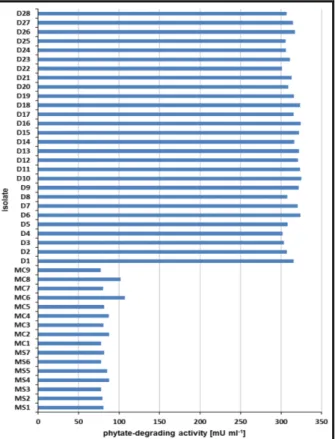
One hundred thirty four single colonies were obtained from the two soil samples taken from Dieng Mountain (D1-D134) and 10 single colonies, each from the soil samples taken from Selo (MS1-MS10) and Cangkringan Yogyakarta (MC1-MC10). The isolates derived from Merapi Mountain exhibited an extracellular phytate-degrading activity between 77.6 and 107.1 mU mL-1 (Fig. 1).
Figure 1 - Extracellular phytate-degrading activity of the positive isolates derived from Merapi Mountain north side (MS), Merapi Mountain south side (MC) and Dieng Mountain (D) after fermentation for 16 hrs at 37°C in liquid LB supplemented with sodium phytate.
Cangkringan Yogyakarta (MC1-MC9) soil sample. The isolates derived from Dieng Mountain (D1-D28), however, showed a significant higher extracellular phytate-degrading activity (300.8 to 323.9 mU mL-1) compared to the isolates from Merapi Mountain.
Identification of phytate-degrading enzyme producing bacteria
Blast analysis of the partial 16S rRNA gene sequences six isolates (MS5, MC6, MC8, D6, D10 and D16) showed high similarities to the reference sequences to the genus Bacillus. Isolates MS5, MC6, D10 and D16 showed 99% sequence identity to B. cereus, while isolate MC8 exhibited 99% sequence identity to B. aryabhatti and D6 99% sequence identity to B. psychrotolerans. Neighbor-joining phylogenetic analysis of the sequence data revealed that the all the six isolates were located in three distinct clusters (Fig. 2). Group 1 consisted of the closely related isolates MS5 and MC6 as well as B. cereus, whereas group 2 comprised the isolate D6 and MC8 as well as B.
aryabhattai. Group 3 consisted of the closely
related isolates D10 and D16 as well as two uncultured bacteria clones.
Figure 2 - ClustalW phylogenetic tree based on 16S rRNA multiple sequence alignment of phytase-producing bacteria.
Properties of the phytate-degrading enzymes
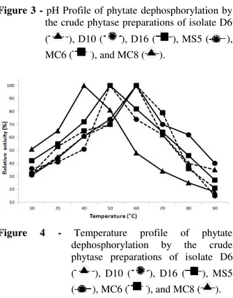
Optimal conditions for phytate dephosphorylation were determined using the crude extracellular phytase preparations from the isolates. The pH optimum of the phytase preparation of isolate D10 was 4.0, which was 5.0 for the isolates MC6 and MS5 and 6.0 for the isolates D6, D16 and MC8 (Fig. 3). For all the isolates, the pH profile for
phytate dephosphorylation was broad, pointing to the presence for more than one phytate-degrading enzyme in the crude enzyme preparations. Isolate MC 8 showed temperature optima at 40°C for its phytase preparation, which was 50°C for the phytase preparation from the isolates D10 and D16 and 60°C for the isolates D6, MS5 and MC6 (Fig. 4).
Figure 3 - pH Profile of phytate dephosphorylation by the crude phytase preparations of isolate D6
( ), D10 ( ), D16 ( ), MS5 ( ), MC6 ( ), and MC8 ( ).
Figure 4 - Temperature profile of phytate dephosphorylation by the crude phytase preparations of isolate D6
( ), D10 ( ), D16 ( ), MS5 ( ), MC6 ( ), and MC8 ( ).
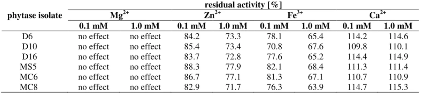
mM Zn2+. A concentration of 0.1 mM Fe3+ resulted in a decrease in phytase activity of 18-24%, whereas a 32-36% reduction was observed in the
presence of 1 mM Fe3+. In the presence of 0.1 mM and 1.0 mM Ca2+ an increase in phytase activity of 10-15% was determined (Table 1).
Table 1 - Effects of potential inhibitors on phytase activity.
phytase isolate
residual activity [%]
Mg2+ Zn2+ Fe3+ Ca2+
0.1 mM 1.0 mM 0.1 mM 1.0 mM 0.1 mM 1.0 mM 0.1 mM 1.0 mM
D6 no effect no effect 84.2 73.3 78.1 65.4 114.2 114.6 D10 no effect no effect 85.4 73.4 70.8 67.6 109.8 110.1 D16 no effect no effect 83.7 72.8 77.6 65.2 114.4 114.9 MS5 no effect no effect 88.3 77.9 82.1 68.4 111.3 111.4 MC6 no effect no effect 86.7 77.1 81.3 67.1 110.7 110.9 MC8 no effect no effect 82.9 71.7 76.3 63.9 114.7 115.3
The data are mean values of three independent experiments.
DISCUSSION
Bacillus sp. has been identified as the predominant
producer of extracellular phytate-degrading activity in the soil samples of different origin (Kerovuo et al. 1998; Kim et al. 1998; Choi et al. 2001; Park 2001; Joseph and Raj 2007; Gulati et al. 2007; Anis Sobirin et al. 2009; Shamna et al. 2012; El-Toukhy et al. 2013; Kumar et al. 2013; Singh et al. 2013; Ushasri et al. 2013; Demirkan et al. 2014; Jain and Chauhan 2014). To our knowledge, B. aryabhattai, however, has been found for the first time as a phytase producer. The ability of the different Bacillus strains investigated to produce extracellular phytate-degrading activity was dependent on the strain itself as well as the culture conditions used. The results obtained in this study (77.6 - 323.9 mU mL-1) were in good agreement with those already reported for Bacillus
sp. KHU-10 (60 mU mL-1) (Choi 2001), B.
laevolacticus (158 - 283 mU mL-1) (Gulati et al
2007) and B. subtilis DR6 (129 - 378 mU mL-1) (Singh et al. 2013). Other studies reported significantly higher extracellular phytase activities such as 720 U mL-1 (B. subtilis MJA) (El-Toukhy et al. 2013), 20 - 150 U mL-1 (B. subtilis) (Powar and Jagannathan 1982), 55 - 88 U mL-1 (B.
subtilis) (Shamna et al. 2012), and 39 - 348 U mL-1
(B. cereus and B. subtilis) (Anis Sobirin et al.
2009). The differences in phytase production might be explained by the difference in the phytase expression levels among the Bacillus
strains studied and in the application of optimised culture conditions in respect to phytase production.
Histidine acid phytases, β--propeller phytases and
cysteine phytases have been identified in bacteria (Greiner and Konietzny 2006). The majority of the
so far known phytases belong to the subfamily of histidine acid phosphatases and do not need any co-factor for optimal activity. Currently, all the phytases used for the animal feed application belong to the class of histidine acid phytases. Cysteine phytases have been reported from the anaerobic ruminal bacteria (Chu et al. 2004) and these enzymes also do not need any co-factor for enzymatic activity. The amino acid sequences of ß-propeller phytases exhibit no homology to the sequences of any other known phosphatase (Kerovuo et al. 1998; Kim et al. 1998; Ha et al. 2000). Initially, ß-propeller phytases were reported from Bacillus species (Kerovuo et al. 1998; Kim et al. 1998; Choi et al. 2001; Tye et al. 2002), but protein sequence identity suggested that ß-propeller phytases were widespread in the aquatic environment (Cheng and Lim 2006). In contrast to the other two phytase subfamilies, ß-propeller phytases need calcium ions for optimal stability and enzymatic activity (Powar and Jagannathan 1982; Shimizu 1992; Kerovuo et al. 1998; Kim et al. 1998; Choi et al. 2001; Oh et al. 2001; Park 2001; Shin et al. 2001; Gulati et al. 2007; Anis Sobirin et al. 2009; El-Toukhy et al. 2013; Kumar et al. 2013; Yu and Chen 2013; Jain and Chauhan 2014). Maximum phytase activity of ß-propeller phytases occurs in the pH range of 6.5 to 8.0 (Choi et al. 2001; Gulati et al. 2007; Jain and Chauhan 2014; Kerovuo et al. 1998; Kim et al. 1998; Oh et al. 2001; Park 2001; Powar and Jagannathan 1982; Shimizu 1992; Yu and Chen 2013). All the six
Bacillus strain included in this study exhibited a
more than one phytate-degrading enzyme in the
Bacillus strains might explain this observation.
This conclusion was further confirmed by the relatively high temperature optimum at pH 5.0 in the absence of Ca2+. Optimal temperature for phytate dephosphorylationy was at 40, 50 and 60°C. In the absence of calcium ions in the assay buffer, temperature optima around 40°C were reported for Bacillus phytases (Powar and Jagannathan 1982; Choi et al. 2001; Park 2001; El Thouky et al. 2013), whereas higher temperature optima (60 - 70°C) were found in the presence of 1 - 5 mM Ca2+ (Kerovuo et al. 1998; Kim et al. 1998; Park 2001; Choi et al. 2001, Gulati et al. 2007; Jain and Chauhan 2014). The Bacillus
phytases investigated in this study exhibited an increased phytase activity in the presence of Ca2+ and decreased phytase activity in the presence of Zn2+ and Fe2+. Mg2+ did not show any effect on enzymatic activity. This was in accordance with already reported data on phytases (Konietzny and Greiner 2002).
REFERENCES
Abelson PH. A potential phosphate crisis. Science
1999; 283: 2015.
Anis Shobirin MH, Farouk A, Greiner R. Potential phytate-degrading enzyme producing bacteria isolated from Malaysian maize plantation. Afr J Biotechnol. 2009; 8: 3540-3546.
Ashle, K, Cordell D, Mavinic D. A brief history of
phosphorus: From the philosopher’s stone to nutrient
recovery and reuse. Chemosphere 2011; 84: 737-746. Cheng C, Lim BL. Beta-propeller phytases in the
aquatic environment. Arch Microbiol. 2006; 185: 1-13.
Choi YM, Suh HJ, Kim JM. (2001) Purification and properties of extracellular phytase from Bacillus sp. KHU-10. J Prot Chem. 2001; 20: 287-292.
Chu HM, Guo RT, Lin TW, Chou CW, Shr HL, Lai HL, et al. Structures of Selenomonas ruminantium
phytase in complex with persulfated phytate: DSP phytase fold and mechanism for sequential substrate hydrolysis. Structure 2004; 12: 2015-2024.
Damiani G, Amedeo P, Bandi C, Fani R, Bellizi D, Sgamarella V. Bacteria identification by PCR-based techniques. In: Adolph KW. Microbial genome methods. Florida: CRC Press, 1996, 167p.
Demirkan E, Baygın E, Ustaet A. Screening of phytate
hydrolysis Bacillus sp. isolated from soil and optimization of the certain nutritional and physical parameters on the production of phytase. Turk J Biochem. 2014; 39: 206-214.
El-Toukhy NMK, Youssef AS, Mikhail MGM. Isolation, purification and characterization of phytase from Bacillus subtilis MJA. Afr J Biotechnol. 2013; 12: 2957-2967.
Greiner R, Konietzny U. Phytase for food application.
Food Technol Biotechnol. 2006; 44: 125-140. Gulati HK, Chadha BS, Saini HS. Production and
characterization of thermostable alkaline phytase from Bacillus laevolacticus isolated from rhizosphere soil. J Ind Microbiol Biotechnol. 2007; 34: 91-98. Ha NC, Oh BC, Shin S, Kim HJ, Oh TK, Kim YO, et
al. Crystal structures of a novel, thermostable phytase in partially and fully calcium-loaded state. Nat Struc Biol. 2000; 7: 147-153.
Heinonen JK, Lahti RJ. A new and convenient colorimetric determination of inorganic orthophosphate and its application to the assay of inorganic pyrophosphatase. Anal Biochem. 1981; 113: 313-317.
Iqbal TH, Lewis KO, Cooper BT. Phytase activity in the human and rat small intestine. Gut 1994; 35: 1233-1236.
Jain U, Chauhan N. Bacillus cereus 10072 Phytase - Detection, purification, characterization and physiological role. Int J Sci Res Develop. 2014; 2: 14-19.
Jorquera MA, Hernandes MT, Rengel Z, Marschner P, and Mora MDL. Isolation of culturable phosphobacteria with both phytase-mineralization and phosphate-solubilization activity from rhizosphere of plant grown in a volcanic soil. Biol Fertil Soils. 2008; 44: 1025-1034.
Joseph I, Raj RP. (2007) Isolation and characterization of phytase producing Bacillus strains from mangrove ecosystem. J Mar Biol Assoc India. 2007; 49: 177-182.
Kembhavi A. Biotechnology application for the Indian animal food industry: Prospect for Growth. [Internet].
2005 [cited 2015 Mar 19]
http://www.fao.org/docrep/ARTICLE/AGRIPPA/660 _en00.htm#TopOfPage.
Kerovuo J, Lauraeus M, Nurminen P, Kalkkinen N, and Apajalahti J. (1998) Isolation, characterization, molecular gene cloning, and sequencing of a novel phytase from Bacillus subtilis. App Environ Microbiol: 1998, 64: 2079-2085.
Kim YO, Kim HK, Bae KS, Yu JH; Oh TK. Purification and properties of a thermostable phytase from Bacillus sp. DS11. Enzyme Microbial Technol.
1998; 22: 2-7.
Konietzny U, Greiner R. Molecular and catalytic properties of phytate-degrading enzymes (phytases).
Int J Food Sci Technol. 2002, 37: 791-812.
Mitchell DB, Vogel K, Weimann BJ, Pasamontes L,. van Loon APGM. The phytase subfamily of histidine acid phosphatases: Isolation of genes for two novel phytases from the fungi Aspergillus terreus and
Myceliophthora thermophile. Microbiol. 1997; 143: 245-252.
Oh BC, Kim MH, Yun BS, Choi WC, Park SC, Bae SC, et al. Ca2+-inositol phosphate chelation mediates the
substrate specificity of β-propeller phytase.
Biochemistry 2006; 45: 9531-9539.
Park YJ. (2001) Expression, characterization, and antifungal activity of phytase from Bacillus subtilis
TS16-111 [PhD Thesis]. Seoul, South Korea: Seoul National University.
Powar VK, Jagannathan V. Purification and properties of phytate-specific phosphatase from Bacillus subtilis. J Bacteriol. 1982; 151: 1102-1108.
Shamna KS, Rajamanikandan KCP, Mukesh Kumar DJ, Balakumaran MD, Kalaichelvan PT. Extracellular production of phytases by a native Bacillus subtilis
strain. Ann Biol Res. 2012; 3: 979-987.
Shimizu M Purification and characterization of a phytase from Bacillus subtilis (natto) N-77. Biosci Biotech Biochem. 1992, 56: 1266-1269.
Shin S, Ha NC, Oh BC, Oh TK, Oh BH. Enzyme mechanism and catalytic property of beta propeller phytase. Structure 2001; 9: 851-858.
Shivange AV, Serwe A, Dennig A, Roccatano D, Haefner S, Schwaneberg U. Directed evolution of a highly active Yersinia mollaretii phytase. App Microbiol Biotechnol. 2012; 95: 405-418.
Singh NK, Joshi DK, Gupta RK. Isolation of phytase producing bacteria and optimization of phytase production parameters. J J Microbiol. 2013, 6: e6419. Tye AJ, Siu FK, Leung TY, Lim BL. Molecular cloning and the biochemical characterization of two novel phytases from B. subtilis 168 and B. licheniformis.
App Microbiol Biotechnol. 2002, 59, 190-197. Ushasri K, Sivaragini P, Vijayalakshmi K. Isolation,
characterization of phytase producing Bacillus sps NBtRS6 from the rhizospere soil of NBt cotton field.
International. J Cur Microbiol Appl Sci. 2013, 2: 142-149.
Weaver CM, Kannan S. Phytate and mineral bioavailability. In: Reddy NR, Sathe SK. Food Phytates. London: CRC Press, 2001, 211p.
Yu P, Chen Y. Purification and characterization of a novel neutral and heat-tolerant phytase from a newly isolated strain Bacillus nealsonii ZJ0702. BMC Biotechnol. 2013; 13: 78-85.