Case Report
Key words
Exercise; physical fitness; hypertension, pulmonary; exercise test.
The 6-minute walk test (T6) is used to assess cardiopulmonary diseases due to its prognosis capacity, easy performance and reproducibility. Pulmonary hypertension (PH) is defined and classified as a hemodynamic consequence leading to increased pulmonary arterial pressure and may result in right ventricular failure and consequent death. This case reports the prescription of T6 by the cardiology team at a university hospital as a means of physical control of a patient with HP, after the introduction and initiation of therapy.
Use of 6-minute Walk Test in Pulmonary Hypertension Management
Elize Fumagalli, Maria Ângela de Oliveira Ribeiro, Mariana Simões Ferreira, Camila Isabel da Silva Santos
Universidade Estadual de Campinas - UNICAMP, Campinas, SP - Brazil
Mailing address: Mariana Simões Ferreira •
Rua Tessália Vieira Camargo 126 - Barão Geraldo - 13084-970 - Campinas, SP - Brazil
E-mail: [email protected], [email protected]
Manuscript received July 27, 2009; revised manuscript received September 16, 2009, accepted October 14, 2009.
viruses, drugs, hypoxia, autoimmune disorders and genetic predisposition4. Its symptoms are: dyspnea (60%), angina, right ventricular hyperfunction, distention of large pulmonary arteries (50%), syncope (8%), cough, hemoptysis, hoarseness, Raymound phenomenon (10%)4.
Methods
Case report
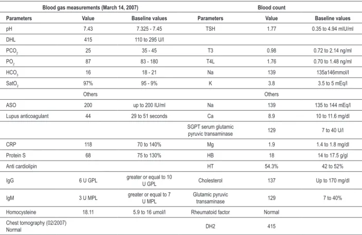
B.M., 13 years old, white male, born on October 20, 1994 uneventful pre-and post-pregnancy, was delivered by cesarean section at 8 months with 2,500 kg. In September 2006, the patient from the city of Jundiaí, state of São Paulo, only child of young non-consanguineous parents and no family history, started to present episodes of morning nausea and constant cough. Within two months, the patient presented lower limb edema and dyspnea on moderate exertion, and required admission. Congestive heart failure and PH were diagnosed. Pu l m o n a r y a n g i o g r a p h y r e v e a l e d p u l m o n a r y thromboembolism and the pressure of 73 mmHg corroborated PH (normality parameter smaller than 25 mmHg). In March 2007, the patient attended the cardiology outpatient clinic of a university hospital for investigation. As the patient reported dyspnea, bed rest was prescribed and tests were performed to confirm the diagnosis (Table 1).
From the results of table 1, other complementary exams were requested: serology for cytomegalovirus, hepatitis B and C, antigliadin antibody and HIV, all of which were found negative.
Pulmonary perfusion scintigraphy performed on July 11, 2007, with chest perfusion images after 5 minutes of intravenous injection of macroaggregated albumin (99mTc) showed homogeneous distribution of radiotracer in the lungs, as well as images of inhalation, with even distribution of radioaerosol. There was no evidence of acute pulmonary thromboembolism. Doppler echocardiogram revealed systolic pulmonary artery pressure of 95 mmHg, revealing PH. The presence of trunk dilatation and pulmonary artery branches, echocardiography revealed interatrial septum with no defect, but with moderate tricuspid insufficiency, dilatation of right chamber and discreet pericardial effusion. PsPa revealed paradoxical dilatation of the right ventricle (95 mmHg), finding moderate tricuspid insufficiency.
In this period, the child was asymptomatic and physical activity remained suspended. Physical exam revealed heart rate (HR) of 88 bpm, pulmonary auscultation with vesicular murmur and no adventitious sounds and lower limbs without edema. For further investigation, ventilation/perfusion
Introduction
The 6-minute walk test (T6) is a simple, easy, safe and unexpensive test, but there are few reports and discussions on the standardization of the technique and factors that interfere with its effectiveness. It consists of walking for 6 minutes along a 30-meter corridor (m) to evaluate patient’s tolerance to physical exertion. Its performance is associated with the Borg scale (Perceived Exertion Scale) before and at the 1st, 6th, 9th and 12th minutes after the test to check patient’ subjective fatigue. Vital signs are also measured (blood pressure, heart frequency, respiratory rate and oxygen saturation)1.T
6 has been used in the evaluation of heart failure (HF) in order to define cardiac involvement intensity, prognosis and drug effectiveness5. It is a simple method for assessing the physical capacity of individuals with pulmonary and cardiac impairments2.
Pulmonary hypertension (PH) is defined and classified as a hemodynamic consequence leading to increased pulmonary arterial pressure and may result in right ventricular failure and eventual death1-3. It is characterized by mean pulmonary artery pressure exceeding 25 mmHg at rest and 30 mmHg during exercise. This pressure is generated by the increase of pulmonary vascular resistance, leading to abnormal function of the right ventricle4. Primary PH may occur due to endothelial aggression caused by pulmonary shear stress,
Case Report
Fumagalli et al Use of T6 in PH management
Arq Bras Cardiol 2010; 95(1) : e10-e13
Table 1 - Laboratory tests
Blood gas measurements (March 14, 2007) Blood count
Parameters Value Baseline values Parameters Value Baseline values
pH 7.43 7.325 - 7.45 TSH 1.77 0.35 to 4.94 mIU/ml
DHL 415 110 to 295 U/l
PCO2 25 35 - 45 T3 0.98 0.72 to 2.14 ng/ml
PO2 87 83 - 180 T4L 1.76 0.70 to 1.48 ng/ml
HCO3 16 18 - 21 Na 139 135a146mmol/l
SatO2 97% 95 - 9% K 3.8 3.5 to 5 mEq/l
Others Others
ASO 200 up to 200 IU/ml Na 139 135 to 144 mEq/l
Lupus anticoagulant 44 29 to 51 seconds Ca 8.9 10 to 11.6 mg/dl
SGPT serum glutamic
pyruvic transaminase 129 7 to 40 U/l
CRP 118 70 to 140% Mg 1.9 1.4 to 1.8 mg/dl
Protein S 68 75 to 130% HB 18 14 to 17.5 g/gl
Anti cardiolipin HT 54.3% 42 to 52%
IgG 6 U GPL greater or equal to 10
U GPL Cholesterol 137 Up to 170 mg/dl
IgM 3 U MPL greater or equal to 7
U MPL
Glutamic pyruvic
transaminase 129 7 to 40%
Homocysteine 18.11 5.9 to 16 umol/l Rheumatoid factor Normal
Chest tomography (02/2007)
Normal DH2 415
mmHg - millimeters of mercury; % - percentage; pH - hydrogen potential; PCO2 - partial pressure of carbon dioxide; PO2 - partial pressure of oxygen; HCO3 - bicarbonate;
SatO2 - oxygen saturation; TSH - thyroid-stimulating hormone; T3 - triiodothyronine; FT4 - free thyroxine; Na - sodium, K - potassium; ASO - antistreptolysin O; Ca - calcium;
GPT - glutamic pyruvic transaminase, Mg - magnesium; HB - hemoglobin; HT - red cells, IgM - immunoglobulin M, IgG - immunoglobulin G; LDH - lactate dehydrogenase.
scintigraphy, myocardial perfusion scintigraphy, (GATED) and T6 were requested.
GATED of May 2007, through angiocardiography radionuclide, revealed decreased clearance of the right ventricle with an ejection fraction of 28% (normal greater than 45%). Synchronized scintigraphy of cardiac chambers revealed increased volume and decreased motility, as well as normal overall left ventricular function and decreased overall right ventricular function at moderate/high level. In view of these results, Sildenafil 25 mg, Furosemide 20 mg and Spironolactone 12.5 mg were prescribed.
A physiotherapy team performed T6 in order to evaluate the improvement of physical abilities before and after the introduction of the drug (Sildenafil 25 mg, Furosemide 20 mg, Spironolactone 25 mg, acetylsalicylic acid 100 mg/day).
Results
T6 was done on June 13, 2007 (Table 2). During the examination, the patient was pale with cold extremities in the 3rd minute of exertion, running 127.5 m at the end of the test, with return of clinical parameters to baseline within 10 minutes. Along with the test, the patient scored the Borg scale in order to check the perceived exertion.
After two months (August 24, 2007), the T6 was repeated and the patient was able to walk faster, running a greater distance (230 m). However, it ended the test reporting tiredness, lump in the throat sensation, cold extremities and pallor. This test was completed in the 3rd minute at patient’s request (table 2). The physiotherapy team requested spirometry test due the possibility of restrictive lung disease associated with the cardiac condition, which was performed in the Pulmonary Physiology Laboratory on June 27, 2007. The examination revealed no changes.
Discussion
This case report depicts a prescription of T6 by the cardiology team of a university hospital as a form of functional control of a patient with primary PH, post-introduction and initiation of therapy.
Since this is an easily executable and reproducible test, therapist’ mastery of the technique, integrating a multidisciplinary team, may help managing patients with cardiopulmonary diseases, as in this case. This collaboration may be both in the investigation period and in the control of different treatments prescribed.
With the T6, the distance run by patients with HF is
Case Report
Fumagalli et al
Use of T6 in PH management
Arq Bras Cardiol 2010; 95(1) : e10-e13
considered a powerful factor of mortality and admission rate. The baseline for a good prognosis involves a distance greater than 450 m, with a worse prognosis between 150 and 300 m1.
Rubim et al5 also aimed to evaluate the usefulness of T 6 as a prognostic indicator and its contribution to clinical practice of patients with HF. After evaluating men and women with aged 58.32, this stuudy revealed a strong correlation between the distance run and the prognosis for mortality in this disease.
Besides heart diseases, studies with individuals with chronic obstructive pulmonary disease (COPD), also noted the importance of the test. Rodrigues and Viegas6 investigated patients with COPD between 48 and 80 (average 66 years). These patients underwent evaluation of spirometric variables, blood gas, respiratory pressures and T6. Test performance was statistically significant related to the variables of forced expiratory volume in the first second, partial pressure of oxygen, peripheral oxygen saturation and maximum expiratory pressure. The authors concluded that T6 can be used as an alternative for the physical assessment of patients with COPD.
In cystic fibrosis, Coelho7 compared the modified version of T6, known as the Shuttle Walk Test in children with the disease and healthy individuals (7 to 15 years), and evaluated its reproducibility in the two cohorts. The heart overload
Table 2 - Walk test values
Date Jun 13, 2007 Aug 24, 2007 Jun 13, 2007 Aug 24, 2007 Jun 13, 2007 Aug 24, 2007 Jun 13, 2007 Aug 24, 2007
Time 1st min 6th min 9th min 12th min
BP (mmHg) 90/60 100/70 110/60 110/70 90/60 100/70 90/60
-HR (bpm) 130 103 117 107 100 110 100
-FR (rpm) 18 17 28 26 19 26 20
-SatO2 (%) 99 97 96 97 97 96 97
-Borg scale 6/0 6/0 12/3 6/0 6/0 6/0 6/0
-Distance run - - 127,50 m 230 m - - -
-imposed by the test did not differ between the groups, which emphasized the importance of T6 as a complement in the management of cardio-respiratory diseases.
Conclusion
Considering that there is a high correlation between respiratory functional tests and T6 in patients with COPD6, cystic fibrosis and other diseases, since it is a simple, objective and reliable method for prognostic assessment of HF5, this case report suggests its application in PH management.
Potential Conflict of Interest
No potential conflict of interest relevant to this article was reported.
Sources of Funding
There were no external funding sources for this study.
Study Association
This study is not associated with any post-graduation program.
References
1. Guimarães Filho FV, Carrasco HVCJ. Hipertensão pulmonar. In: Nobre F, Serrano Jr CV. Tratado de cardiologia SOCESP. Barueri: Manole; 2005. p. 1114-31.
2. Oliveira Jr MT, Guimarães GV, Barretto ACP. Teste de 6 minutos em insuficiência cardíaca. Arq Bras Cardiol. 1996; 67 (6): 373-4.
3. Souza R, Cardoso AP, Pedra CA, Jardim C, Watge D, Campos FT, et al. Diretrizes brasileiras para o manejo da hipertensão pulmonar. J Bras Pneumol. 2005; 31 (2): S1-S31.
4. Arroliga AC. Doença vascular pulmonar. In: Stoller KJ, Wilkins LR, Scanlan LC. Fundamentos da terapia respiratória de Egan. São Paulo: Manole; 2000. p. 519-23.
5. Rubim VSM, Drumond Neto C, Romeo JLM, Montera MW. Valor prognóstico do teste de caminhada de seis minutos na insuficiência cardíaca. Arq Bras Cardiol. 2006; 86 (2): 120-5.
6. Rodrigues SL, Viegas CAA. Estudo de correlação entre provas funcionais respiratórias e o teste de caminhada de seis minutos em pacientes portadores de doença pulmonar obstrutiva crônica. J Pneumol. 2002; 28 (6): 324-8.
7. Coelho CC, Aquino ES, Almeida DC, Oliveira GC, Pinto RC, Rezende IMO, et al. Análise comparativa e reprodutibilidade do teste de caminhada com carga progressiva (modificado) em crianças normais e em portadoras de fibrose cística. J Pneumol. 2007; 33 (2): 168-74.