Capacity Occur during Chronic
Mycobacterium
tuberculosis
Infection
Joshua C. Cyktor1,3, Bridget Carruthers3, Gillian L. Beamer2,3¤, Joanne Turner1,3*
1Department of Microbial Infection and Immunity, The Ohio State University, Columbus, Ohio, United States of America,2Department of Veterinary Biosciences, The Ohio State University, Columbus, Ohio, United States of America,3Center for Microbial Interface Biology, The Ohio State University, Columbus, Ohio, United States of America
Abstract
The exact role of CD8+T cells duringMycobacterium tuberculosis(Mtb) infection has been heavily debated, yet it is generally accepted that CD8+T cells contribute to protection againstMtb. In this study, however, we show that theMtb-susceptible CBA/J mouse strain accumulates large numbers of CD8+T cells in the lung as infection progresses, and that these cells display a dysfunctional and immunosuppressive phenotype (PD-1+, Tim-3+, CD122+). CD8+T cell expansions from the lungs ofMtb-infected CBA/J mice were also capable of secreting the immunosuppressive cytokine interleukin-10 (IL-10), although in vivoCD8+T cell depletion did not significantly alterMtbburden. Further analysis revealed that pulmonary CD8+T cells fromMtb-infected CBA/J mice were clonally expanded, preferentially expressing T cell receptor (TcR) Vbchain 8 (8.2, 8.3) or Vb14. Although Vb8+CD8+T cells were responsible for the majority of IL-10 production,in vivodepletion of Vb8+did not significantly change the outcome ofMtbinfection, which we hypothesize was a consequence of their dual IL-10/IFN-c
secreting profiles. Our data demonstrate that IL-10-secreting CD8+T cells can arise during chronic
Mtbinfection, although the significance of this T cell population in tuberculosis pathogenesis remains unclear.
Citation:Cyktor JC, Carruthers B, Beamer GL, Turner J (2013) Clonal Expansions of CD8+
T Cells with IL-10 Secreting Capacity Occur during Chronic
Mycobacterium tuberculosisInfection. PLoS ONE 8(3): e58612. doi:10.1371/journal.pone.0058612
Editor:Francesco Dieli, University of Palermo, Italy
ReceivedDecember 20, 2012;AcceptedFebruary 5, 2013;PublishedMarch 5, 2013
Copyright:ß2013 Cyktor et al. This is an open-access article distributed under the terms of the Creative Commons Attribution License, which permits unrestricted use, distribution, and reproduction in any medium, provided the original author and source are credited.
Funding:Support was provided by the National Institutes of Health (NIH) R01 (AI-064522; JT). The funders had no role in study design, data collection and analysis, decision to publish, or preparation of the manuscript.
Competing Interests:The authors have declared that no competing interests exist.
* E-mail: [email protected]
¤ Current address: Department of Biomedical Sciences, Tufts University, North Grafton, Massachusetts, United States of America
Introduction
The factors that are responsible for the reactivation of latent Mtb infection are not well understood, but likely involve contributions from both the host and the pathogen. To appreciate the role that the host immune system plays inMtbreactivation, we used relatively resistant (C57BL/6) or susceptible (CBA/J) mice, whose susceptibility phenotype is most apparent during late stages of infection, to represent differences in the natural progression of TB between different human populations. CBA/J mice have low numbers of antigen-specific CD4 T cells that produce relatively small amounts of IFN-c [1–4]. CBA/J mice also have elevated amounts of IL-10 duringMtbinfection [5,6], contributing to their increased susceptibility to infection. However, the importance of CD8+
T cells during Mtbinfection in this mouse strain remains unclear.
CD8+ T cells are an important component of the protective immune response toMtb, as defined by studies showing that mice deficient in CD8+T cells had impaired control ofMtbinfection [7– 10]. Although there is no consensus on the specific requirement for CD8+T cells duringMtbinfection, CD8+T cells can contribute to Mtbcontrol by secretion of IFN-c[11,12] and cytotoxic lysis of host cells [13,14], yet their ability to maintain maximal effector function is dependent on CD4+T cells [15–17]. Studies have also
reported that CD8+
T cells are most important during latentMtb infection in mice, and that CD8+
T cell depletion early after infection had little effect on disease outcome [18]. Conversely, other studies suggest that CD8+
T cells are dispensable duringMtb infection [19–21].
In chronic viral infection models, CD8+
T cells can become dysfunctional after chronic antigenic stimulation, characterized by a lack of functional or proliferative capability, secretion of IL-10 [22–24] and surface expression of inhibitory molecules, such as programmed cell death-1 (PD-1) and T cell immunoglobulin and mucin protein-3 (Tim-3) [25,26]. PD-1 has classically been used as a marker of T cell exhaustion in viral infection and in cancer [27– 30], while other studies have found that cells expressing Tim-3 are dysfunctional and lack regulation [31,32], and that coexpression of PD-1 and Tim-3 leads to extensive dysfunction of CD8+
T cells [33]. Furthermore, CD8+
supporting an investigation into the IL-10-producing properties of CD8+
T cells duringMtbinfection in CBA/J mice.
In this study we show that Mtb-susceptible CBA/J mice accumulated large numbers of CD8+
T cells in their lungs as Mtbinfection progressed that could not be fully accounted for by an expansion of IFN-c-producing CD8+ T cells. CD8+ T cell expansions expressed the inhibitory molecules PD-1, Tim-3, and/ or CD122, and were capable of secreting IL-10. CD8+
T cells from CBA/J mice also preferentially expressed TcR Vb8 and Vb14, severely limiting the diversity of the CD8+T cell repertoire. Although Vb8 CD8+
T cells could secrete IL-10,in vivodepletion of this specific T cell clonal population during chronic infection did not overtly change the Mtb burden in the lungs in the timeframe tested, although the amount of IL-10 in the lung was reduced indicating some biological impact of depletion. Compar-ing mouse strains that are relatively resistant and susceptible to Mtbhas enabled us to uncover a previously unappreciated role for CD8+
T cells in Mtb susceptibility, and links the poor T cell function previously described by us [4,6,36] with increased production of IL-10 in the CBA/J mouse strain.
Materials and Methods
Ethics Statement
This study was carried out in strict accordance with the recommendations in the Guide for the Care and Use of Laboratory Animals of the National Institutes of Health. The protocol was approved by the Institutional Animal Care and Use Committee of The Ohio State University.
Mice
Specific pathogen-free, age/sex-matched CBA/J wild-type (National Cancer Institute, NIH, Frederick, MD), C57BL/6 wild-type (Jackson Laboratories, Bar Harbor, Maine), or CBA/J IL-102/2 mice were maintained in ventilated cages inside a
biosafety level 3 (BSL3) facility and provided with sterile food and waterad libitum. To generate CBA/J IL-102/2mice, CBA/J mice (Jackson laboratories, Bar Harbor, Maine) were crossed with C57BL/6 IL-102/2mice (Jackson) for eight generations. At each
cross progeny mice were ear-punched and DNA was screened for the presence of a neomycin cassette at theil10gene locus. IL-10+/
2 mice were selected for further breeding. At the eighth
generation, heterozygotes were crossed and IL-10-deficient homozygote CBA/J mice were selected. A homozygous breeder colony of CBA/J IL-102/2 mice was maintained thereafter. All
protocols were approved by The Ohio State University’s Institutional Laboratory Animal Care and Use Committee.
MtbInfection and Colony Forming Unit Enumeration MtbErdman (ATCC 35801) was obtained from the American Type Culture Collection (Manassas, VA). Stocks were grown in Proskauer-Beck liquid medium containing 0.05% Tween 80 to mid-log phase and frozen in 1 mL aliquots at280uC. Mice were infected with MtbErdman using an inhalation exposure system (Glas-Col) calibrated to deliver 50–100 CFU to the lungs of each
mouse, as previously described [38]. At specific time points post Mtb infection mice were sacrificed and lungs were aseptically removed into sterile saline. Organs were homogenized and serial dilutions plated onto 7H11 agar supplemented with OADC as previously described [4]. Plates were incubated at 37uC for 21 days in order to enumerate bacterial colonies and calculate the bacterial burden.
Cell Isolation
Mice were euthanized by CO2asphyxiation and lungs perfused
with cold phosphate buffered saline containing 50 Units/mL of heparin through the right ventricle of the heart. Lungs from individual mice were mechanically disrupted using a GentleMACS dissociator (Miltenyi Biotec, Boston, MA) followed by collagenase A (type XI) (0.7 mg/mL, Sigma) and type IV bovine pancreatic DNAse (30mg/mL, Sigma) digestion at 37uC for 30 minutes in GentleMACS C-tubes. Lung cell suspensions were passed through a 70mm nylon cell screen and residual erythrocytes were lysed with Gey’s solution. Viable cells were determined by trypan blue exclusion.
Cell Purification
Single lung cell suspensions were adhered to sterile tissue culture dishes for 1 hr at 37uC. Non-adherent cells were washed and removed from the plates. CD4+
and CD8+
T cells were obtained from the non-adherent cell fraction by magnetic cell separation (BD IMAG anti-CD4+
particles GK1.5, anti-CD8+
particles 53– 6.7) and either placed directly into TRIzol reagent (Invitrogen, Grand Island, NY), homogenized, and frozen at280uC or used for culture as described below. CD8+
Vb8+
T cells were obtained from the CD4negfraction after treatment with anti-CD4 magnetic
beads (BD), then stained with PE anti-Vb8 (eBioscience) and purified using anti-PE magnetic particles (BD). Purity of all CD4+ and CD8+
T cell populations was determined to be greater than 90% for all experiments by flow cytometry using an LSRII flow cytometer (BD Biosciences, San Jose, CA).
Cytokine Assays
For ELISpot, bone marrow-derived dendritic cells (BMDCs) were obtained from the tibiae and femora of age and sex matched non-infected wild-type or IL-102/2 C57BL/6 or CBA/J mice.
Cells were differentiated into dendritic cells using complete DMEM supplemented with 10% conditioned media derived from GM-EL4 cells, a GM-CSF-producing clone kindly provided by Arthur A. Hurwitz (NCI). 26106bone marrow cells were plated at 37uC in 1 ml of GM-EL4 conditioned media in sterile 24-well tissue culture plates. GM-EL4 conditioned media was replaced on days 2, 4 and 6. 36104BMDCs were infected overnight withMtb Erdman at an MOI of 1:1 then fixed in 2% paraformaldehyde (CBA/J IL-102/2BMDCs were used unfixed). Infected BMDCs
were cultured with 26105 CD8+ T cells or CD8negT cells for 72 hr at 37uC in media containing either tissue culture media alone or 10mg/mL anti-CD3 (145-2C11) and 1mg/mL anti-CD28 (37.51). ELISpot reagents were obtained from eBioscience
Figure 1. Accumulation and characterization of CBA/J CD8+T cells.
C57BL/6, CBA/J, and CBA/J IL-102/2 mice were infected with an aerosolized dose ofMtb, and at various times post-infection lungs were removed. (a, b) CBA/J and C57BL/6 lung cell were analyzed by flow cytometry for CD4+
and CD8+
T cells. (c) Ratio of CBA/J CD4+ to CD8+
T cells representative of four independent experiments with 5 mice per group, per timepoint. (d) C57BL/6 and CBA/J lungs were homogenized and plated on 7H11 plates for CFU enumeration. (e, f) 24 hr prior to necropsy mice were injected with BrdU, and lung cells were analyzed for expression of BrdU+CD4+or CD8+T cells. (g, h) Absolute numbers of CD4+or CD8+T cells in wild-type or IL-102/2CBA/J mice as determined by flow cytometry. Results representative of at least three independent experiments with 5 mice per group, per timepoint. * p,0.05, ** p,0.01, *** p,0.001 as obtained by Student’sttest. (b, h)+p
,0.05,++p
,0.01,+++p
,0.001 as obtained by two-way analysis of variance comparing day 90 to day 150 post-infection.
Ready-Set-GO! Spot-forming units (SFU) were enumerated with an ELISpot plate counter (C.T.L.).
For ELISA, 16105 purified pulmonary CD8+
T cells were cultured with 10mg/mL anti-CD3 (145-2C11) and 1mg/mL anti-CD28 (37.51) for 72 hr at 37uC with 5% CO2. After incubation,
plates were frozen at280uC until all timepoints were completed. ELISA antibodies and standards were obtained from BD Biosciences and processed as previously described [38]. Color-metric reactions were read on a SpectraMax plate reader (Molecular Devices, Sunnyvale, CA).
Flow Cytometry
Isolated lung cells or MLN were suspended in deficient RPMI (Mediatech, Manassas, VA) supplemented with 0.1% sodium
azide (Sigma-Aldrich). Surface targets were detected as previously described. Specific antibodies and isotype controls were purchased from BD Biosciences: PerCP-Cy5.5 anti-CD3e (145-2C11), allophycocyanin-Cy7 anti-CD4+
(GK1.5), PE-Cy7 anti-CD8+ (53–6.7), PerCP-Cy5.5 anti-CD8+
(53–6.7), PE-Cy7 anti-IFN-c
(XMG1.2), PE anti-PD-1 (J43), FITC anti-CD122 (TM-Beta 1), and FITC Vbscreening kit. PE anti-Tim-3 (HAVCR2) antibody was purchased from eBiosciences. Cytokine levels were deter-mined according to the manufacturer’s instructions for intracel-lular cytokine staining (Cytofix/Cytoperm fixation/permeabiliza-tion solufixation/permeabiliza-tion kit with BD GolgiStop, BD Biosciences), following a 4 hr incubation with 1mg/mL anti-CD3 (145-2C11) and 0.1mg/ mL anti-CD28 (37.51). Samples were read using an LSRII flow
Figure 2. Surface phenotype of pulmonary T cells in CBA/J and C57BL/6 mice.C57BL/6 and CBA/J mice were infected with an aerosolized dose ofMtband at various timepoints post-infection lungs were removed and processed for flow cytometry. Absolute numbers of IFN-c+CD4+(a) or CD8+
(b) T cells after 4 hrex vivostimulation with anti-CD3/CD28/GolgiSTOP, with representative flow plots at day 150 post-infection. Absolute numbers of CD8+
T cells expressing CD69 (c), Tim3 (d), or PD-1 (e) afterMtbinfection. (f) Absolute number of CD8+
T cells expressing both PD-1 and CD122. Data representative of at least two independent experiments with 4 mice per group per timepoint. * p,0.05, ** p,0.01, *** p,0.001 as obtained by Student’sttest. (c, d)+
p,0.05,++
p,0.01,+++
p,0.001 obtained by two-way analysis of variance comparing only CBA/J mice across all timepoints.
doi:10.1371/journal.pone.0058612.g002
Figure 3. IL-10 production by CD8+T cells from CBA/J mice.C57BL/6 and CBA/J mice were infected with an aerosolized dose ofMtband at various times post-infection lungs were removed and cell populations were purified. Spot-forming units (SFU) representing the absolute number of IL-10+CD8+T cells (a) or CD8negT cells (b) per lung after 72 hr culture with anti-CD3/CD28. (c) Supernatants from (a) were analyzed for IFN-clevels by
ELISA. (d) SFU per lung of CBA/J IL-10+ CD8+
or CD8negT cells cultured withMtb-infected BMDCs for 72 hr. Data representative of two independent experiments with 4 mice per group per timepoint, * p,0.05, ** p,0.01, *** p,0.001 as obtained by Student’sttest.
cytometer and analyzed with FACSDiva software (BD Bioscienc-es).
Cell Depletion Anti-CD8+
(53.6.72) depletion antibody (BioXCell) or whole rat IgG2a (2A3) (BioXCell), were diluted to 2.5 mg/ml in PBS and stored at –80uC. Thawed stocks were maintained at 4uC for up to 1 month. Anti-Vb8 (F23.1) antibody-secreting cell line [39] was kindly provided by Dr. Michael Bevan. Antibody administration was modified based on Silva et al. [40] as follows: at day 90 post-infection, 0.5 mg of anti-CD8+ (53.6.72), anti-Vb8 (F23.1), or control antibody was injected into the peritoneal cavity of each mouse, followed by 0.5 mg at weekly intervals thereafter until the designated experimental time points. At the initiation of antibody treatment CBA/J mice consistently harbor 6.596 SE 0.12 log10
MtbCFU [4].
Statistics
Statistical analysis performed using GraphPad Prism software for the Studentsttest per individual time point of each graph. Any comparisons between timepoints of the same experiment utilize a two-way analysis of variance test with Bonferonni post-tests for multiple comparisons. * p,0.05, ** p,0.01, *** p,0.001.
Results
CD8+ T cells accumulate in the lungs of CBA/J mice as
Mtbinfection progresses
Following aerogenic infection of CBA/J and C57BL/6 mice withMtbwe observed a gradual accumulation of CD4+
T cells in the lungs of C57BL/6 mice and significantly fewer CD4+
T cells within the lungs of CBA/J mice (Fig. 1a), as we have previously described [3,4]. In contrast to the reduced number of CD4+
T cells, CBA/J mice demonstrated a significant late accumulation of CD8+
T cells within the lungs as Mtb infection progressed (Fig. 1b), eventually reaching or surpassing the number of pulmonary CD8+
T cells observed in C57BL/6 mice. This late accumulation was absent from C57BL/6 mice which reached a plateau at day 60. Skewing of T cell subset proportions in CBA/J mice could be better appreciated by determination of CD4:CD8 ratios throughout the course of infection (Fig. 1c), where a significant decline in the ratio of CD4+ to CD8+ T cells was observed by day 90 of Mtb infection, preceding the increasing CFU within the lungs of CBA/J mice (Fig. 1d) [4,6]. BrdU staining of pulmonary CD4+(Fig. 1e) and CD8+T (Fig. 1f) cells from CBA/J mice was not dramatically altered throughout infection, suggesting that the increased numbers of CD8+T cells that were evident in CBA/J mice may not be due to local proliferation, but a consequence of enhanced cellular recruitment. CBA/J mice are known to produce abundant IL-10 in their lungs as Mtb infection progresses [5] and IL-10 can stimulate the proliferation of CD8+
T cells [41]. Therefore, we examined the numbers of CD4+
and CD8+
T cells present in the lungs of CBA/J IL-102/2mice over the course ofMtbinfection (Fig. 1g, h) and did not observe any differences between wild-type and IL-102/2
CBA/J mice in CD8+
T cell accumulations at late stages of infection, demonstrating that CD8+
T cell expansions occur independent of IL-10. A significant increase in CD4+
and CD8+ T cells was observed in CBA/J IL-102/2mice at day 30, reflecting
enhanced TH1 immunity in these mice (unpublished observations).
These data demonstrate that the accumulation of CD8+
T cells in the lungs of CBA/J mice during chronic infection is not mediated by IL-10.
CD8+T cell expansions fromMtb-infected CBA/J mice do
not align with IFN-cproducing capacity
We examined the number of T cells that were capable of secreting IFN-c after ex vivo TcR stimulation. As expected, C57BL/6 mice had increasing numbers of CD4+[4] and CD8+ T cells that could produce IFN-cas infection progressed (Fig. 2a), which paralleled the increasing numbers of T cells within the lung (Fig. 1). CBA/J showed an equivalent increase in IFN-c -producing CD4+
T cells (Fig. 2b), albeit at a significantly lower number compared to C57BL/6, as we have previously described [3,4]. Where the data differed, however, was in the finding that the number of IFN-c-producing CD8+T cells reached a plateau as early as day 30 post-infection and did not increase any further, despite a significant accumulation of CD8+
T cells within the lungs of CBA/J mice. AsMtbinfection progressed we also observed a significant increase in the number of CD8+
T cells from CBA/J mice that could express CD69 (Fig. 2c), with over 40% of all CD8+T cells in the lungs of CBA/J mice being activated during chronic infection. CD8+T cells also expressed the suppressor of TH1 responses Tim-3 (Fig. 2d), PD-1 (Fig. 2e), with a small
population expressing both PD-1 and CD122 (Fig. 2f). Therefore, although highly activated, a proportion of CD8+
T cell expansions from chronicMtbinfected CBA/J mice had a phenotype that was not associated with TH1 cytokine secretion but was instead linked
to immuno-suppressive properties including IL-10 production [34]. Because IL-10 production has been closely linked to Mtb susceptibility of CBA/J mice [5], we next examined the capacity of CD8+
T cells to produce IL-10.
CD8+T cells fromMtb-infected CBA/J mice are capable of
secreting IL-10 CD8+
and CD8neg cells were purified from the lungs of Mtb -infected CBA/J and C57BL/6 mice and cultured in IL-10 ELISpot plates for 72 hours with autologous bone marrow-derived dendritic cells (BMDCs) that had been infected with Mtb for 24 hours, in the presence or absence of anti-CD3 and anti-CD28. CD8+
cells from CBA/J mice, and not C57BL/6 mice, were capable of secreting IL-10 in response to TcR cross-linking (Fig. 3a). In contrast, CD8negcells from both mouse strains were capable of secreting IL-10 under these same conditions (Fig. 3b). IL-10-secreting CD8neg cells were predominantly CD4+
as adherent cells were removed during cell purification and we have failed to detect IL-10 within B cells and neutrophils in both mouse strains (not shown). We also observed that CD8+
T cells from CBA/J and C57BL/6 mice were capable of secreting IFN-cunder the same culture conditions (measured in the IL-10 ELISPOT supernatants) (Fig. 3c). Culture of purified CD8+and CD8neg
cells fromMtbinfected CBA/J mice withMtb-infected BMDCs in the absence of TcR cross-linking, indicative of an Mtb-specific response, resulted in the secretion of IL-10 from both CD8neg and CD8+
cells (Fig. 3d). Interestingly, the capacity of CD8+ T cells to produce IL-10 increased over time, in parallel to the increasing CFU and CD8+T cell numbers in the lung. The low SFU, relative to TcR cross-linking likely reflects the challenges of delivering ofMtbantigen into the appropriate processing pathway for presentation to CD8+
T cells.
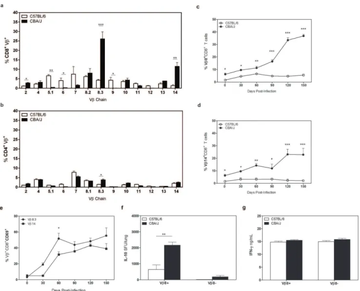
CD8+T cells from CBA/J mice are clonally expanded
We determined the clonal repertoire of the CD8+T cells that accumulated in the lungs of CBA/J mice duringMtbinfection to determine whether the reactivity of CD8+
Mtbinfection, we observed that CD8+
T cells from CBA/J mice primarily expressed two variable regions of the TcR beta chain (Vb). Vb8 (8.2,8.3) and Vb14 were expressed by approximately 45% of all CD8+
T cells in CBA/J mice at day 120 after Mtb infection, compared to a less restrictive repertoire in C57BL/6 mice (Fig. 4a). Vbexpression was comparable on CD4+
T cells from both mouse strains (Fig. 4b), indicating a unique expansion within the CD8+
T cell pool in CBA/J mice as Mtb infection progressed. More detailed examination over the course of Mtb infection showed that the percentage of CD8+T cells expressing Vb8 or Vb14 was always significantly higher throughout Mtb infection in CBA/J mice, and this population significantly expanded further at late time points of Mtbinfection (Fig. 4c,
d). Similar to our findings with CD8+
T cell expansions, we found that both Vb8 and Vb14 CD8+
T cells were highly activated (Fig. 4e).
We determined the IL-10 producing capacity of CD8+ Vb8+
T cells using magnetic bead separation, which purified the dominant
Vb8 expressing CD8+population. CD8+Vb8+and CD8+Vb8neg
T cells were isolated fromMtb-infected CBA/J mice or C57BL/6 mice (controls) at day 150 postMtbinfection and cultured in IL-10 ELISpot plates with autologous IL-10 deficient BMDCs in the presence of anti-CD3/CD28. Significantly more CD8+ Vb8+T cells from Mtb-infected CBA/J mice were capable of producing IL-10 than CD8+Vb8neg
T cells from CBA/J mice or from CD8+ Vb8+and CD8+Vb8neg
T cells from C57BL/6 mice (Fig. 4f). These data indicate a dominant role for CD8+Vb8+expressing T cells in the IL-10 production we previously observed in purified CD8+T cell cultures. Culture supernatants were also assayed for IFN-cby ELISA (Fig. 4g) and our data indicate that CD8+Vb8+ T cells can have dual secretion of IL-10 and IFN-c, or represent a mixed population, as has been described by others [44].
Figure 4. VbTcR expression in CBA/J and C57BL/6 mice.C57BL/6 and CBA/J mice were infected with an aerosol dose ofMtband at various timepoints post-infection lungs were removed and processed for flow cytometry. Percentages of CD8+
(a) or CD4+
(b) T cells expressing specific Vb TcRs at day 120 post-infection. Percentages of CD8+T cells expressing Vb8 (c) or Vb14 (d) TcR over the course ofMtbinfection. (e) Percentages of Vb8+
or Vb14+ CD69+
CD8+
pulmonary T cells over time. (f) Vb8+
and Vb8negT cells from day 120 post-infection were cultured for 72 hr with anti-CD3/
CD28 and IL-10 SFU determined by ELISpot. (g) Supernatants from (a) were analyzed for IFN-clevels by ELISA. Data representative of at least two independent experiments with 4 mice per group per timepoint. * p,0.05, ** p,0.01, *** p,0.001 as obtained by Student’sttest.
In vivodepletion of IL-10 producing CD8+T cells during
chronic Mtbinfection alters pro-inflammatory responses but fails to modify the bacterial load
CD8+
T cells or Vb8+
cells were depleted from wild-type CBA/ J mice from day 90–120 afterMtbinfection, a time when IL-10
and Vb8+ CD8+
T cells were increasing in the lungs of Mtb -infected CBA/J mice [5]. Following CD8+
depletion, MtbCFU (Fig. 5a), total CD4+
T cell numbers (Fig. 5b) and IFN-c -producing CD4+
T cells (Fig. 5c) were all moderately altered but data did not reach statistical significance. Interestingly, CD8+
T cell depletion led to a significant decrease in the total amount of
Figure 5. CD8+T cell depletion in CBA/J mice.CBA/J mice were infected with an aerosolized dose ofMtband from day 90–120 were treated weekly with depletion antibody via intraperitoneal injection then sacrificed at day 125 post-infection. (a) Lungs of anti-CD8+
T cell depleted or control mice were homogenized and plated on 7H11 plates and CFU enumerated after 21 days. Absolute number of total CD4+
T cells (b) or IFN-c+ CD4+
T cells (c) after CD8+
T cell depletion as determined by flow cytometry. (d) Levels of pulmonary IL-10 after CD8+
T cell depletion as determined by ELISA. (e) Lungs of anti-Vb8 depleted or control mice were homogenized and plated onto 7H11 plates and CFU enumerated. Control group = no treatment and isotype control. Results representative of at least two independent experiments with 5–10 mice per group. Depletion resulted in 95% reduction in cell number, as determined by flow cytometry. * p,0.05, ** p,0.01, *** p,0.001 as obtained by Student’sttest.
IL-10 in the lungs of CBA/J mice (Fig. 5d), reflecting the IL-10-secreting capacity of CD8+
T cells we observedin vitro. Depletion of CD4+ T cells led to significantly increased Mtb burden and mortality before day 120 (not shown) showing that depletion of a known protective T cell subset increased susceptibility. Specific depletion of Vb8+cells also failed to significantly impact theMtb burden (Fig. 5e), although a modest reduction in CFU was similarly observed. This modest reduction, similar to our findings with CD8+ T cell depletion, suggests that although IL-10 producing CD8+
T cells may not negatively impact the growth ofMtbduring the timeframe we investigated, they do not provide protection.
Discussion
We have demonstrated that clonal expansions of CD8+ T cells from CBA/J mice accumulate in the lung over the course ofMtb infection. This accumulation did not yield an equivalent increase in IFN-c+
CD8+
T cells and, upon further phenotypic examination, we discovered that highly activated CD8+ T cells from CBA/J mice expressed the T cell dysfunction markers PD-1 and Tim-3, as well as co-expression of PD-1 and CD122 suggesting possible immunosuppressive activity. Afterex vivopurification and culture, it was shown that CD8+T cells from CBA/J mice were capable of secreting IL-10 after TcR stimulation and in response to Mtb infected BMDCs. Depletion of Vb8+
cells or the entire CD8+ T cell population inMtb-infected CBA/J mice led to a significant reduction in pulmonary IL-10 levels, however, we observed no significant change in pulmonary CFU.
Using anMtb-susceptible mouse strain, we reveal that CD8+T cells that are capable of producing IL-10 can accumulate within the lung during Mtb infection. CD8+
T cells within the lung expressed CD69, indicative of activation and functional capacity, yet failed to show a concomitant enhancement of IFN-c-producing capacity. These findings indicate that highly activated CD8+
T cells within the lung have alternate function, which we show here to be the capacity to secrete IL-10, measured by ELISPOT due to the known difficulties of measuring IL-10 by intracellular flow cytometry. Altered function was associated with the co-expression of a variety of receptors know for negative regulation of cell function (PD-1, CD122, Tim-3) [25,28,33,34]. In support of our findings, previous studies have shown that in chronic murineMtb infection PD-1+T cells can proliferate but fail to secrete IFN-c unless this inhibition is overcome by direct TcR stimulation [45,46]. It is unclear at this time why CD8+
T cells with inhibitory properties arise in CBA/J mice asMtbinfection progresses but we can hypothesize that this is a consequence of enhanced immune activation and subsequent exhaustion due to increasing bacterial loads in this mouse strain (Fig. 1) [4,6].
Our failure to observe any significant change in CFU following CD8+
or Vb8+
T cell depletion can be interpreted in several ways, the simplest being that IL-10-producing CD8+
T cells have no biological influence on the control ofMtbinfection. While this is a possibility, we would reason that at the least the presence of IL-10-producing CD8+
T cells can be a putative biomarker of TB disease progression as they are only observed in a mouse strain of Mtb
susceptibility, and IL-10 producing T cells have been previously described in the blood of TB patients [37]. In support of a negative role for CD8+
T cells in CBA/J mice,in vivo depletion led to a moderate, albeit not significant, decrease in CFU. Thus is in contrast to CD4+T cell depletion in which all the mice were either dead or moribund before the necropsy time point (data not shown). These findings indicate that some CD8+
T cells from CBA/J mice fail to contribute to protection in a similar manner than might be expected from studies of other mouse strains [47– 49]. One possibility for our failure to detect significant changes in CFU following CD8+
T cell depletion is our observation that CD8+
T cells can secrete both IL-10 and IFN-c. Depletion of all CD8+
T cells would also remove a protective population, leading to a neutral effect. Previous studies from our laboratory have shown that blocking the action of IL-10 in CBA/J mice duringMtb infection provides enhanced protection [5], and we consider that blocking the action of IL-10 from IL-10/IFN-c-secreting CD8+
T cells contributed to this phenotype.
In addition to IL-10 production, we also observed that CD8+ T cells from CBA/J mice had severely restricted diversity of their TcR repertoire which would significantly limit the breadth of antigens that CBA/J CD8+T cells can recognize. CBA/J mice have an endogenous mouse mammary tumor virus (MTV-6) that selectively deletes various TcR Vbchains such as Vb8.1, and Vb
17a [50,51] however this alone is not sufficient to explain the limited TcR diversity in CBA/J mice. It is possible that Mtb infection leads to specific deletion of certain subsets of protective CD8+
T cells or is driving clonal expansion of less protective cells (Vb8, Vb14). Alternatively Vb8/Vb14 CD8+
T cells in CBA/J mice may respond to a dominantMtbantigen(s) expressedin vivo, where persistent responsiveness leads to immune exhaustion and subsequent regulation through receptors such as PD-1 and Tim-3. Clonal CD8+
expansions have been described in TB patients [52], including pediatric TB [42], supporting the relevance of our findings to TB in man. Clonal CD8+
expansions are also common in the elderly and in many viral models [42,43,53–58], yet their importance inMtbinfection is still unclear.
In summary, we show using an Mtb-susceptible mouse strain (CBA/J) that clonal expansions of CD8+T cells with the capacity to produce IL-10 can arise during chronic Mtb infection. In combination with our previous finding that blocking the action of IL-10 can improve outcome ofMtbinfection [5], these data further support a negative role for IL-10 in the generation and maintenance of protective immunity against Mtbinfection. Our findings are of significance because vaccines that specifically stimulate CD8+
T cells are currently under development [59–63]. Given that IL-10-secreting CD8+
T cells with the potential to negatively impact control of Mtb infection can naturally arise, generating such cells in response to vaccination should be considered.
Author Contributions
Conceived and designed the experiments: JT JC GB. Performed the experiments: JC GB BC JT. Analyzed the data: JC GB. Contributed reagents/materials/analysis tools: JT. Wrote the paper: JC JT.
References
1. Beamer GL, Cyktor J, Flaherty DK, Stromberg PC, Carruthers B, et al. (2012) CBA/J mice generate protective immunity to soluble Ag85 but fail to respond efficiently to Ag85 during natural Mycobacterium tuberculosis infection. Eur J Immunol 42: 870–879.
2. Beamer GL, Cyktor J, Carruthers B, Turner J (2011) H-2 alleles contribute to antigen 85-specific interferon-gamma responses during Mycobacterium tuber-culosis infection. Cell Immunol 271: 53–61.
3. Turner J, Gonzalez-Juarrero M, Saunders BM, Brooks JV, Marietta P, et al. (2001) Immunological basis for reactivation of tuberculosis in mice. Infect Immun 69: 3264–3270.
5. Beamer GL, Flaherty DK, Assogba BD, Stromberg P, Gonzalez-Juarrero M, et al. (2008) Interleukin-10 promotes Mycobacterium tuberculosis disease progres-sion in CBA/J mice. J Immunol 181: 5545–5550.
6. Turner J, Gonzalez-Juarrero M, Ellis DL, Basaraba RJ, Kipnis A, et al. (2002) In vivo IL-10 production reactivates chronic pulmonary tuberculosis in C57BL/6 mice. J Immunol 169: 6343–6351.
7. Flynn JL, Goldstein MM, Triebold KJ, Bloom BR (1993) Major histocompat-ibility complex class I-restricted T cells are necessary for protection against M. tuberculosis in mice. Infect Agents Dis 2: 259–262.
8. Sousa AO, Mazzaccaro RJ, Russell RG, Lee FK, Turner OC, et al. (2000) Relative contributions of distinct MHC class I-dependent cell populations in protection to tuberculosis infection in mice. Proc Natl Acad Sci U S A 97: 4204– 4208.
9. D’Souza CD, Cooper AM, Frank AA, Ehlers S, Turner J, et al. (2000) A novel nonclassic beta2-microglobulin-restricted mechanism influencing early lympho-cyte accumulation and subsequent resistance to tuberculosis in the lung. Am J Respir Cell Mol Biol 23: 188–193.
10. Muller I, Cobbold SP, Waldmann H, Kaufmann SH (1987) Impaired resistance to Mycobacterium tuberculosis infection after selective in vivo depletion of L3T4+and Lyt-2+T cells. Infect Immun 55: 2037–2041.
11. Cho S, Mehra V, Thoma-Uszynski S, Stenger S, Serbina N, et al. (2000) Antimicrobial activity of MHC class I-restricted CD8+ T cells in human tuberculosis. Proc Natl Acad Sci U S A 97: 12210–12215.
12. Shams H, Wizel B, Weis SE, Samten B, Barnes PF (2001) Contribution of CD8(+) T cells to gamma interferon production in human tuberculosis. Infect Immun 69: 3497–3501.
13. Lalvani A, Brookes R, Wilkinson RJ, Malin AS, Pathan AA, et al. (1998) Human cytolytic and interferon gamma-secreting CD8+ T lymphocytes specific for Mycobacterium tuberculosis. Proc Natl Acad Sci U S A 95: 270–275. 14. Lewinsohn DA, Heinzel AS, Gardner JM, Zhu L, Alderson MR, et al. (2003)
Mycobacterium tuberculosis-specific CD8+ T cells preferentially recognize heavily infected cells. Am J Respir Crit Care Med 168: 1346–1352. 15. Bold TD, Ernst JD (2012) CD4+T cell-dependent IFN-gamma production by
CD8+effector T cells in Mycobacterium tuberculosis infection. J Immunol 189: 2530–2536.
16. Shedlock DJ, Shen H (2003) Requirement for CD4 T cell help in generating functional CD8 T cell memory. Science 300: 337–339.
17. Serbina NV, Lazarevic V, Flynn JL (2001) CD4(+) T cells are required for the development of cytotoxic CD8(+) T cells during Mycobacterium tuberculosis infection. J Immunol 167: 6991–7000.
18. van Pinxteren LA, Cassidy JP, Smedegaard BH, Agger EM, Andersen P (2000) Control of latent Mycobacterium tuberculosis infection is dependent on CD8 T cells. Eur J Immunol 30: 3689–3698.
19. Schaible UE, Collins HL, Priem F, Kaufmann SH (2002) Correction of the iron overload defect in beta-2-microglobulin knockout mice by lactoferrin abolishes their increased susceptibility to tuberculosis. J Exp Med 196: 1507–1513. 20. Urdahl KB, Liggitt D, Bevan MJ (2003) CD8+T cells accumulate in the lungs of
Mycobacterium tuberculosis-infected Kb2/2Db2/2 mice, but provide minimal protection. J Immunol 170: 1987–1994.
21. Mogues T, Goodrich ME, Ryan L, LaCourse R, North RJ (2001) The relative importance of T cell subsets in immunity and immunopathology of airborne Mycobacterium tuberculosis infection in mice. J Exp Med 193: 271–280. 22. Brooks DG, Walsh KB, Elsaesser H, Oldstone MB (2010) IL-10 directly
suppresses CD4 but not CD8 T cell effector and memory responses following acute viral infection. Proc Natl Acad Sci U S A 107: 3018–3023.
23. Abel M, Sene D, Pol S, Bourliere M, Poynard T, et al. (2006) Intrahepatic virus-specific IL-10-producing CD8 T cells prevent liver damage during chronic hepatitis C virus infection. Hepatology 44: 1607–1616.
24. Trandem K, Zhao J, Fleming E, Perlman S (2011) Highly activated cytotoxic CD8 T cells express protective IL-10 at the peak of coronavirus-induced encephalitis. J Immunol 186: 3642–3652.
25. Blackburn SD, Shin H, Haining WN, Zou T, Workman CJ, et al. (2009) Coregulation of CD8+T cell exhaustion by multiple inhibitory receptors during chronic viral infection. Nat Immunol 10: 29–37.
26. Baitsch L, Baumgaertner P, Devevre E, Raghav SK, Legat A, et al. (2011) Exhaustion of tumor-specific CD8(+) T cells in metastases from melanoma patients. J Clin Invest 121: 2350–2360.
27. Mumprecht S, Schurch C, Schwaller J, Solenthaler M, Ochsenbein AF (2009) Programmed death 1 signaling on chronic myeloid leukemia-specific T cells results in T-cell exhaustion and disease progression. Blood 114: 1528–1536. 28. Kaufmann DE, Walker BD (2008) Programmed death-1 as a factor in immune
exhaustion and activation in HIV infection. Curr Opin HIV AIDS 3: 362–367. 29. Hofmeyer KA, Jeon H, Zang X (2011) The PD-1/PD-L1 (B7-H1) pathway in chronic infection-induced cytotoxic T lymphocyte exhaustion. J Biomed Biotechnol 2011: 451694.
30. Watanabe T, Bertoletti A, Tanoto TA (2010) PD-1/PD-L1 pathway and T-cell exhaustion in chronic hepatitis virus infection. J Viral Hepat 17: 453–458. 31. Sharma S, Sundararajan A, Suryawanshi A, Kumar N, Veiga-Parga T, et al.
(2011) T cell immunoglobulin and mucin protein-3 (Tim-3)/Galectin-9 interaction regulates influenza A virus-specific humoral and CD8 T-cell responses. Proc Natl Acad Sci U S A 108: 19001–19006.
32. Koguchi K, Anderson DE, Yang L, O’Connor KC, Kuchroo VK, et al. (2006) Dysregulated T cell expression of TIM3 in multiple sclerosis. J Exp Med 203: 1413–1418.
33. Fourcade J, Sun Z, Benallaoua M, Guillaume P, Luescher IF, et al. (2010) Upregulation of Tim-3 and PD-1 expression is associated with tumor antigen-specific CD8+T cell dysfunction in melanoma patients. J Exp Med 207: 2175– 2186.
34. Dai H, Wan N, Zhang S, Moore Y, Wan F, et al. (2010) Cutting edge: programmed death-1 defines CD8+CD122+ T cells as regulatory versus memory T cells. J Immunol 185: 803–807.
35. Junqueira-Kipnis AP, Kipnis A, Henao Tamayo M, Harton M, Gonzalez Juarrero M, et al. (2005) Interleukin-10 production by lung macrophages in CBA xid mutant mice infected with Mycobacterium tuberculosis. Immunology 115: 246–252.
36. Beamer GL, Turner J (2005) Murine models of susceptibility to tuberculosis. Arch Immunol Ther Exp (Warsz) 53: 469–483.
37. Boussiotis VA, Tsai EY, Yunis EJ, Thim S, Delgado JC, et al. (2000) IL-10-producing T cells suppress immune responses in anergic tuberculosis patients. J Clin Invest 105: 1317–1325.
38. Vesosky B, Flaherty DK, Turner J (2006) Th1 cytokines facilitate CD8-T-cell-mediated early resistance to infection with Mycobacterium tuberculosis in old mice. Infect Immun 74: 3314–3324.
39. Staerz UD, Rammensee HG, Benedetto JD, Bevan MJ (1985) Characterization of a murine monoclonal antibody specific for an allotypic determinant on T cell antigen receptor. J Immunol 134: 3994–4000.
40. Silva RA, Pais TF, Appelberg R (2001) Blocking the receptor for IL-10 improves antimycobacterial chemotherapy and vaccination. J Immunol 167: 1535–1541. 41. Rowbottom AW, Lepper MA, Garland RJ, Cox CV, Corley EG (1999)
Interleukin-10-induced CD8 cell proliferation. Immunology 98: 80–89. 42. Jacobsen M, Detjen AK, Mueller H, Gutschmidt A, Leitner S, et al. (2007)
Clonal expansion of CD8+effector T cells in childhood tuberculosis. J Immunol 179: 1331–1339.
43. Du G, Chen CY, Shen Y, Qiu L, Huang D, et al. (2010) TCR repertoire, clonal dominance, and pulmonary trafficking of mycobacterium-specific CD4+and CD8+T effector cells in immunity against tuberculosis. J Immunol 185: 3940– 3947.
44. Chen J, Liu XS (2009) Development and function of IL-10 IFN-gamma-secreting CD4(+) T cells. J Leukoc Biol 86: 1305–1310.
45. Bennett F, Luxenberg D, Ling V, Wang IM, Marquette K, et al. (2003) Program death-1 engagement upon TCR activation has distinct effects on costimulation and cytokine-driven proliferation: attenuation of ICOS, IL-4, and IL-21, but not CD28, IL-7, and IL-15 responses. J Immunol 170: 711–718.
46. Keir ME, Butte MJ, Freeman GJ, Sharpe AH (2008) PD-1 and its ligands in tolerance and immunity. Annu Rev Immunol 26: 677–704.
47. Winau F, Weber S, Sad S, de Diego J, Hoops SL, et al. (2006) Apoptotic vesicles crossprime CD8 T cells and protect against tuberculosis. Immunity 24: 105–117. 48. Tascon RE, Stavropoulos E, Lukacs KV, Colston MJ (1998) Protection against Mycobacterium tuberculosis infection by CD8+T cells requires the production of gamma interferon. Infect Immun 66: 830–834.
49. Sud D, Bigbee C, Flynn JL, Kirschner DE (2006) Contribution of CD8+T cells to control of Mycobacterium tuberculosis infection. J Immunol 176: 4296–4314. 50. Jouvin-Marche E, Cazenave PA, Voegtle D, Marche PN (1992) V beta 17 T-cell deletion by endogenous mammary tumor virus in wild-type-derived mouse strain. Proc Natl Acad Sci U S A 89: 3232–3235.
51. Behlke MA, Chou HS, Huppi K, Loh DY (1986) Murine T-cell receptor mutants with deletions of beta-chain variable region genes. Proc Natl Acad Sci U S A 83: 767–771.
52. Tully G, Kortsik C, Hohn H, Zehbe I, Hitzler WE, et al. (2005) Highly focused T cell responses in latent human pulmonary Mycobacterium tuberculosis infection. J Immunol 174: 2174–2184.
53. Charles ED, Brunetti C, Marukian S, Ritola KD, Talal AH, et al. (2011) Clonal B cells in patients with hepatitis C virus-associated mixed cryoglobulinemia contain an expanded anergic CD21low B-cell subset. Blood 117: 5425–5437. 54. Kang HS, Kim BS (2010) Predominant clonal accumulation of CD8+T cells
with moderate avidity in the central nervous systems of Theiler’s virus-infected C57BL/6 mice. J Virol 84: 2774–2786.
55. Dalakas MC, Rakocevic G, Shatunov A, Goldfarb L, Raju R, et al. (2007) Inclusion body myositis with human immunodeficiency virus infection: four cases with clonal expansion of viral-specific T cells. Ann Neurol 61: 466–475. 56. Aichele P, Unsoeld H, Koschella M, Schweier O, Kalinke U, et al. (2006) CD8
T cells specific for lymphocytic choriomeningitis virus require type I IFN receptor for clonal expansion. J Immunol 176: 4525–4529.
57. Itoh S, Sugawara T, Enomoto S, Ono Y, Numaoka H, et al. (2002) Clonal evolution of blasts in an elderly patient with CD56(+) relapsed acute promyelocytic leukemia. Am J Hematol 69: 59–63.
58. Huang D, Chen CY, Zhang M, Qiu L, Shen Y, et al. (2012) Clonal immune responses of Mycobacterium-specific gammadelta T cells in tuberculous and non-tuberculous tissues during M. tuberculosis infection. PLoS One 7: e30631. 59. Begum D, Umemura M, Yahagi A, Okamoto Y, Hamada S, et al. (2009) Accelerated induction of mycobacterial antigen-specific CD8+T cells in the Mycobacterium tuberculosis-infected lung by subcutaneous vaccination with Mycobacterium bovis bacille Calmette-Guerin. Immunology 128: 556–563. 60. Elvang T, Christensen JP, Billeskov R, Thi Kim Thanh Hoang T, Holst P, et al.
61. Boom WH (2007) New TB vaccines: is there a requirement for CD8 T cells? J Clin Invest 117: 2092–2094.
62. Wang J, Santosuosso M, Ngai P, Zganiacz A, Xing Z (2004) Activation of CD8 T cells by mycobacterial vaccination protects against pulmonary tuberculosis in the absence of CD4 T cells. J Immunol 173: 4590–4597.