ABSTRACT
Aim The aim of the present study was to report two cases with Crohn’s disease in whom dental implants successfully osseointegrated and remained functionally stable up to 13 and 12 months of follow-up, respectively.
Cases presentation in cases 1 (age 35 years) and 2 (age 36 years), tooth 24 and 14, respectively, were atraumatically extracted and a particulated bone grafting material (buccal and palatal aspect of the defect) and a Trabecular Metal implant (11.5 mm length, 4.7 mm diameter) were inserted in each extraction socket. after implant placement and abutment connection with the inal torque (25 Ncm), the provisional restoration was adapted in the oral cavity creating the emergence proile. The provisional crown was screw-retained and had slight occlusal contacts in the centric occlusion (intercuspation position). a periapical radiograph was taken as a control radiograph at the baseline. Postoperatively, antibiotics were prescribed as well as analgesics and an oral rinse was recommended. in both cases, the provisional restoration was removed after 2 weeks and replaced with a full ceramic restoration. Case-1 and case-2 were followed up after 13 months and 12 months respectively. in both cases postoperative healing was uneventful and radiographs taken at follow-up showed no evidence of crestal bone loss. implants in both cases demonstrated an excellent clinical condition at follow-up.
Conclusion Trabecular Metal implants can osseointegrate and remain functionally stable in patients with Crohn’s disease.
Crohn’s disease and Trabecular Metal implants: a report
of two cases and literature review
INTRODUCTION
Systemic health status of the patient and implant design and surface characteristics are among the list of factors that influence implant osseointegration (1-4). It is well known that systemic conditions, such as poorly controlled diabetes, osteoporosis and oral cancer, can jeopardize osseointegration and long-term survival of dental implants (1-4). Crohn’s disease (CD) is an idiopathic chronic inflammatory disease of the gastrointestinal tract that may also affect the oral cavity (5). CD is characterized by the presence of several antigen-antibody-complexes, which tend to induce an autoimmune inflammatory process in many parts of the body, including enteritis, recurrent oral ulceration, vasculitis, arthritis or keratoconjunctivitis. Results from retrospective studies (1, 5-9) have labeled CD as a significant risk factor of early dental implant failure. In the past two decades, various implants with porous surfaces (Trabecular Metal, TM, implants; Zimmer, Carlsbad, USA) have been used to obtain fixation of bone ingrowth in medical prosthesis (10, 11). Results from histologic studies have shown that TM implants support tissue ingrowth and ongrowth and effectively supplement implant stability by biological fixation (10-13). Likewise, a multicenter prospective study, Schlee et al. (14) investigated the survival of highly porous TM dental implants placed in an uncontrolled patient population. In this study, 105 patients with 57 maxillary and 88 mandibular implants were observed over a period of 1-year. Smokers and patients with bruxism, periodontitis and osteoporosis were included. Follow-up results demonstrated that TM implants had a cumulative implant survival rate of 95.2% (14). The study concluded that TM dental implants were clinically effective under various clinical conditions in an uncontrolled patient population with and without associated health conditions. It was therefore speculated that TM implants can osseointegrate and remain functionally stable in patients with CD. However, to our knowledge from indexed literature, there are no studies that have assessed the survival of TM dental implants in patients with CD.
1 Private Practice, Torrazza Piemonte (Turin), italy
2 eastman institute for Oral Health, dept. of General dentistry, university of rochester, rochester, NY, usa 3 school of dental Medicine, dept. of Periodontology, stony Brook university, stony Brook, NY, usa
KeYwOrds Crohn’s disease; dental implants; systemic diseases; Trabecular Metal implants.
TO CiTe THis arTiCle
The aim of the present study was to present two clinical cases with CD in whom dental implants successfully osseointegrated and remained functionally stable up to 13 and 12 months of follow-up, respectively.
CASES PRESENTATION
Case 1
A 35 years old male smoker was diagnosed with CD in 2001 and had underwent ileocecal recectomy in the year 2014. From the year 2001 to 2007, the patient was prescribed the following medications for the treatment of CD: (a) Pentasa® 500 (mesalazine- Ferring SpA, Milan, Italy), (b) Asamax® 500 (mesalazine - Astellas Pharma SpA, Carugate, Italy), (c) Deltacortene (Prednisone - Bruno Farmaceutici SpA, Rome, Italy) and (d) later on his medication was only Infliximab (Remicade® - Janssen Biologics B.V., Leiden, The Netherlands) once every week. General manifestations in the patient were diarrhea, abdominal pain and weight loss (Table 1). The patient reported to smoke 10-15 cigarettes daily since 18 years. The patient was presented in the private practice in Torrazza Piemonte (Turin, Italy) with deep decay at tooth 24, which was diagnosed as “hopeless” and the potential implant treatment options were discussed with the patient. The patient agreed for implant treatment and immediate functional loading protocol and signed the informed consent with this treatment plan.
After a thorough comprehensive clinical as well as radiographical examination, a dental impression was taken using silicone impression material to fabricate a provisional crown in the dental laboratory.
Using local anesthetic infiltration (Articaine 1:100,000 - Ubistesin®, 3M Espe, St. Paul, MN, USA) the tooth was atraumatically extracted using piezosurgery (Piezosurgery, Mectron, Carasco, Italy) and periotoms (Hu Friedy Mfg., Chicago, USA). Implant placement (palatally 3-4 mm apical to the gingival margin) was performed using piezosurgical tips and according to the
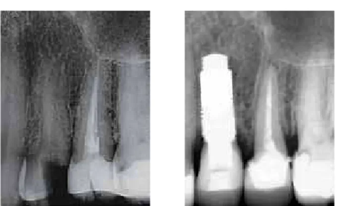
FiG. 1 G-H The radiographical evaluation before the tooth extraction (G) and after 13 months of loading demonstrates crestal bone stability around the TM immediate implant (H).
FiG. 1 a Fresh extraction socket after atraumatic tooth extraction before implant placement (a).
FiG. 1 B-C a TM dental implant was placed more palatally oriented (B) and the socket was grafted simultaneously (C).
FiG. 1 d a provisional crown was made and placed in occlusion with the opposing dentition (d).
FiG. 1 e-F The inal result after 13 months of loading presents an excellent tissue thickness (e) and optimal esthetic result (F).
General manifestations Case 1 Case 2
diarrhea Yes Yes
abdominal Pain Yes Yes
Fever No Occasional
Fatigue No No
stomatitis No Yes
weight loss Yes Yes
Vasculitis No No
recurrent Oral ulceration No No
arthritis No No
manufacturer protocol the osteotomy was completed with the drills. Particulated bone grafting material (Puros cancellous®, Zimmer, Carlsbad, USA) was placed in the socket (buccal and palatal aspect of the defect) and a TM implant (11.5 mm length, 4.7 mm diameter) (Zimmer, Carlsbad, USA) was inserted. After implant placement and abutment connection with the final torque (25 Ncm), the provisional restoration was adapted in the oral cavity creating the emergence profile. The provisional crown was screw-retained and had slight occlusal contacts in the centric occlusion, also known as intercuspation position (ICP). A periapical radiograph was taken as a control radiograph at the baseline.
Postoperatively, antibiotics (Amoxicillin 500 mg/day for 7 days) (Amoxicillin; Pfizer Manufacturing, Puurs, Belgium) were prescribed as well as analgesics (Ibuprofen 600 mg/ 3 times a day) (Brufen; BGP Products Limited, Abbott House, Maidenhead, UK) and an oral rinse (Listerine; Johnson & Johnson, New Brunswick, USA) was recommended. The patient was advised to consume soft/ liquid diet for 6-8 weeks after surgery.
After 2 weeks of healing, the provisional restoration was removed and an impression coping was used for final
impression for a definitive restoration. The impression coping was modified (within the sulcus) using light-cure resin to capture the soft tissue profile. The definitive restoration was full ceramic restoration in lithium di-silicate basis (E-MAX Press; Ivoclar, Armhest, USA), screwed onto the implant.
A final radiographic examination was performed after delivery of the final prosthesis. The patient was examined clinically and radiographically once every six months in the conventional recall program. The latest radiograph shows no crestal bone loss and an excellent clinical condition after 13 months of loading (Fig. 1).
This patient had another implant (Tapered Screw Vent, Zimmer Carlsbad, USA) (11.5 mm length, 3.75 mm diameter) in the oral cavity for a period of two years loaded with conventional loading protocol (delayed loading). The clinical and radiographic findings of this implant presented no signs of periimplant infection (Fig. 2).
Case 2
A 36-year old female non-smoker diagnosed with CD since 2004 reported for the restoration of a deep carious lesion at tooth 14. General manifestations were diarrhea, abdominal pain, stomatitis and weight loss as well as occasional episodes of fever (Table 1).
In the first year of the disease, the following medications for the treatment of CD were prescribed: (a) Pentasa® 500 (mesalazine - Ferring SpA, Milan, Italy), (b) Corticosteroids (Deltacortene 25 mg - Bruno Farmaceutici SpA, Rome, Italy), and (c) Infliximab (Remicade®; Janssen Biologics
FiG. 2 a radiographical evaluation before implant placement.
FiG. 2 B radiographical evaluation immediately after implant placement.
FiG. 2 C radiographical evaluation after two years of loading.
FiG. 2 d after two years of loading (delayed loading protocol) stable periimplant hard and soft tissue conditions.
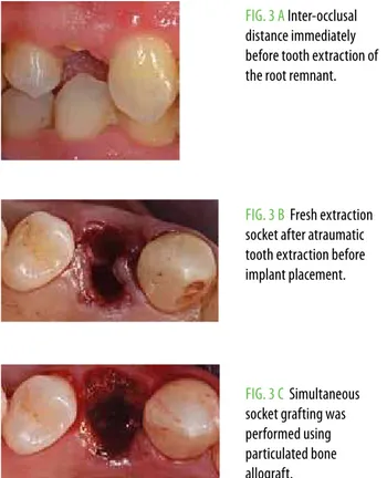
FiG. 3 a inter-occlusal distance immediately before tooth extraction of the root remnant.
FiG. 3 B Fresh extraction socket after atraumatic tooth extraction before implant placement.
nearly 50% of recipients either fail to respond (steroid-resistant) or will be steroid dependent at 1 year (17). To our knowledge newer alternatives to corticosteroids are not yet available.
In both cases, successful osseointegration of TM dental implants was achieved and the TM implants remained aesthetically and functionally stable for up to 12-months of follow-up. The present results are in accordance with an experimental study (10) in which TM implants obtained promising bone ingrowth and mechanical fixation. The TM implants represent a novel implant material (tantalum) and were initially developed for potential applications in reconstructive orthopedics and other surgical disciplines (18). It has been reported that tantalum enhances the process of osteoblastic differentiation (19). In addition, implant surface characteristics and the coatings’ pore size are also essential parameters that influence the overall percentage of bone attachment and ingrowth (20-22). A pore size of 100 µm is normally suitable to facilitate bone ingrowth (23). Therefore, replacement of threads in the midsection of a conventional titanium-based implant body with a tantalum material has been proposed to enhance the interconnected porosity of TM implants thereby augmenting secondary stability through a high volume of bone ingrowth (24).
Results from the present case studies showed that there was no significant difference in crestal bone heights between baseline and following one-year of follow-up. This is most likely associated with the microgrooved collar design of TM implants. It has been reported that B.V., Leiden, The Netherlands) once every week. Following
clinical and radiographic examination, the tooth 14 was considered hopeless and an implant placement with immediate restoration was recommended. The surgical and prosthetic protocol was similar with the first case. The implant was loaded for one year without complications (Fig. 3).
DISCUSSION AND CONCLUSION
The present report presents two clinical cases with CD. Both cases presented with classical general manifestations of CD which included prolonged diarrhea with abdominal pain, and weight loss. Patients with CD may also present with complaints indicating intestinal obstruction (15). Initially, the obstruction is secondary to inflammatory edema and spasm of the bowel and manifests as postprandial bloating, cramping pains (lower right quadrant), and borborygmi. It is important to note that colonic CD may be clinically indistinguishable from ulcerative colitis, with symptoms of bloody mucopurulent diarrhea, cramping abdominal pain, and urgency to defecate. Once the bowel lumen becomes chronically narrowed from fibrosis, patients may complain of constipation and obstipation (15). These symptoms generally do not improve with anti-inflammatory agents (16). Corticosteroids and immunosuppressant drugs, when used correctly, are a highly effective, well tolerated, cheap and generally safe treatment for active CD (16, 17). However, it has also been reported that
FiG. 3 d-F Provisional restoration was fabricated by laboratory (d) and screwed onto the implant (e) with slight occlusal contacts (F).
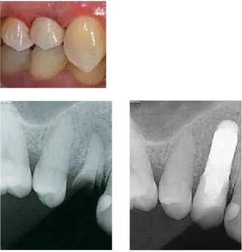
FiG. 3 G-H deinitive full ceramic restoration in lithium di-silicate was delivered at 2 weeks. The inal result presents an excellent tissue thickness (G) and optimal esthetic result after 12 months of loading (H).
microgrooved implant collars provide more promising conditions for the attachment of hard and soft tissues and reduce the level of marginal bone resorption as well as soft tissue recession (25). In an in vitro study, Fillies et
al. (26) investigated osteoblastic behavior on different sandblasted anD acid-etched (SLA) and microgrooved implant surfaces under standardized conditions. The results showed that proliferation rate of osteoblasts and the synthesis of bone-specific proteins were significantly higher on microgrooved surfaces than on SLA surfaces (26).
Javed and Romanos (3), in a systematic review, concluded that an immunocompromised state (poorly-controlled diabetes mellitus) is a significant risk factor for early implant failure. However, results from the present study showed that implants can remain functionally stable in immunosuppressed individuals (such as those with CD). An explanation in this regard may be derived from the fact that amongst the studies included in the systematic review by Javed and Romanos (3), conventional titanium were used. It is therefore tempting to speculate, on at least a local level, TM implant design exhibits the potential to attract osteoblasts and sustain crestal bone heights regardless of an immunocompromised status. The present results support the study by Schlee et al. (14) that reported a cumulative implant survival of 95.2% for TM implants. However, the present results as well as those reported by Schlee et al. (14) were based on rather short-term (12 months) follow-up periods.
A limitation of the present study is that the conclusion was based on results from two clinical cases. Moreover, the follow-up durations of both cases investigated in the present study were relatively short-term (12 and 13 months). It is yet to be determined whether or not TM dental implants can sustain crestal bone heights and demonstrate high survival and success rates over long-term periods (for example 5 years or longer). Further long-term prospective randomized controlled based clinical trials based on a large sample size are needed to test this hypothesis.
Within the limits of the present investigation, it is concluded that TM implants can osseointegrate and be functionally stable in patients with CD.
REFERENCES
1. alsaadi G, Quirynen M, Komarek a, van steenberghe d. impact of local and systemic factors on the incidence of oral implant failures, up to abutment connection. J Clin Periodontol 2007; 34: 610-617.
2. Cao Y, weischer T. Comparison of maxillary implant-supported prosthesis in irradiated and non-irradiated patients. J Huazhong univ sci Technolog Med sci 2003; 23: 209-212. 3. Javed F, romanos Ge. impact of diabetes mellitus and glycemic control on the
osseointegration of dental implants: a systematic literature review. J Periodontol 2009; 80:
1719-1730.
4. von wilmowsky C, stockmann P, Harsch i, amann K, Metzler P, lutz r, Moest T, Neukam Fw, schlegel Ka. diabetes mellitus negatively afects peri-implant bone formation in the diabetic domestic pig. J Clin Periodontol 2011; 38: 771-779.
5. alsaadi G, Quirynen M, Komarek a, van steenberghe d. impact of local and systemic factors on the incidence of late oral implant loss. Clin Oral implants res 2008; 19: 670-676. 6. alsaadi G, Quirynen M, Michiles K, Teughels w, Komarek a, van steenberghe d. impact
of local and systemic factors on the incidence of failures up to abutment connection with modiied surface oral implants. J Clin Periodontol 2008; 35: 51-57.
7. Carr aB. implant location and radiotherapy are the only factors linked to 2-year implant failure. J evid Based dent Pract 2010; 10: 49-51.
8. Carr aB. implant location and radiotherapy are the only factors linked to 2-year implant failure. J evid Based dent Pract 2012; 12: 217-219.
9. van steenberghe d, Jacobs r, desnyder M, Mafei G, Quirynen M. The relative impact of local and endogenous patient-related factors on implant failure up to the abutment stage. Clin Oral implants res 2002; 13: 617-622.
10. Bobyn Jd, stackpool GJ, Hacking sa, Tanzer M, Krygier JJ. Characteristics of bone ingrowth and interface mechanics of a new porous tantalum biomaterial. J Bone Joint surg Br 1999; 81: 907-914.
11. Cook sd, Barrack rl, Thomas Ka, Haddad rJ Jr. Tissue growth into porous primary and revision femoral stems. J arthroplasty 1991; 6 suppl: s37-46.
12. Collier JP, Mayor MB, Chae JC, surprenant Va, surprenant HP, dauphinais la. Macroscopic and microscopic evidence of prosthetic ixation with porous-coated materials. Clin Orthop relat res 1988; 173-180.
13. engh Ca, Hooten JP Jr., Zettl-schafer KF, Ghafarpour M, McGovern TF, Bobyn Jd. evaluation of bone ingrowth in proximally and extensively porous-coated anatomic medullary locking prostheses retrieved at autopsy. J Bone Joint surg am 1995, 77, 903-910.
14. schlee M, Pradies G, Mehmke wu, Beneytout a, stamm M, Meda rG, Kamm T, Poiroux F, weinlich F, del Canto Pingarron M, Crichton e, Poulet JB, Bousquet P. Prospective, multicenter evaluation of trabecular metal-enhanced titanium dental implants placed in routine dental practices: 1-year interim report from the development period (2010 to 2011). Clin implant dent relat res 2014; 10.1111/cid.12232.
15. d'Haens G, Baert F, van assche G, Caenepeel P, Vergauwe P, Tuynman H, de Vos M, van deventer s, stitt l, donner a, Vermeire s, Van de Mierop FJ, Coche JC, van der woude J, Ochsenkuhn T, van Bodegraven aa, Van Hootegem PP, lambrecht Gl, Mana F, rutgeerts P, Feagan BG, Hommes d. early combined immunosuppression or conventional management in patients with newly diagnosed Crohn's disease: an open randomised trial. lancet 2008; 371: 660-667.
16. Panes J, Gomollon F, Taxonera C, Hinojosa J, Clofent J, Nos P. Crohn's disease: a review of current treatment with a focus on biologics. drugs 2007; 67: 2511-2537.
17. irving PM, Gearry rB, sparrow MP, Gibson Pr. review article: appropriate use of corticosteroids in Crohn's disease. aliment Pharmacol Ther 2007; 26: 313-329.
18. Bobyn Jd, Toh KK, Hacking sa, Tanzer M, Krygier JJ. Tissue response to porous tantalum acetabular cups: a canine model. J arthroplasty 1999; 14: 347-354.
19. stiehler M, lind M, Mygind T, Baatrup a, dolatshahi-Pirouz a, li H, Foss M, Besenbacher F, Kassem M, Bunger C. Morphology, proliferation, and osteogenic diferentiation of mesenchymal stem cells cultured on titanium, tantalum, and chromium surfaces. J Biomed Mater res a 2008; 86: 448-458.
20. Javed F, almas K, Crespi r, romanos Ge. implant surface morphology and primary stability: is there a connection? implant dent 2011; 20: 40-46.
21. Javed F, Vohra F, Zafar s, almas K, signiicance of osteogenic surface coatings on implants to enhance osseointegration under osteoporotic-like conditions. implant dent 2014; 23: 679-686.
22. roberts ew, Poon lC, smith rK. interface histology of rigid endosseous implants. J Oral implantol 1986; 12: 406-416.
23. Hulbert sF, Cooke Fw, Klawitter JJ, leonard rB, sauer Bw, Moyle dd, skinner HB. attachment of prostheses to the musculoskeletal system by tissue ingrowth and mechanical interlocking. J Biomed Mater res 1973; 7: 1-23.
24. Bencharit s, Byrd wC, altarawneh s, Hosseini B, leong a, reside G, Morelli T, Ofenbacher s. development and applications of porous tantalum trabecular metal-enhanced titanium dental implants. Clin implant dent relat res 2013; 10.1111/cid.12059.
25. shin sY, Han dH. inluence of a microgrooved collar design on soft and hard tissue healing of immediate implantation in fresh extraction sites in dogs. Clin Oral implants res 2010; 21: 804-814.