Metaproteomics of saliva identifies
human protein markers specific for
individuals with periodontitis and
dental caries compared to orally
healthy controls
Daniel Belstrøm1,*, Rosa R. Jersie-Christensen2,*, David Lyon3, Christian Damgaard1,4, Lars J. Jensen3, Palle Holmstrup1and Jesper V. Olsen2
1Section of Periodontology and Microbiology, Department of Odontology, Faculty of Health and Medical Sciences, University of Copenhagen, Copenhagen, Denmark
2Proteomics Program, Novo Nordisk Foundation Center for Protein Research, Faculty of Health and Medical Sciences, University of Copenhagen, Copenhagen, Denmark
3Disease Systems Biology Program, Novo Nordisk Foundation Center for Protein Research, Faculty of Health and Medical Sciences, University of Copenhagen, Copenhagen, Denmark 4Institute for Inflammation Research, Center for Rheumatology and Spine Diseases,
Rigshospitalet, Copenhagen University Hospital, Copenhagen, Denmark *These authors contributed equally to this work.
ABSTRACT
Background:The composition of the salivary microbiota has been reported to differentiate between patients with periodontitis, dental caries and orally healthy individuals. To identify characteristics of diseased and healthy saliva we thus wanted to compare saliva metaproteomes from patients with periodontitis and dental caries to healthy individuals.
Methods:Stimulated saliva samples were collected from 10 patients with
periodontitis, 10 patients with dental caries and 10 orally healthy individuals. The proteins in the saliva samples were subjected to denaturing buffer and digested enzymatically with LysC and trypsin. The resulting peptide mixtures were cleaned up by solid-phase extraction and separated online with 2 h gradients by nano-scale C18reversed-phase chromatography connected to a mass spectrometer through an electrospray source. The eluting peptides were analyzed on a tandem mass
spectrometer operated in data-dependent acquisition mode.
Results: We identified a total of 35,664 unique peptides from 4,161 different proteins, of which 1,946 and 2,090 were of bacterial and human origin, respectively. The human protein profiles displayed significant overexpression of the complement system and inflammatory markers in periodontitis and dental caries compared to healthy controls. Bacterial proteome profiles and functional annotation were very similar in health and disease.
Conclusions:Overexpression of proteins related to the complement system and inflammation seems to correlate with oral disease status. Similar bacterial proteomes in healthy and diseased individuals suggests that the salivary microbiota
predominantly thrives in a planktonic state expressing no disease-associated characteristics of metabolic activity.
Submitted29 June 2016
Accepted12 August 2016
Published14 September 2016
Corresponding author
Daniel Belstrøm, [email protected]
Academic editor
Melissa Grant
Additional Information and Declarations can be found on page 12
DOI10.7717/peerj.2433
Copyright
2016 Belstrøm et al.
Distributed under
Subjects Microbiology, Dentistry, Immunology
Keywords Metaproteomics, Saliva, Periodontitis, Dental caries, Bacteria, Proteins, Immune response
INTRODUCTION
Saliva is a biological fluid critically involved in maintenance of oral homeostasis (Marsh et al., 2016), as qualitative and quantitative changes of saliva associates with increased frequency and severity of diseases in the oral cavity (Dawes et al., 2015; Almstahl & Wikstrom, 1999). Furthermore, saliva is easily and non-invasively collected (Giannobile et al., 2011), making it interesting to screen for biomarkers associated with oral and general health and disease status (Baum et al., 2011;Zhang et al., 2016).
In the last decade, salivary biomarkers of periodontitis and dental caries have been intensively investigated (Yoshizawa et al., 2013;Miller et al., 2010). These include salivary bacterial profiles that differentiate in patients with periodontitis (Paju et al., 2009; Belstrøm et al., 2014b;Belstrøm et al., 2016b), dental caries (Yang et al., 2012;Belstrøm et al., 2014a;Belstrøm et al., 2015) and orally healthy individuals. Furthermore, increased salivary levels of inflammatory protein biomarkers such as interleukin-1b(IL-1b), IL-6 and matrix metalloproteinase-8 (MMP-8) have been described to be associated with periodontal disease status (Kinney et al., 2011;Ebersole et al., 2013;Rathnayake et al., 2013; Ebersole et al., 2015). Recently, the salivary transcriptome has been assessed (Spielman et al., 2012), and some transcriptomic characteristics of saliva have been reported in patients with dental caries (Do et al., 2015). Collectively, these reports conclude that biomarkers of different biological origin may be adequately assessed in saliva samples and support the concept that the biological composition of saliva reflects individual oral health status.
Mass spectrometry-based proteomics enables characterization of the protein content in any sample, including proteins of human and bacterial origin. It thus provides the possibility for simultaneous characterization of bacterial and host specific differences of saliva associated with oral health and disease. Only three studies have so far attempted to perform metaproteomic analysis of saliva in oral health (Rudney et al., 2010;Jagtap et al., 2012;Grassl et al., 2016). To the best of our knowledge, no study has so far compared metaproteomic profiles of saliva from patients with periodontitis and dental caries to orally healthy individuals.
The aim of the present study was to characterize the salivary metaproteome in 30 saliva samples, and compare human and bacterial proteome profiles between patients with periodontitis, dental caries and orally healthy individuals. The hypothesis was that both bacterial and human subsets of salivary metaproteome would differentiate between individuals with different oral health status.
MATERIALS AND METHODS
Study population and sample collection
chewing on a tasteless paraffin gum, and chewing-stimulated saliva samples were collected from 10 patients with periodontitis, 10 patients with dental caries and 10 orally
healthy individuals following a standardized protocol (Kongstad et al., 2013). Immediately after collection saliva samples were divided into four aliquots and stored at -80C
for further analysis. One aliquot has previously been analyzed by next-generation sequencing (the Human Oral Microbe Identification using Next Generation Sequencing, HOMINGS) (Belstrøm et al., 2016b). All participants signed an informed consent prior to participation, and the study was approved by the regional ethical committee
(H-15000856-53175) and reported to the Danish Data Authorization (2015-54-0970).
Sample preparation
The saliva proteome samples were prepared as described in (Jersie-Christensen, Sultan & Olsen, 2016) with a few modifications. Briefly, 1 ml of saliva was mixed with 1.5 ml lysis buffer (9 M Guanidine hydrochloride, 10 mM Chloroacetamide, 5 mMtris
(2-carboxyethyl)phosphine in 100 mM Tris pH 8.5) and heated for 10 min (99C) followed
by 4 min of sonication.
Protein concentration was measured with Bradford protein assay and ranged from 1–2.5 mg/ml. All samples were digested with the same amount of Lysyl Endoproteinase (Wako, Osaka, Japan) in a ratio of 1:100 w/w calculated from the highest concentration for 2 h. Samples were diluted to a final volume of 10 ml with 25 mM Tris pH8 and digested overnight with Trypsin (modified sequencing grade; Sigma) in a 1:100 w/w ratio.
Digestion was quenched by adding 1 ml of 10% trifluoroacetic acid and centrifuged at 2,000 g for 5 min. The resulting soluble peptides in the supernatant were desalted and concentrated on Waters Sep-Pak reversed-phase C18cartridges (one per sample) and the tryptic peptide mixtures were eluted with 40% acetonitrile (ACN) followed by 60% ACN. Peptide concentrations were determined by NanoDrop (Thermo, Wilmington, DE, USA) measurement.
Mass spectrometry analysis
A total of 1.5mg peptide mixture from each sample was analyzed by online nano-scale liquid chromatography tandem mass spectrometry (LC-MS/MS) in turn. Peptides were separated on an in-house packed 50 cm capillary column with 1.9 mm Reprosil-Pur C18beads using an EASY-nLC 1000 system (Thermo Scientific). The column temperature
was maintained at 50C using an integrated column oven (PRSO-V1; Sonation
GmbH, Biberach, Germany). Buffer A consisted of 0.1% Formic acid, and buffer B of 80% ACN, 0.1% Formic acid. The flow rate of the gradient was 200 nl/min and started at 5% buffer B, going to 25% buffer B in 110 min, followed by a 25 min step going to 40% buffer B and continuing to 80% buffer B in 5 min for a 5 min wash and returning to 5% in 5 min and continuing for re-equilibration for 5 min.
60,000 at m/z 200 and the scan target was 3106with a maximum fill time of 20 ms.
Full-scan MS mass range was set to 300–1,750 and dynamic exclusion to 20 s. Target value
for HCD-MS/MS scans was set at 1 105with a resolution of 30,000 and a maximum
fill time of 60 ms. Normalized collision energy was set at 28.
Data analysis
All 30 raw LC-MS/MS data files were processed together using MaxQuant version 1.5.0.38 (Cox & Mann, 2008) with default settings and match between runs. The integrated Andromeda peptide search engine and a reversed database approach applying a 1% FDR at both peptide and protein level was used. The data was searched in two iterations analogous to a previously described metaproteomics database search strategy (Jagtap et al., 2013). First, the search space consisted of the full SwissProt protein database (The UniProt Consortium, 2015) and the Human Oral Microbiome database (Chen et al., 2010) (both downloaded August 2014). The resulting search output was then used for reduction of the search space after filtering on different parameters. As a quality control measure, proteins with less than two unique peptides were removed. Furthermore, we required proteins to be detected in at least five out of 30 samples. Accession numbers from the Majority protein IDs column in the proteinGroups.txt were used to retrieve information about Lowest Common Ancestor (LCA) for each protein group entry. To find the LCA of a protein group, accession numbers with the most peptide-associations were selected, mapped to species and their full taxonomic lineage. The lowest taxonomic rank of the intersection of the latter yielded the LCA. All LCA searches resulting in the parvorder Catarrhini (primates) were set to be human. LCAs at taxonomic rank of species and genera, as well as all of their descendants were used to create a new, reduced search space. The latter was used for the second iteration of MS data identification and quantification and all accession numbers within a protein group were used to perform LCA searches. The above functionality was achieved using the Python programming language. Species names from SwissProt and HOMD were mapped to NCBI taxonomic identifiers using UniProt (http://www.uniprot.org/docs/ speclist) and NCBI resources (http://www.ncbi.nlm.nih.gov/Taxonomy/TaxIdentifier/ tax_identifier.cgi), respectively. Full taxonomic lineages were retrieved from NCBI Taxonomy database dump files (ftp://ftp.ncbi.nlm.nih.gov/pub/taxonomy/). Taxonomic comparison at genus- and species level was performed using Mann-Whitney U test with Benjamini-Hochberg correction for multiple testing.
Protein intensities based on summed peptide MS signal intensities were quantile normalized using the limma package version 3.24.15 under R version 3.2.2. Only proteins identified with more than one peptide (“razor + unique”) and present in more than five out of the 30 samples were considered for further analysis. The mass spectrometry proteomics data have been deposited to the ProteomeXchange
Consortium via the PRIDE (Vizcaı´no et al., 2016) partner repository with the dataset identifier PXD004319.
replaced with the constant value 19, representing the lowest protein intensity value measured. Analysis of significance (ANOVA) between groups was performed with the software package Perseus (http://www.perseus-framework.org). The resulting differentially expressed proteins were clustered using Euclidian distance after scaling the data by subtracting the mean intensity value. All p-values were corrected for multiple comparisons.
Functional annotation of bacterial proteins
Bacterial proteins from HOMD were searched against Hidden Markov Models (HMMs) of bacterial Nested Orthologous Groups (NOGs) from eggNOG (Huerta-Cepas et al., 2016) using HMMscan version 3.1 (http://hmmer.org) (Eddy, 2009). For each protein query the resulting hits were restricted by two criteria. E-values had to be equal or lower than 1e-4 and a maximum overlap of eight amino acids of HMMs was allowed (selecting hits with the lowest e-value). All corresponding NOG-names were used to retrieve Gene Ontology (GO)-terms as well as KEGG pathways from eggNOG.
KEGG pathway enrichment and characterization
To gain insights into differences between the three sample groups, KEGG pathway enrichment was performed using a modified version of AGOtool (Scho¨lz et al., 2015). Individual samples were grouped to sample categories and the three paired combinations used for the enrichment analysis. All bacterial protein groups with an LCA at rank genus or below were selected. Benjamini-Hochberg correction (FDR) of p-values was applied to correct for multiple testing. The FDR was set to 1%. The following additional filter criterion was applied. The fold change had to be equal or higher than 2 or equal or lower than 0.5.
To get a functional overview of the bacterial proteins, we characterized each individual sample group by counting the number of protein groups associated with each KEGG pathway. For visualization purposes (Fig. S2), we selected the most highly associated terms. Within each group the number of associations was converted to percentages, and the most highly associated terms retained, until a cumulative sum of 90% was reached. This reduced the number of KEGG terms from 135 to 50.
RESULTS
General findings
Human protein profiling
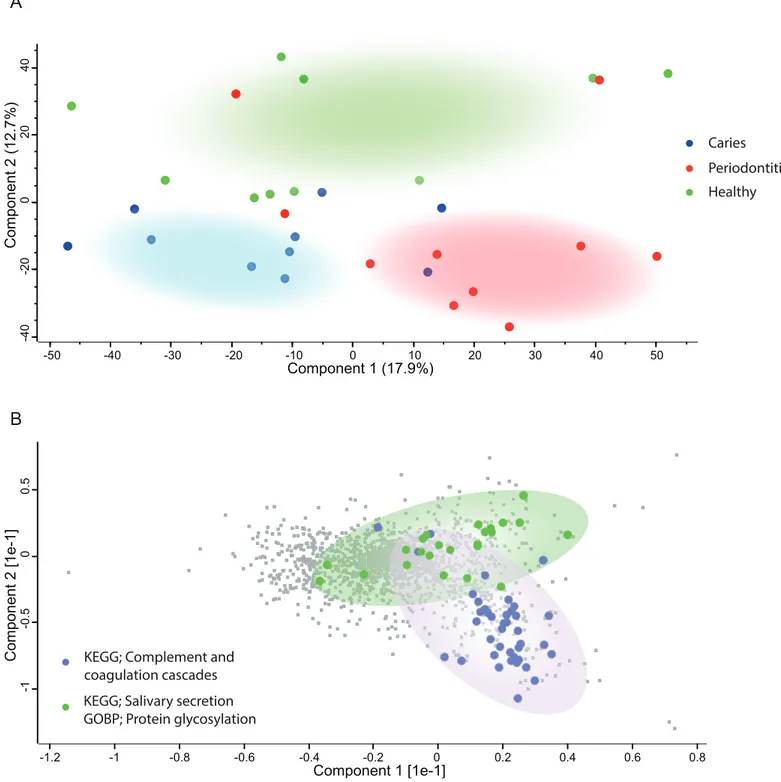
Principal component analysis (Fig. 2A) of the human proteins in saliva showed decent separation of samples from patients with periodontitis and dental caries from orally healthy individuals, based on the most decisive component of the dataset, accounting for 17.9% of the variation. The most enriched KEGG pathway in component 1 and 2 was ‘Complement and coagulation cascades’ (Fig. 2B). Component 2 also separated samples from patients with dental caries and periodontitis patients from orally healthy individuals with the component explaining 12.7% of the variation. Two of the most enriched terms in component 2 in the direction of the orally healthy individuals were KEGG pathway ‘Salivary secretion’ and GOBP ‘protein glycosylation.’
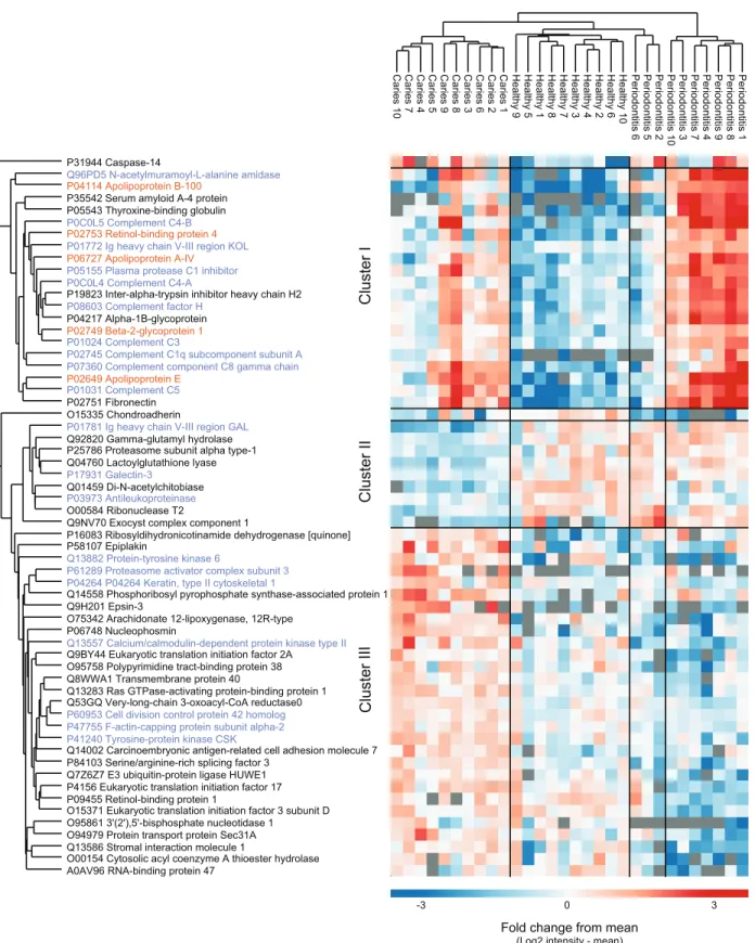
From a total of 2,090 identified proteins of human origin, 60 proteins were significantly differentially expressed when performing multiple sample test (ANOVA, p < 0.05). Hierarchical cluster analysis of the proteins nicely separated the three sample groups,
%
0 10 20 30 40 50 60 70 80 90 100
Biomass Proteins
Bacteria
Human
Other
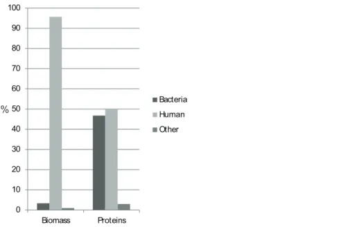
Figure 1 Protein biomass and abundance across sample groups.Relative distribution as a measure of summed intensity and protein count.
Table 1 Overview of proteins identified.
Number of proteins
Caries Healthy Periodontitis Total*
Other** 125 115 120 125
Human 2,084 2,079 2,084 2,090
Bacteria 1,861 1,926 1,924 1,946
-mapped to genus level 1,710 (91.9%) 1,765 (91.6%) 1,762 (91.6%) 1,784 (91.7%) -mapped to species level 594 (31.9%) 602 (34.1%) 609 (34.6%) 616 (34.5%)
Total 4,070 4,120 4,128 4,161
Notes:
*Unique proteins.
although three periodontitis individuals cluster together with the healthy group (Fig. 3). We identified three main protein clusters. Cluster I contains human proteins that are higher expressed in both disease groups compared to controls, and 10 of 20 proteins in
-40
-20
0
20
40
&RPSRQHQW
Loading...
-50 -40 -30 -20 -10 0 10 20 30 40 50
&RPSRQHQW
-1
-0
.
5
0
0
.
5
&RPSRQHQW>H@
-1.2 -1 -0.8 -0.6 -0.4 -0.2 0 0.2 0.4 0.6 0.8
&RPSRQHQW>H@
Caries
Periodontitis
Healthy
KEGG; Complement and coagulation cascades
KEGG; Salivary secretion GOBP; Protein glycosylation A
B
Caries 10 Caries 7 Caries 4 Caries 5 Caries 9 Caries 8 Caries 3 Caries 6 Caries 2 Caries 1 Healthy 9 Healthy 5 Healthy 1 Healthy 8 Healthy 7 Healthy 3 Healthy 4 Healthy 2 Healthy 6 Healthy 10 Perio d ontitis 6 Perio d ontitis 5 Perio d ontitis 2 Perio d ontitis 10 Perio d ontitis 3 Perio d ontitis 7 Perio d ontitis 4 Perio d ontitis 9 Perio d ontitis 8 Perio d ontitis 1
P31944 Caspase-14
Q96PD5 N-acetylmuramoyl-L-alanine amidase
P04114 Apolipoprotein B-100
P35542 Serum amyloidA-4 protein P05543 Thyroxine-bindingglobulin
P0C0L5 Complement C4-B
P02753 Retinol-binding protein 4
P01772 Ig heavy chain V-III region KOL P06727 Apolipoprotein A-IV
P05155 Plasma protease C1 inhibitor P0C0L4 Complement C4-A
P19823 Inter-alpha-trypsin inhibitor heavy chain H2
P08603 Complement factor H
P04217 Alpha-1B-glycoprotein
P02749 Beta-2-glycoprotein 1
P01024 Complement C3
P02745 Complement C1q subcomponent subunit A
P07360 Complement component C8 gamma chain
P02649 Apolipoprotein E
P01031 Complement C5
P02751 Fibronectin O15335 Chondroadherin
P01781 Ig heavy chain V-III region GAL
Q92820 Gamma-glutamyl hydrolase P25786 Proteasome subunit alpha type-1 Q04760 Lactoylglutathione lyase
P17931 Galectin-3
Q01459 Di-N-acetylchitobiase
P03973 Antileukoproteinase
O00584 Ribonuclease T2
Q9NV70 Exocyst complex component 1
P16083 Ribosyldihydronicotinamide dehydrogenase [quinone] P58107 Epiplakin
Q13882 Protein-tyrosine kinase 6
P61289 Proteasome activator complex subunit 3 P04264 P04264 Keratin, type II cytoskeletal 1
Q14558 Phosphoribosyl pyrophosphate synthase-associated protein 1 Q9H201 Epsin-3
O75342 Arachidonate 12-lipoxygenase, 12R-type P06748 Nucleophosmin
Q13557 Calcium/calmodulin-dependent protein kinase type II
Q9BY44 Eukaryotic translation initiation factor 2A
O95758 Polypyrimidine tract-binding protein 38 Q8WWA1 Transmembrane protein 40
Q13283 Ras GTPase-activating protein-binding protein 1 Q53GQ Very-long-chain 3-oxoacyl-CoA reductase0
P60953 Cell division control protein 42 homolog
P47755 F-actin-capping protein subunit alpha-2 P41240 Tyrosine-protein kinase CSK
Q14002 Carcinoembryonic antigen-related cell adhesion molecule 7 P84103 Serine/arginine-rich splicing factor 3
Q7Z6Z7 E3 ubiquitin-protein ligase HUWE1 P4156 Eukaryotic translation initiation factor 17 P09455 Retinol-binding protein 1
O15371 Eukaryotic translation initiation factor 3 subunit D O95861 3'(2'),5'-bisphosphate nucleotidase 1
O94979 Protein transport protein Sec31A
Q13586 Stromal interaction molecule 1
O00154 Cytosolic acyl coenzyme A thioester hydrolase
A0AV96 RNA-binding protein 47
0
-3 3
Fold change from mean (Log2 intensity - mean)
Cluster III
Cluster II
Cluster I
this cluster are associated with the GO term ‘innate immune response’ (protein name in purple). Cluster II consist of nine proteins that distinguish the individuals with caries from the other groups. In cluster III the protein intensities in the caries group are higher than the mean, for the orally healthy group it is around the mean and for the individuals with periodontitis lower.
0 10 20 30 40 50 60 70 80 90 100
Caries Healthy Periodontitis
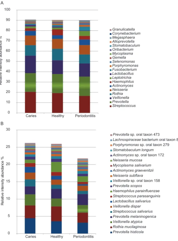
Re la tiv e i n te ns ity ab u n d a n ce % Granulicatella Corynebacterium Megasphaera Alloprevotella Stomatobaculum Oribacterium Mycoplasma Gemella Selenomonas Porphyromonas Fusobacterium Lactobacillus Leptotrichia Haemophilus Actinomyces Neisseria Rothia Veillonella Prevotella Streptococcus 0 5 10 15 20 25 30
Caries Healthy Periodontitis
R ela tiv e in te n s ity a b u n d an c e %
Prevotella sp. oral taxon 473
Lachnospiraceae bacterium oral taxon 82 Porphyromonas sp. oral taxon 279 Stomatobaculum longum Actinomyces sp. oral taxon 172 Neisseria mucosa
Mycoplasma salivarium Actinomyces graevenitzii Neisseria subflava
Veillonella sp. oral taxon 158 Prevotella scopos Haemophilus parainfluenzae Streptococcus parasanguinis Lactobacillus salivarius Veillonella dispar Streptococcus salivarius Prevotella melaninogenica Veillonella atypica Rothia mucilaginosa Prevotella histicola A B
Bacterial protein profiling
Of the 1,946 proteins of bacterial origin identified, approximately 92% and 34% could be assigned to genus and species level, respectively (Table 1). A total of 29 different
bacterial genera and 81 species were identified. The five most predominant bacterial genera wereStreptococcus,Prevotella, Veillonella,RothiaandNeisseriacollectively
representing approx. 70% of the total bacterial mass. The five most predominant bacterial species identified wereRothia mucilanginosa,Veillonella atypica,Prevotella histicola,
Prevotella melaninogenica andStreptococcus salivarius. Abundances of the 20 most
predominant bacterial genera and species are displayed inFigs. 4Aand4B. No statistically significant differences were observed between groups at genus or species level. However, at genus level there is a trend of higher proportion of Veillonellaand lower proportion of
Haemophiluswere associated with dental caries and higher proportions ofFusobacterium,
LeptotrichiaandSelenomonasand lower proportions ofStreptococcus,Rothia and
Haemophiluswere associated with periodontitis, when compared to orally healthy
individuals. The same trend is seen at species level where higher proportion ofVeillonella
atypicaand lower proportion ofHaemophilus parainfluenzaewere associated with
dental caries, and higher proportions of Fusobacterium periodonticumandLeptotrichia
wadeiand lower proportions ofHaemophilus parainfluenzaewere associated with
periodontitis, when compared to orally healthy individuals. A full list of all bacterial genera and species identified are presented inTable S1.
KEGG pathway enrichment for bacterial proteins
KEGG pathway enrichment analysis of bacterial proteins resulted in no significant differences with the application of the previously mentioned fold-change and FDR filter criteria. The characterization of functional associations of bacterial proteins is shown in Fig. S2.
DISCUSSION
The purpose of the present study was to compare metaproteome profiles of saliva from patients with periodontitis or dental caries to that of orally healthy individuals, as we hypothesized that the composition of the salivary metaproteome would associate with oral health status. To the best of our knowledge, this is the first study to characterize both human and bacterial parts of the salivary metaproteome in patients with periodontitis and dental caries.
In this study, proteins of bacterial origin constituted 46% of the proteome diversity, despite only 3% of the total biomass being bacterial. This agrees with the previously reported approx. 1% of DNA in saliva being of bacterial origin (Lazarevic et al., 2012).
metaproteomic analysis of saliva (Rudney et al., 2010;Jagtap et al., 2012). Streptococcus,
Prevotella,Veillonella,RothiaandNeisseriawere the most predominant genera
identified, constituting approx. 70% of the biomass across all samples (Fig. 4A). This phylogenetic distribution is in line with analysis of the same samples using next-generation sequencing (Belstrøm et al., 2016b), and with previous metaproteomic analysis of saliva in oral health (Rudney et al., 2010;Jagtap et al., 2012;Grassl et al., 2016). By contrast, an analysis of 17 plaque samples from patients with dental caries and healthy controls by metagenomics, metatranscriptomics and metaproteomics found different bacterial compositions in dental plaque at DNA, mRNA and protein level (Belda-Ferre et al., 2015). This may reflect differences between studying the metabolically active dental plaque biofilm and the planktonic, metabolically inactive state of the salivary microbiota, and it is in concordance with the functional annotation analysis performed (Fig. S2). Moreover, the finding of higher proportions ofVeillonellain saliva samples from patients with caries and higher proportions of Fusobacteriumin samples from periodontitis patients confirms findings from 16S analysis of the same samples (Belstrøm et al., 2016b). Interestingly, specific oral bacterial species such asVeillonella parvulaandFusobacterium
periodonticumhave been reported to associate with dental caries and periodontitis,
respectively (Takahashi & Nyvad, 2011;Colombo et al., 2009).
Furthermore, 2,090 different proteins of human origin were identified, which is more than in metaproteome profiling of dental plaque (Belda-Ferre et al., 2015) and less than a recent study that identified more than 3,700 different human proteins in a mouth swab analysis (Grassl et al., 2016). The higher number of human protein identifications in mouth swabs is probably due to swabbing the inside of the complete oral cavity including the inside of the cheek. In this study, we used stimulated saliva samples, which may have diluted the concentration of proteins within the samples compared to that of unstimulated saliva (Yakob et al., 2014; Schafer et al., 2014). This will of course also affect number of identifications. Based on this finding, unstimulated saliva samples may be preferred for in-depth analysis of the salivary proteome. However, as collection of unstimulated saliva samples is considerably more intricate and time-consuming than collection of stimulated saliva samples, the feasibility of using unstimulated saliva samples for population-based biomarker screening approaches may be limited (Belstrøm et al., 2016b). In addition, we have recently compared the salivary microbiota in unstimulated and stimulated saliva samples, collected from the same individuals, and reported that comparable microbiotas could be identified using the two types of samples (Belstrøm et al., 2016a). Consequently, stimulated saliva samples were used in this study.
Data on the human profile of the salivary metaproteome showed differences between oral health and disease, as proteins involved in innate immunity and inflammatory proteins were more abundantly expressed in saliva samples from patients with
(Cole et al., 1981; Aurer et al., 1999). Likewise, it has been reported that active components of the complement system in the gingival crevicular fluid associates with both periodontitis (Schenkein & Genco, 1977;Courts et al., 1977) and gingivitis (Patters, Niekrash & Lang, 1989; Attstro¨um et al., 1975). Increased local activation of the complement system in the periodontal tissues increases vascular permeability, vasodilatation and recruitment of inflammatory cells, resulting in excessive release of reactive oxygen species, proteolytic enzymes and interleukins (Okada & Silverman, 1979; Okuda & Takazoe, 1980; Watanabe et al., 1997). Furthermore, serum levels of
complement proteins, has been suggested to express a linear relationship with the degree of periodontal inflammation (Henry et al., 1987). Gingivitis is a mild form of gum disease that results in irritation, redness and swelling caused by inflammation of the gums. Thus, the abundant expression of complement proteins and inflammatory mediators in saliva might reflect either a spillover from the gingival crevicular fluid, or alternatively, mirror increased serum levels of these proteins. Notably, while the complement system has been acknowledged to have a profound role in the pathogenesis of periodontitis (Damgaard et al., 2015), the complement system seems to have limited impact on development of dental caries. The expression of complement proteins and other inflammatory proteins in saliva from patients with dental caries is most likely associated with gingivitis in the periodontal tissues adjacent to approximal and gingival caries lesions, and presumably not directly associated with presence of dental caries as such.
CONCLUSION
Quantitative proteomics data from the present investigation suggest that the salivary microbiota predominantly thrives in a planktonic state with limited metabolic activity, as comparable microbial compositions of the salivary microbiota were obtained based on different omics analysis. Thus, the bacterial part of the metaproteome seems to be inadequate for biomarker analysis of periodontitis and caries. Conversely, a set of human proteins hold the potential to be used as future biomarkers of oral disease status. However, the cross-sectional study design obviously hampers the possibility to address causality of this observation. Thus, future large-scale longitudinal studies of human saliva proteome changes are warranted to reveal the full potential of
quantitative proteomics of saliva as a technique to discover biomarkers of oral health and disease.
ADDITIONAL INFORMATION AND DECLARATIONS
Funding
Grant Disclosures
The following grant information was disclosed by the authors: University of Copenhagen: KU2016 programme.
Novo Nordisk Foundation: NNF14CC0001.
Competing Interests
The authors declare that they have no competing interests.
Author Contributions
Daniel Belstrøm conceived and designed the experiments, performed the experiments,
analyzed the data, wrote the paper.
Rosa R. Jersie-Christensen conceived and designed the experiments, performed the
experiments, analyzed the data, wrote the paper, prepared figures and/or tables.
David Lyon analyzed the data, prepared figures and/or tables, reviewed drafts of the
paper.
Christian Damgaard reviewed drafts of the paper.
Lars J. Jensen contributed reagents/materials/analysis tools, reviewed drafts of the paper. Palle Holmstrup contributed reagents/materials/analysis tools, reviewed drafts of the
paper.
Jesper V. Olsen contributed reagents/materials/analysis tools, reviewed drafts of the
paper.
Human Ethics
The following information was supplied relating to ethical approvals (i.e., approving body and any reference numbers):
All participants signed an informed consent prior to participation, and the study was approved by the regional ethical committee (H-15000856-53175) and reported to the Danish Data Authorization (2015-54-0970).
Data Deposition
The following information was supplied regarding data availability:
ProteomeXchange Pride:https://www.ebi.ac.uk/pride/archive/projects/PXD004319/ files.
Supplemental Information
Supplemental information for this article can be found online athttp://dx.doi.org/ 10.7717/peerj.2433#supplemental-information.
REFERENCES
Almstahl A, Wikstrom M. 1999.Oral microflora in subjects with reduced salivary secretion.
Journal of Dental Research78(8):1410–1416DOI 10.1177/00220345990780080601.
Attstro¨um R, Laurel A-B, Lahsson U, Sjo¨uholm A. 1975.Complement factors in gingival crevice material from healthy and inflamed gingiva in humans.Journal of Periodontal Research
Aurer A, Aurer-Kozˇelj J, Stavljenic-Rukavina A, Kalenic S, Ivic-Kardum M, Haban V. 1999.
Inflammatory mediators in saliva of patients with rapidly progressive periodontitis during war stress induced incidence increase.Collegium Antropologicum23(1):117–124.
Baum BJ, Yates JR III, Srivastava S, Wong DTW, Melvin JE. 2011.Scientific frontiers: emerging technologies for salivary diagnostics.Advances in Dental Research23(4):360–368
DOI 10.1177/0022034511420433.
Belda-Ferre P, Williamson J, Simo´n-Soro A´, Artacho A, Jensen ON, Mira A. 2015.The human oral metaproteome reveals potential biomarkers for caries disease.Proteomics15(20):3497–3507
DOI 10.1002/pmic.201400600.
Belstrøm D, Fiehn N-E, Nielsen CH, Holmstrup P, Kirkby N, Klepac-Ceraj V, Paster BJ, Twetman S. 2014a.Altered bacterial profiles in saliva from adults with caries lesions: a case-cohort study.Caries Research48(5):368–375DOI 10.1159/000357502.
Belstrøm D, Fiehn N-E, Nielsen CH, Kirkby N, Twetman S, Klepac-Ceraj V, Paster BJ, Holmstrup P. 2014b.Differences in bacterial saliva profile between periodontitis patients and a control cohort.Journal of Clinical Periodontology41(2):104–112DOI 10.1111/jcpe.12190.
Belstrøm D, Fiehn N-E, Nielsen CH, Klepac-Ceraj V, Paster BJ, Twetman S, Holmstrup P. 2015.
Differentiation of salivary bacterial profiles of subjects with periodontitis and dental caries.
Journal of Oral Microbiology7:27429DOI 10.3402/jom.v7.27429.
Belstrøm D, Holmstrup P, Bardow A, Kokaras A, Fiehn N-E, Paster BJ. 2016a.Comparative analysis of bacterial profiles in unstimulated and stimulated saliva samples.Journal of Oral Microbiology8:30112DOI 10.3402/jom.v8.30112.
Belstrøm D, Paster BJ, Fiehn N-E, Bardow A, Holmstrup P. 2016b.Salivary bacterial fingerprints of established oral disease revealed by the Human Oral Microbe Identification using Next Generation Sequencing (HOMINGS) technique.Journal of Oral Microbiology8:30170
DOI 10.3402/jom.v8.30170.
Chen T, Yu W-H, Izard J, Baranova OV, Lakshmanan A, Dewhirst FE. 2010.The Human Oral Microbiome Database: a web accessible resource for investigating oral microbe taxonomic and genomic information.Database2010:baq013DOI 10.1093/database/baq013.
Cole MF, Hsu SD, Baum BJ, Bowen WH, Sierra LI, Aquirre M, Gillespie G. 1981.Specific and nonspecific immune factors in dental plaque fluid and saliva from young and old populations.
Infection and Immunity31(3):998–1002.
Colombo APV, Boches SK, Cotton SL, Goodson JM, Kent R, Haffajee AD, Socransky SS, Hasturk H, Van Dyke TE, Dewhirst F, Paster BJ. 2009.Comparisons of subgingival microbial profiles of refractory periodontitis, severe periodontitis, and periodontal health using the human oral microbe identification microarray.Journal of Periodontology80(9):1421–1432
DOI 10.1902/jop.2009.090185.
Courts FJ, Boackle RJ, Fudenberg HH, Silverman MS. 1977.Detection of functional complement components in gingival crevicular fluid from humans with periodontal diseases.Journal of Dental Research56(3):327–331DOI 10.1177/00220345770560032001.
Cox J, Mann M. 2008.MaxQuant enables high peptide identification rates, individualized p.p.b.-range mass accuracies and proteome-wide protein quantification.Nature Biotechnology
26(12):1367–1372DOI 10.1038/nbt.1511.
Damgaard C, Holmstrup P, Van Dyke TE, Nielsen CH. 2015.The complement system and its role in the pathogenesis of periodontitis: current concepts.Journal of Periodontal Research
50(3):283–293DOI 10.1111/jre.12209.
human saliva: a review sponsored by the World Workshop on Oral Medicine VI.Archives of Oral Biology60(6):863–874DOI 10.1016/j.archoralbio.2015.03.004.
Do T, Sheehy EC, Mulli T, Hughes F, Beighton D. 2015.Transcriptomic analysis of three
Veillonellaspp. present in carious dentine and in the saliva of caries-free individuals.Frontiers in Cellular and Infection Microbiology5:25DOI 10.3389/fcimb.2015.00025.
Ebersole JL, Nagarajan R, Akers D, Miller CS. 2015.Targeted salivary biomarkers for discrimination of periodontal health and disease(s).Frontiers in Cellular and Infection Microbiology5:62DOI 10.3389/fcimb.2015.00062.
Ebersole JL, Schuster JL, Stevens J, Dawson D III, Kryscio RJ, Lin Y, Thomas MV, Miller CS. 2013.Patterns of salivary analytes provide diagnostic capacity for distinguishing chronic adult periodontitis from health.Journal of Clinical Immunology33(1):271–279
DOI 10.1007/s10875-012-9771-3.
Eddy SR. 2009.A new generation of homology search tools based on probabilistic inference.
Genome Informatics23(1):205–211DOI 10.1142/9781848165632_0019.
Giannobile WV, McDevitt JT, Niedbala RS, Malamud D. 2011.Translational and clinical applications of salivary diagnostics.Advances in Dental Research23(4):375–380
DOI 10.1177/0022034511420434.
Grassl N, Kulak NA, Pichler G, Geyer PE, Jung J, Schubert S, Sinitcyn P, Cox J, Mann M. 2016.
Ultra-deep and quantitative saliva proteome reveals dynamics of the oral microbiome.Genome Medicine8(1):44DOI 10.1186/s13073-016-0293-0.
Henry CA, Ungchusri T, Charbeneau TD, Winford TE. 1987.Relationships of serum opsonins and complement in human experimental gingivitis.Journal of Periodontology58(3):177–186
DOI 10.1902/jop.1987.58.3.177.
Huerta-Cepas J, Szklarczyk D, Forslund K, Cook H, Heller D, Walter C, Rattei T, Mende DR, Sunagawa S, Kuhn M, Jensen LJ, von Mering C, Bork P. 2016.eggNOG 4.5: a hierarchical orthology framework with improved functional annotations for eukaryotic, prokaryotic and viral sequences.Nucleic Acids Research44(D1):D286–D293DOI 10.1093/nar/gkv1248.
Jagtap P, Goslinga J, Kooren JA, McGowan T, Wroblewski MS, Seymour SL, Griffin TJ. 2013.A two-step database search method improves sensitivity in peptide sequence matches for metaproteomics and proteogenomics studies.Proteomics13(8):1352–1357
DOI 10.1002/pmic.201200352.
Jagtap P, McGowan T, Bandhakavi S, Tu ZJ, Seymour S, Griffin TJ, Rudney JD. 2012.Deep metaproteomic analysis of human salivary supernatant.Proteomics12(7):992–1001
DOI 10.1002/pmic.201100503.
Jersie-Christensen RR, Sultan A, Olsen JV. 2016.Simple and reproducible sample preparation for single-shot phosphoproteomics with high sensitivity.Methods in Molecular Biology
1355:251–260DOI 10.1007/978-1-4939-3049-4_17.
Kinney JS, Morelli T, Braun T, Ramseier CA, Herr AE, Sugai JV, Shelburne CE, Rayburn LA, Singh AK, Giannobile WV. 2011.Saliva/pathogen biomarker signatures and periodontal disease progression.Journal of Dental Research90(6):752–758DOI 10.1177/0022034511399908.
Kongstad J, Ekstrand K, Qvist V, Christensen LB, Cortsen B, Grønbæk M, Holm-Pedersen P, Holmstrup P, Bardow A, Twetman S, Fiehn NE. 2013.Findings from the oral health study of the Danish Health Examination Survey 2007–2008.Acta Odontologica Scandinavica
71(6):1560–1569DOI 10.3109/00016357.2013.776701.
Lazarevic V, Whiteson K, Gaı¨a N, Gizard Y, Hernandez D, Farinelli L, Østera˚s M, Franc¸ois P, Schrenzel J. 2012.Analysis of the salivary microbiome using culture-independent techniques.
Marsh PD, Do T, Beighton D, Devine DA. 2016.Influence of saliva on the oral microbiota.
Periodontology 200070(1):80–92DOI 10.1111/prd.12098.
Miller CS, Foley JD, Bailey AL, Campell CL, Humphries RL, Christodoulides N, Floriano PN, Simmons G, Bhagwandin B, Jacobson JW, Redding SW, Ebersole JL, McDevitt JT. 2010.
Current developments in salivary diagnostics.Biomarkers in Medicine4(1):171–189
DOI 10.2217/bmm.09.68.
Okada H, Silverman MS. 1979.Chemotactic activity in periodontal disease. I. The role of complement in monocyte chemotaxis.Journal of Periodontal Research14(1):20–25
DOI 10.1111/j.1600-0765.1979.tb00214.x.
Okuda K, Takazoe I. 1980.Activation of complement by dental plaque.Journal of Periodontal Research15(3):232–239DOI 10.1111/j.1600-0765.1980.tb00280.x.
Paju S, Pussinen PJ, Suominen-Taipale L, Hyvo¨nen M, Knuuttila M, Kononen E. 2009.
Detection of multiple pathogenic species in saliva is associated with periodontal infection in adults.Journal of Clinical Microbiology47(1):235–238DOI 10.1128/JCM.01824-08.
Patters MR, Niekrash CE, Lang NP. 1989.Assessment of complement cleavage in gingival fluid during experimental gingivitis in man.Journal of Clinical Periodontology16(1):33–37
DOI 10.1111/j.1600-051X.1989.tb01609.x.
Rathnayake N, A˚ kerman S, Klinge B, Lundegren N, Jansson H, Tryselius Y, Sorsa T, Gustafsson A. 2013.Salivary biomarkers of oral health: a cross-sectional study.Journal of Clinical Periodontology40(2):140–147DOI 10.1111/jcpe.12038.
Rudney JD, Xie H, Rhodus NL, Ondrey FG, Griffin TJ. 2010.A metaproteomic analysis of the human salivary microbiota by three-dimensional peptide fractionation and tandem mass spectrometry.Molecular Oral Microbiology25(1):38–49DOI 10.1111/j.2041-1014.2009.00558.x.
Schafer CA, Schafer JJ, Yakob M, Lima P, Camargo P, Wong DTW. 2014.Saliva diagnostics: utilizing oral fluids to determine health status.Monographs in Oral Science24:88–98
DOI 10.1159/000358791.
Schenkein HA, Genco RJ. 1977.Gingival fluid and serum in periodontal diseases. I. Quantitative study of immunoglobulins, complement components, and other plasma proteins.Journal of Periodontology48(12):772–777DOI 10.1902/jop.1977.48.12.772.
Scho¨lz C, Lyon D, Refsgaard JC, Jensen LJ, Choudhary C, Weinert BT. 2015.Avoiding abundance bias in the functional annotation of posttranslationally modified proteins.Nature Methods
12(11):1003–1004DOI 10.1038/nmeth.3621.
Spielman N, Ilsley D, Gu J, Lea K, Brockman J, Heater S, Setterquist R, Wong DTW. 2012.
The human salivary RNA transcriptome revealed by massively parallel sequencing.Clinical Chemistry58(9):1314–1321DOI 10.1373/clinchem.2011.176941.
Takahashi N, Nyvad B. 2011.The role of bacteria in the caries process: ecological perspectives.
Journal of Dental Research90(3):294–303DOI 10.1177/0022034510379602.
The UniProt Consortium. 2015.UniProt: a hub for protein information.Nucleic Acids Research
43(D1):D204–D212DOI 10.1093/nar/gku989.
Vizcaı´no JA, Csordas A, del-Toro N, Dianes JA, Griss J, Lavidas I, Mayer G, Perez-Riverol Y, Reisinger F, Ternent T, Xu Q-W, Wang R, Hermjakob H. 2016.2016 update of the PRIDE database and its related tools.Nucleic Acids Research44(D1):D447–D456
DOI 10.1093/nar/gkv1145.
Watanabe K, Blew B, Scherer M, Burke J, Koh G, Block C, Ramakrishnan V,
Frommel TO. 1997.CD11b mRNA expression in neutrophils isolated from peripheral blood and gingival crevicular fluid.Journal of Clinical Periodontology24(11):814–822
Yakob M, Fuentes L, Wang MB, Abemayor E, Wong DTW. 2014.Salivary biomarkers for detection of oral squamous cell carcinoma: current state and recent advances.Current Oral Health Reports1(2):133–141DOI 10.1007/s40496-014-0014-y.
Yang F, Zeng X, Ning K, Liu K-L, Lo C-C, Wang W, Chen J, Wang D, Huang R, Chang X, Chain PS, Xie G, Ling J, Xu J. 2012.Saliva microbiomes distinguish caries-active from healthy human populations.ISME Journal6(1):1–10DOI 10.1038/ismej.2011.71.
Yoshizawa JM, Schafer CA, Schafer JJ, Farrell JJ, Paster BJ, Wong DTW. 2013.Salivary biomarkers: toward future clinical and diagnostic utilities.Clinical Microbiology Reviews
26(4):781–791DOI 10.1128/CMR.00021-13.