UNIVERSIDADE FEDERAL DO CEARÁ
FACULDADE DE FARMÁCIA, ODONTOLOGIA E ENFERMAGEM PROGRAMA DE PÓS-GRADUAÇÃO EM ODONTOLOGIA
SARAH FLORINDO DE FIGUEIREDO GUEDES
PROTEÍNAS SALIVARES COMO BIOMARCADORES PARA CÁRIE DENTÁRIA
SARAH FLORINDO DE FIGUEIREDO GUEDES
PROTEÍNAS SALIVARES COMO BIOMARCADORES PARA CÁRIE DENTÁRIA
Dissertação apresentada ao Programa de Pós-Graduação em Odontologia da Universidade Federal do Ceará, como requisito parcial à obtenção do título de mestre em Odontologia. Área de concentração: Clínica Odontológica.
Orientadora: Profa. Dra. Lidiany Karla Azevedo Rodrigues.
Dados Internacionais de Catalogação na Publicação Universidade Federal do Ceará
Biblioteca de Ciências da Saúde
G957p Guedes, Sarah Florindo de Figueiredo.
Proteínas salivares como biomarcadores para cárie dentária / Sarah Florindo de Figueiredo
Guedes. – 2014. 55 f. : il.
Dissertação (Mestrado) – Universidade Federal do Ceará, Faculdade de Farmácia, Odontologia e Enfermagem, Departamento de Clínica Odontológica, Programa de Pós-Graduação em Odontologia, Mestrado em Odontologia, Fortaleza, 2014.
Área de Concentração: Clínica Odontológica.
Orientação: Profa. Dra. Lidiany Karla Azevedo Rodrigues. 1. Cárie Dentária. 2. Saliva. 3. Proteínas. 4. Peptídeos. I. Título.
A Deus.
Aos meus pais, Izabel e Luiz.
AGRADECIMENTOS ESPECIAIS
A DEUS, por sua eterna presença em minha vida.
Aos meus pais LUIZ GUEDES DE FIGUEIREDO ALCOFORADO e IZABEL FLORINDO DE FIGUEIREDO GUEDES, e irmãos LUIZ GONZAGA DE FIGUEIREDO ALCOFORADO e ISABELA FLORINDO DE FIGUEIREDO GUEDES, pela imensa dedicação à minha formação como pessoa, por todo amor e carinho, pelo apoio incondicional em todos os momentos e pelos inúmeros bons conselhos.
Ao meu marido ANDERSON LEITE BRITO, por todo amor, incentivo, por compreender a minha ausência em diversos momentos e pelo apoio incondicional.
À minha professora orientadora, Dra. LIDIANY KARLA AZEVEDO RODRIGUES, não só pela orientação deste trabalho, mas por ter contribuído para minha formação científica desde a graduação. Agradeço pelos ensinamentos, pela paciência e por toda sua dedicação profissional.
À BEATRIZ GONÇALVES NEVES e DANIELA DA SILVA BEZERRA, amigas e companheiras de pesquisa. Obrigada pelo carinho, companheirismo, apoio e pela compreensão nos momentos estressantes.
À MYRNA MARIA ARCANJO FROTA pelo suporte constante e pela amizade tão valiosa.
Aos meus amigos do curso de graduação e mestrado JACQUELINE DE SANTIAGO NOJOSA e PAULO GOBERLÂNIO BARROS SILVA pela amizade e apoio ao longo desses anos.
AGRADECIMENTOS
Á Universidade Federal do Ceará, na pessoa do reitor JESUALDO PEREIRA FARIAS.
À Faculdade de Farmácia, Odontologia e Enfermagem (FFOE/UFC), na pessoa de sua diretora Profa. Dra. MARIA GORETTI RODRIGUES DE QUEIROZ.
Ao Curso de Odontologia, na pessoa do seu coordenador Prof. Dr. FABRÍCIO BITU SOUSA.
À coordenadora do Programa de Pós-Graduação em Odontologia da Faculdade de Farmácia, Odontologia e Enfermagem (FFOE/UFC) Profa. Dra. LIDIANY KARLA AZEVEDO RODRIGUES.
“A mente que se abre a uma nova idéia jamais voltará ao seu tamanho original.”
RESUMO
O número de estudos que correlacionam proteínas salivares e cárie dentária têm aumentado consideravelmente, no entanto, não há elementos suficientes que permitam qualificá-las como biomarcadores específicos para a doença. O objetivo do estudo foi realizar uma análise comparativa das proteínas da saliva de pacientes com cárie precoce da infância (CPI) em diferentes níveis de severidade, visando à identificação de potenciais biomarcadores salivares específicos para diagnóstico desta patologia. Metodologia: 1) busca eletrônica foi realizada na base de dados PubMed Medline aplicando os termos: "cárie" e "proteínas e peptídeos salivares". Cento e vinte estudos foram identificados e 10 selecionados para a revisão; 2) Cento e vinte e seis crianças (24-71 meses de idade) foram selecionadas. As crianças foram examinadas de acordo com o índice visual ICDAS II e divididas em 3 grupos (n = 42): Grupo livre de cárie (CF) (ICDAS 0), Grupo cárie de esmalte (EC) (ICDAS 1, 2 e 3) e Grupo cárie dentina (DC) (ICDAS 5 e 6). As amostras foram digeridas e analisadas por espectrometria de massa de varrimento multiplexado alternativo (MSE). As análises foram realizadas com
TransOmics Software, Anova e Volcano plot v-plot (p<0,05). Resultados: 1) Noventa por cento de artigos mostraram uma relação entre proteínas salivares e presença ou ausência de cárie; 2) Trezentos e uma proteínas foram identificadas e quantificadas. Destas, 122 proteínas foram expressas com diferença estatística significante em pelo menos um grupo: 84 foram identificados nos 3 grupos; uma proteína foi expressa apenas no CF e outro no EC; 11 foram expressas apenas no CF e EC, 15 no CF e DC, 10 no EC e DC. Das 122 proteínas, 54 proteínas foram, pelo menos, 5 vezes mais expressas na comparação entre os grupos e foram consideradas como candidatas a biomarcadores. Destas 54, 18 proteínas com potencial mecanismo de ação relacionado a cárie dentária foram selecionadas e analisadas. Conclusão: A relação entre proteínas salivares e presença ou ausência de cárie é notória e já está estabelecida. Neste estudo,18 proteínas apresentaram uma relação com a CPI, e a SAA1, AIMP1, IL36A, IL36G e CAH1 possuem um maior potencial para serem consideradas biomarcadores da cárie dentária. No entanto, estudos longitudinais ainda precisam ser realizados, a fim de estabelecer o papel de cada uma das proteínas estudadas e consolidar o seu papel como um biomarcador.
ABSTRACT
The number of studies that correlate salivary proteins and dental caries have increased considerably, however, there is not sufficient evidence to classify them as specific biomarkers for the disease. The aim of the study was to conduct a comparative analysis of saliva proteins of patients with early childhood caries (CPI) at different levels of severity, in order to identify specific potential salivary biomarkers for diagnosis of this pathology. Methodology: 1) electronic search was performed in PubMed Medline database by applying the terms "carie" and "protein and salivary peptides." One hundred and twenty studies were identified and 10 selected for review; 2) One hundred twenty-six children (24-71 months old) were selected. The children were examined in accordance with the visual index ICDAS II and divided into 3 groups (n = 42): Group caries-free (CF) (ICDAS 0), Group enamel caries (EC) (ICDAS 1, 2 and 3) and Group caries dentin (DC) (ICDAS 5 and 6).Then, the saliva was collected, processed and stored. The samples were digested and analyzed for mass spectrometry alternative multiplexed scanning (MSE). Analyses were performed with TransOmics Software, ANOVA and Volcano plot v-plot (p<0.05). Results: 1) ninety percent out of articles showed a relationship between salivary proteins and the presence or absence of caries; 2) three hundred one proteins were identified and quantified, among them, 122 proteins were statistically significant different at least in one group: 84 were identified in all 3 groups; one protein was expressed only in CF and other in EC; 11 were expressed only in CF and EC, 15 in CF and DC, 10 in EC and DC. Of the 122 proteins, 54 proteins were at least 5 times more expressed in the comparison between the groups and were considered as candidates for biomarkers.From these 54, 18 proteins with potential mechanism of action related to dental caries were selected and analyzed. Conclusion: The relationship between salivary proteins and the presence or absence of caries is notorious and is already established. In this study, 18 proteins showed a relationship with the CPI, and SAA1, AIMP1, IL36A, IL36G and CAH1 have a greater potential to be considered biomarkers of dental caries. However, longitudinal studies have yet to be performed in order to establish the role of each protein studied and consolidate its role as a biomarker.
SUMÁRIO
RESUMO ABSTRACT
1 INTRODUÇÃO GERAL 12
2 PROPOSIÇÃO 16
3 CAPÍTULOS 17
3.1 CAPÍTULO 1 18
Dental caries and salivary proteins – A systematic review
3.2 CAPÍTULO 2 33
Comparative proteome analysis of the saliva of children with caries at
different stages of severity and without caries
4 CONCLUSÃO GERAL 51
REFERÊNCIAS GERAIS
1 INTRODUÇÃO GERAL
A cárie dentária é caracterizada pela desmineralização dos tecidos duros dentários e alguns fatores são determinantes para o seu aparecimento: o acúmulo de biofilme na superfície do dente, o frequente consumo de carboidratos fermentáveis e a susceptibilidade do indivíduo, após um período de tempo (LOESCHE, 1986; MARSH, 1991). Em crianças, apresenta-se como a doença crônica mais prevalente da infância (de SILVA-SANIGORSKI et al., 2010). Apesar de haver disponibilidade de medidas preventivas como o uso de fluoretos, de selantes e conhecimento a respeito da etiologia bacteriana da doença, há uma grande necessidade de descobertas que permitam gerar imunidade, prevenção e/ou propiciar tratamento da doença cárie na área científica.
A cárie precoce da infância (CPI), conhecida mundialmente pelo termo “Early
Childhood Caries’’ (ECC), é definida como a presença de uma ou mais superfícies cariadas
(lesões cavitadas ou não-cavitadas), perdidas (devido à cárie) ou restauradas em qualquer dente decíduo em uma criança com menos de seis anos de idade. A presença de padrões atípicos, progressivos, agudos ou rampantes desta doença é designada cárie precoce da infância severa. Em crianças de pouca idade, a grande quantidade de bactérias cariogênicas como estreptococos mutans e lactobacilos, a presença de biofilme dental, práticas inapropriadas de alimentação e condição sócio-econômica têm sido identificadas como fatores predisponentes ao desenvolvimento de cárie (BERKOWITZ, 2003; AAPD, 2009).
Com o advento de métodos e sistemas de diagnósticos para a cárie dentária ao longo dos últimos anos, houve a inserção do ICDAS II (International Caries Detection & Assessment System), que é um sistema de detecção de cárie baseado na inspeção visual e inclui avaliação de lesões cariosas cavitadas e não-cavitadas. Esse sistema foi desenvolvido para uso na prática clínica, bem como em pesquisas clínicas e epidemiológicas (PITTS, 2004). O método provou ser bem aceito e praticável, baseado na sua padronização de critérios e possibilidade de comparação com outros índices (SHOAIB et al., 2009).
apresenta várias funções, sendo a mais relevante à ação física de limpeza que realiza na cavidade oral, além de lubrificar os tecidos bucais, permitindo a fonética, a alimentação e a deglutição. Também atua como protetora do esmalte e da mucosa oral.
Alguns aspectos da saliva são comumente relacionados com a doença cárie, dentre eles, o pH, o fluxo e a capacidade tampão (KRASSE, 1988). No entanto, ela possui diversas proteínas antimicrobianas que estão diretamente relacionadas com a prevenção dessa doença: sistema peroxidase e mieloperoxidase (inibe o metabolismo da célula e diminui a capacidade das bactérias em produzir ácidos), lisozima (quebra da parede celular de algumas bactérias), lactoferrina (extrai ferro que algumas bactérias necessitam para crescer), aglutininas: glicoproteínas, mucinas, IgA secretora, β2-microglobulina e fibronectina (promovem aglutinação e remoção física de bactérias), histidina (atividade antibacteriana e antifúngica) e imunoglobulinas: IgG, IgA secretora e IgM (previnem adesão microbiana; ativam o sistema complemento) (TENOVUO & LAGERLOF, 1994).
Como o sangue, a saliva é um fluido complexo que contém uma variedade de enzimas, hormônios, anticorpos, constituintes antimicrobianos e citocinas (ZELLES et al., 1995; REHAK et al., 2000). Assim, a maioria dos compostos encontrados no sangue está também presente na saliva, podendo esta refletir o estado fisiológico do corpo, inclusive variações emocionais, endócrinas, nutricionais e metabólicas. Consequentemente, esse líquido é uma fonte para o monitoramento da saúde sistêmica e também bucal (SPIELMANN & WONG, 2011).
A análise da saliva pode potencialmente auxiliar no diagnóstico precoce, monitoramento da progressão e/ou a resposta ao tratamento de muitas doenças, como a doença periodontal, e avaliar o risco de cárie dentária (KORNMAN et al., 1997; BAUGHAN
Estudos envolvendo proteínas salivares (GHAFOURI, et al., 2003; PERRY, et al., 2007; ZHANG et al., 2009) têm mostrado que, além das principais famílias de proteínas salivares, a saliva contém centenas de pequenas proteínas ou peptídeos e ácidos nucléicos que estão presentes em baixas concentrações, mas podem desempenhar um papel importante no diagnóstico de doenças. Com os avanços biotecnológicos das últimas décadas, houve o desenvolvimento de tecnologias capazes de analisar esses peptídeos disponíveis em baixas concentrações, o que trouxe vantagens inestimáveis em todos os campos de conhecimento da área da saúde. A maioria desses esforços têm se concentrado em avanço tecnológico e desenvolvimento de estudos, a fim de identificar biomarcadores que sinalizem determinadas doenças e atendam aos critérios de diagnósticos salivares.
A importância de se analisar as proteínas diretamente é que elas desempenham as funções fisiológicas e patológicas nas células, além disso, estão sujeitas às modificações pós-traducionais, as quais vão influenciar o enovelamento correto para formação da estrutura tridimensional, a localização, a interação com outras proteínas, formando a estrutura quaternária, muitas vezes fundamental para se tornarem funcionais.
A identificação de proteínas em um meio biológico complexo, como a saliva, pode ser realizada por diferentes métodos, dentre eles a espectrometria de massa (MS) merece destaque devido à sua alta sensibilidade (HU et al., 2006; RAMACHANDRAN et al., 2006). O desenvolvimento da tecnologia de MS levou a uma nova era na fabricação de biomarcadores, o que potencialmente terá um enorme impacto sobre diagnóstico e terapia de doenças. A MS permite examinar detalhadamente um proteoma salivar em um curto período de tempo. A presença ou a ausência de biomarcadores múltiplos num proteoma salivar, alterado por doenças ou intervenções, pode ser detectada com a MS (LEE & WONG, 2009; CAPOROSSI
et al., 2010; AL-TARAWNEH et al., 2011;). O resultado da análise de MS é um mapa peptídico ou "fingerprinting", expresso em relação as suas razões de m/z (massa/carga), que quando comparado com banco de dados, fornece dados sobre a identificação e modificações pós-traducionais da proteína em questão.
2 PROPOSIÇÃO
Essa dissertação será apresentada em capítulos, tendo como objetivos:
Capítulo 1: Revisar, a partir da literatura, a relação entre cárie dentária e proteínas salivares, comparando indivíduos com e sem experiência de cárie.
Capítulo 2: Realizar uma análise comparativa da expressão do proteoma da saliva de
3 CAPÍTULOS
REGIMENTO INTERNO
Esta dissertação está baseada no Artigo 46 do Regimento Interno do Programa de Pós-Graduação em Odontologia da Universidade Federal do Ceará que regulamenta o formato alternativo para dissertações de Mestrado e teses de Doutorado e permite a inserção de artigos científicos de autoria ou co-autoria do candidato. Por se tratar de pesquisa envolvendo seres humanos, o projeto de pesquisa do trabalho referente ao capítulo 2 foi submetido à apreciação do Comitê de Ética em Pesquisa da Universidade Federal do Ceará, tendo sido aprovado (ANEXO A). Assim sendo, esta dissertação é composta de dois capítulos contendo artigos a serem submetidos para publicação em revistas científicas, conforme descrito abaixo:
Capítulo 1
“Salivary proteins as a tool for diagnostic of dental caries – A systematic review” Sarah F. F. Guedes, Beatriz G. Neves, Daniela S. Bezerra, Myrna M. A. Frota, Lidiany K. A. Rodrigues. Este artigo será submetido à publicação no periódico “Brazilian Oral Research”.
Capítulo 2
3.1 CAPÍTULO 1
Salivary proteins as a tool for diagnostic of dental caries– A systematic review
Sarah F. F. Guedes1, Beatriz G. Neves1, Daniela S. Bezerra1, Myrna M. A. Frota1, Lidiany K. A. Rodrigues1.
1Post-graduation Program, Faculty of Pharmacy, Dentistry and Nursing, Federal University of Ceará, Brazil, w/n Cap Francisco Pedro Street - Rodolfo Teófilo, Zip Code: 60430-170,
Phone-+558533668410, Fax- +558533668232.
Correspondence: Lidiany K. A. Rodrigues (Email: [email protected]), Professor, Coordinator, Postgraduate Program in Dentistry, Faculty of Pharmacy, Dentistry and Nursing,
ABSTRACT
The number of studies trying to correlate salivary proteins and dental caries has considerably increased, but until now there are no sufficient elements to establish salivary proteins as biomarkers for this disease. Thus, we can evaluate the possibility whether salivary proteins can be considered biomarkers for dental caries or not and if there is already identified salivary proteins that have the potential to become, in the near future, biomarkers of dental caries. The present study aimed to develop a systematic review based on the relationship between dental caries and salivary proteins by comparing subjects with and without caries experience. Electronic search was performed in the PubMed Medline database applying the following combinations of MeSH (Medical Subject Heading) terms: “dental caries” and “salivary proteins and peptides”. From 120 identified studies, 10 were included in this systematic review. Ninety percent out of articles showed a relationship between salivary proteins and the presence or absence of caries. Although a notorious relationship between salivary proteins and the presence or absence of caries is established, there is insufficient evidence to make some salivary protein eligible as biomarker for caries.
INTRODUCTION
Dental caries is the demineralization of dental hard tissues and factors such as the biofilm accumulation of on the tooth surface and the frequent consumption of fermentable carbohydrates are involved in its origin[1,2] Although, there is availability of preventive measures such as the use of fluorides, sealants and knowledge about the bacterial etiology of the disease, there is a great need for new discoveries that allow generating immunity, prevention and/or provide treatment of caries in science.
Saliva is corporal fluid very associated to tooth integrity due to some aspects, such as pH, flow and buffering capacity.[3] Besides, as blood, saliva is a complex liquid that contains a variety of enzymes, hormones, antibodies, cytokines and antimicrobial constituents.[4,5] Therefore, most of the compounds found in the blood are also present in saliva, which may reflect the physiological state of the body, including emotional, endocrine, nutritional and metabolic changes, being an important source for monitoring systemic and oral health.[6] Several antimicrobial proteins are directly related to the prevention of this disease for example, peroxidase and myeloperoxidase system, which inhibit cell metabolism and decrease the ability of bacteria to produce acids; lysozyme ( that breaks cell wall of some bacteria; lactoferrin related to releasing ofiron, a necessary ion for growing some kinds of bacteria need; agglutinins, glycoproteins, mucins, secretory IgA, β2-microglobulin and fibronectin, which promote agglutination and physical removal of bacteria; antibacterial and antifungal histidine, and immunoglobulins as IgG, secretory IgA and IgM associated with prevention of microbial adhesion and activation of the complement system.[7]
The analysis of saliva can potentially aid in obtaining early diagnosis, monitoring the progression and/or the response to treatment of many diseases such as periodontal disease, as well as to evaluate the risk of dental caries.[8,9,10] Furthermore, the noninvasive nature of the salivary sample collection reduces patient anxiety and discomfort. These advantages have attracted the attention of researchers seeking to simplify the diagnostic procedure in bodily fluids. [11,12,13]
The number of studies trying to correlate salivary proteins and dental caries has considerably increased, however there is no enough scientific evidence to establish salivary proteins as biomarkers for caries.[15] Therefore, the present study aimed to develop a systematic review based on the relation between dental caries and salivary proteins by comparing persons without and with caries. Thus, we can evaluate the possibility of whether salivary proteins can be considered biomarkers for dental caries or not and if there is already identified salivary proteins that have the potential to become, in the near future, biomarkers of dental caries.
METHODS
This systematic review included controlled observational studies that evaluated the relation between salivary proteins and dental caries.
Search strategy
Electronic research was performed in the PubMed Medline database applying the following combinations of MeSH (Medical Subject Heading) terms: “dental caries” and “salivary proteins and peptides”.
The database research was performed by one reviewer who included studies published between 1976 and March, 2014. All studies were limited to “humans”. The reviewer checked, independently, the title and abstract of each identified study. Those studies which could not be included in selection criteria were discarded from the research. Then, the full text of each remaining study was analyzed. The relevant studies were selected and those that fulfilled the inclusion criteria were selected for this systematic review.
Study selection criteria
RESULTS
The electronic search resulted in 120 non-duplicate records. After checking title, abstract and full texts of the identified studies, 110 articles were excluded (Figure 1) and 10 articles were selected for this systematic review (Table 1). The studies selected for this review were those that best correlated tooth decay and proteins present in saliva.
Figure 1. Stages of the studies selection process.
Table 1. Papers selected for the systematic review Authors and
year Han et al., 2013 Ribeiro et al., 2013 Zehetbauer et al., 2009 Jonasson et al., 2007
Objective
To determine the association of salivary glutathione with dental caries and cariogenic bacteria
To evaluate the salivary
peptide profile of children
with and without ECC and its association with caries experience
To perform a qualitative comparison of major
salivary protein profiles
between children with ECC and caries-free controls
To examine the association of the gp-340 I to III polymorphisms with caries experience and adhesion of
S. mutans
Sample size (n) 257 106 50 38
Age 6-14 years 10–71 months Not reported 12 years
Caries index DMF-T DMF-T
DMF-S Not reported DMF-S
Groups Not reported
Children who had (CE, DMFT > 0, n = 48) and had not experienced dental caries (CF, DMFT =0, n = 58)
Children who had been diagnosed with ECC (ECC group, n=30) and children who were free of carious lesions (CF group, n=20)
Children with high (mean baseline DMFS = 5.0, n = 19) or low (baseline DMFS = 0, n = 19) caries experiences
Type of saliva SWS UWS
Saliva collection and preparation
All the subjects were instructed to refrain from eating or drinking for a minimum of 2 h before the saliva samples were collected Individuals rinsed their mouths with water, and SWS samples were collected using 50 ml, ice-chilled, Falcon screw cap tubes. SWS samples were obtained after subjects had chewed for 5 min on wax blocks provided in the commercial kits
UWS was collected during 60 s, and supernatants were lyophilized and stored at 80°C for later peptide analysis. SWS was collected by asking patients to chew on a standard piece
of Parafilm® during 60 s,
stored in sterile
microcentrifuge tubes, and used for microbiological analysis
Whole saliva was collected by expectoration into a sterile polypropylene vial at the time-point of caries examination. For children who were not able to expectorate into a vial, saliva was collected by manual suction with the use of sterile disposable polypropylene Pasteur pipettes. No intake of food had taken place for at least 2 h before the collection of the saliva sample. Samples were stored on ice and further processed not later than 30 min after collection
Parotid saliva, collected on ice using Lashley cups and 3 % citric acid stimulation, was stored frozen (-80°C) in aliquots for subsequent Db and gp-340 phenotyping and the ability to mediate adhesion of S. mutans to saliva-coated
hydroxyapatite
Salivary
parameters Glutathione Salivary peptide profile Salivary peptide profile gp-340 I to III
Data analysis X2
T-test
ANOVA and linear regression
Binary logistic regression
Student’s t-test
The comparison of patterns was performed using the image master 1D data- base software, v. 4.0.
Similarity coefficients were
calculated by the software according to the method of Pearson
Student's unpaired t test (2 groups) or
ANOVA followed by Tukey's test (>2 groups)
Results
Salivary GSH level was significantly associated with salivary LB level, DMFT and DT, and the relationships were positively linear
Three peptides were significantly related with caries experience. HNP-3
(α-defensin 3) (P = 0.019)
and HBD-3 (β-defensin 3)
(P = 0.034) reduced the chances of experiencing ECC. Proline-rich peptides
IB-4 significantly increased
caries experience (P = 0.035). Age (P = 0.020) and MS counts (P = 0.036) increased caries experience; however, gender was not associated with dental caries (P = 0.877)
The results attest a uniform expression of the major protein components in
children’s saliva, regardless
of the clinical manifestation of ECC, and thus pave the way for further detailed investigations of more
subtle differences in the
salivary proteome
The results reveal a positive association of gp-340 I with both caries experience and saliva adhesion of S. mutans, and that purified gp-340 I protein mediates increased AgI/II-mediated adhesion of S. mutans
Other considerations
More comprehensive and detailed prospective studies are needed to clarify the causality between salivary glutathione levels and dental caries
Future in-depth studies to evaluate the function of
specific salivary peptides in
ECC may help elucidate host-related aspects associated with this disease
It is now indicated to test specifically for differential expression of relevant salivary proteins in relation to ECC by employing advanced proteomics techniques
Further studies on host polymorphisms and their evaluation in disease profiling and risk assessment using larger clinical samples may reveal the usefulness of the gp-340 and other host polymorphisms in risk assessment of caries in a clinical setting
Authors and
year Vitorino et al., 2006 Tao et al., 2005 Vitorino et al., 2005 Farias et al., 2003
Objective
Evaluate the influence of
salivary protein composition on in vitro dental pellicle formation and its possible correlation with dental caries
Determine a possible correlation between caries prevalence in children and salivary concentrations of the antimicrobial peptides human beta-defensin-3 (hBD-3), the cathelicidin LL37, and the alpha-defensins HNP1-3 (a mixture of HNP1, 2, 3)
Correlate peptide saliva composition with dental caries susceptibility through the analysis of saliva and hydroxyapatite-adsorbed salivary peptides samples
Analyze the organic composition of saliva from children without dental caries and children with ECC
Sample size (n) 32 149 20 40
Caries index DMF-T Not reported DMF-T DMF-S
Groups
Caries-free (CF, n=16, DMFT=0) and caries susceptible (CS, n=16, DMFT ranging from 3 to 12) with treated caries
Examiners were instructed to rank subjects separately for active caries and for
filled surfaces as follows: 0,
no decayed or filled
surfaces (n=53); 1, mild (one or two affected
surfaces – n=37); 2,
moderate (three to six
affected surfaces – n=39);
3, severe (more than six
affected surfaces – n=20)
Caries-free (DMFT = 0) and caries- susceptible (DMFT > 0)
Group I, caries-free children (n=20); Group II, children with ECC (n=20)
Type of saliva UWS UWS UWS UWS
Saliva collection and preparation
Each volunteer washed his mouth twice with water before saliva collection. Collection of UWS was performed by direct draining into a saliva collection tube. Immediately after collection, UWS was centrifuged at 13,200 g for 20 min (4°C ) to remove insoluble material and divided in two fractions for 2D gel electrophoresis analysis and for in vitro assays
UWS was collected (3 to 5 ml), the detergent Nonidet P-40 (Sigma, St. Louis, MO)
was added to a final
concentration of 0.1%, and the sample was frozen for later analysis
After collection UWS was divided into two fractions. To fraction 1, 5 µL of a protease inhibitor cocktail were added to avoid proteolytic activity with 300 µL of guanidine and the mixture was incubated for 2 h at 37°C under continuous stirring; the second fraction was centrifuged at 12,000 g for 20 min at 4°C to remove particulate matter
Two samples of whole saliva, 1.5 ml each, were collected from the floor of the mouth of each child using a soft, disposable plastic pipette for aspiration. All samples were collected between 8:00 and 11:00 a.m., and the time spent on the procedure did not exceed 25 min. The samples were codified, stored in Eppendorf plastic tubes in an icebox at 0 °C, and carried immediately to the laboratory, where the immunological and biochemical assays were performed
Salivary parameters
Salivary peptide profile in
vitro
HBD-3 LL37
HNP1-3 Salivary peptide profile
IgA, IgG and IgM Amylase activity
Total protein concentrations
Data analysis Student’s t-test
Spearman correlation
Kruskal-Wallis nonparametric test
MassLynx version 3.5 Software (MaxEnt 1 and MaxEnt 3)
U Mann-Whitney test
Results
The results obtained from the WS and HA
experiments were similar. From the matrix of correlations, it can be seen that DMFT presents a
significantly negative
statistical correlation (between 60 and 80%) with cystatins (S, SN2 and SAIII), acidic PRPs and lipocalin-1 for WS. A similar trend is also observed for HA pellicle. A positive
significant correlation was
found for IgA, amylase, and lactoferrin in saliva and HA pellicle
Levels of LL37 and hBD-3 did not correlate with caries experience and HNP1-3
levels were significantly
higher in children with no caries than in children with caries
Data analysis has allowed us to verify a strong correlation between large amounts phosphopeptides (PRP1/3, histatin 1 and statherin), and the absence of dental caries, which reinforces the importance of these peptides in the maintenance of tooth integrity
Children with ECC presented significantly higher levels of total salivary IgA and IgG, while the mean values of amylase activity, total protein concentrations and total IgM were similar between the groups
Other
considerations Not reported
Low salivary levels of HNP1-3 may represent a biological factor that contributes to caries susceptibility
In the caries-susceptible group a high number of peptide fragments was observed, suggesting a high proteolytic activity
The presence of ECC was associated with an increase in total salivary IgA
Authors and
year Sikorska et al., 2002 Ayad et al., 2000
Objective
Determine the potential connection between
certain agents of the salivary defence system and the level of dental caries in a group of teenagers
Investigate the association between the peptide composition of human parotid saliva and dental decay (caries)
experience
Sample size (n) 83 18
Age 15 years DMFS = 0 (mean age = 59.2) and DMFS > 0 (mean age =
Caries index DMF-S DMF-S
Groups Not reported Caries-free (DMFS = 0; n = 9) and caries- susceptible
(DMFS > 0; n= 9)
Type of saliva UWS SWS
Saliva collection and preparation
The subjects had been instructed to refrain from eating (particularly sweets), drinking or using breath fresheners for a minimum of 2h before the saliva was collected.
Unstimulated whole saliva was collected into sterile test tubes on ice in the morning and maintained at 4°C during centrifugation. The saliva was not collected in fractions. The collected samples were centrifuged at 5000 g for 15min, transferred to tubes in 0.5 ml amounts, and frozen at -70°C
All samples were collected between 9 a.m. and 12. The subjects were requested not to eat 2 hrs prior to their appointment. Gustatory-stimulated secretions were obtained by means of sugar-free lemon drops. Secretions collected during the first 2 min were discarded. Saliva was collected by means of a modified Lashley cup that fits over the duct of the parotid glands and is retained by means of a vacuum drawn seal, into a polypropylene tube set in crushed ice. Subjects were asked to collect at least 25 mL of saliva, and for most subjects, this took approximately 30 min. The specimens were returned to the laboratory and immediately frozen at -20°C until the time of analysis
Salivary parameters
Lactoferrin
Secretory IgA (SIgA)
α1 proteinase inhibitor
Proline-rich Peptides
Data analysis
Kruskal–Wallis test
Spearman test
Mann-Whitney test
Results
A significant relationship was found between the decayed surface index and the levels of lactoferrin, SIgA and a lactoferrin; saliva; SIgA 1 proteinase inhibitor in the UWS
Sequence analysis identified 18 peptides that appear to be proteolytic cleavage products of the basic prolinerich proteins IB-4, IB-5, IB-7, IB-8b, and P-B. The peptides that were more abundant in saliva obtained from the caries-free group differed from those isolated from the
caries-susceptible group. The median peptide concentration of one possible precursor protein, IB-7, was found to be higher in saliva collected from caries-free individuals than in that from caries-susceptible individuals
Other considerations
The decayed surface index rose with increases in the levels
of lactoferrin, SIgA and α1 proteinase inhibitor, the
relationship with α1 proteinase inhibitor being strongest
Collectively, our results indicate that proteolytic processing of
parotid salivary proteins differs among individuals who have remained caries-free and those who have experienced dental decay
DMF-T = decayed, missing or filled teeth; DMFS = decayed, missing and filled surfaces; SWS = stimulated whole saliva; UWS = unstimulated whole saliva; WS= whole saliva; LB = Lactobacilli; DT= decayed tooth; ECC = early childhood caries.
DISCUSSION
support the relationship of the mechanism of action of GSH with the protection of Lactobacillus spp. nor with increased odds of progression of caries.
Ribeiro et al.[17] evaluated the salivary peptide profile of 106 children with and without early childhood caries (ECC) and its association with caries experience (dmft > 0). Identification of molecular masses suggested the presence of three peptides significantly related with caries experience: HNP-3 (α-defensin 3) and HBD-3 (β-defensin 3) reduced the chances of experiencing ECC and proline-rich peptides IB-4 (PRPIB-4) significantly increased caries experience. They concluded that specific salivary peptides of CF (children with-out caries experience) or CE (children with caries experience) children in early childhood predispose to a higher or lower risk of caries experience. Already described in the literature that defensins present in saliva (HNP-3 and HBD-3) are antimicrobial peptides that play a role in innate immunity, and most likely are protective factors of caries.[18] However, the results of the relationship between PRPIB-4 and caries are contradictory because Ayad et al.[19] said PRPIB-4 were over expressed in individuals caries-free.
Zehetbauer et al.[20] made a qualitative comparison of major salivary protein profiles between children with ECC and caries-free controls. Saliva was collected from 30 children with ECC and, after separation by sodium dodecyl sulphate–polyacrylamide gel electrophoresis, was compared with saliva from 20 caries-free controls for the general composition of proteins by means of silver staining, glycoprotein staining, and lectin blotting. Gels and blots were analysed using densitometry, and the protein-banding patterns resulting from the individuals’ samples were compared by image analysis for the presence or absence of protein bands. The results attest a uniform expression of the major protein components in children’s saliva, regardless of the clinical manifestation of ECC, and thus pave the way for further detailed investigations of more subtle differences in the salivary proteome.
Jonasson et al.[21] examined the association of the receptor cysteine-rich glycoprotein gp-340, which mediates adhesion of Streptococcus mutans (microorganism implicated in caries) gp-340 I to III polymorphisms with caries experience and S. mutans adhesion. The results reveal a positive association of gp-340 I with both caries experience and saliva adhesion of S. mutans, and that purified gp-340 I protein mediates increased antigen I/II-mediated adhesion of S. mutans. Thus, Gp-340 I was considered as a protein related to caries susceptibility.
Vitorino et al.[22] through a proteomic approach to evaluate the influence of salivary protein composition on in vitro dental pellicle formation and its possible correlation with dental caries. Whole saliva, collected from caries-free and caries- susceptible subjects, was analyzed by two-dimensional electrophoresis, and protein spots were identified by mass spectrometry. Data analysis of salivary protein composition showed a statistically significant correlation between the quantity of acidic proline-rich proteins (PRPs), lipocalin, cystatin SN and cystatin S, and samples from the caries-free group of subjects [decayed, missing or filled teeth (DMFT) = 0]. Samples from subjects with a high DMFT index appear to be correlated with high levels of amylase, immunoglobulin A, and lactoferrin. As cystatins are known physiological inhibitors of cathepsins, the higher quantities of lipocalin, and cystatins S and SN found in the samples from the caries-free subjects suggest that inhibition of proteolytic events on other salivary proteins may indirectly provide tooth protection. The correlation between higher levels of the phosphorylated acidic PRPs 1/2 with samples from the caries-free group also suggests a protective role for these proteins.
correlation by the different techniques have been used for peptide analysis, immunoassays used Tao et al. while the Ribeiro used liquid chromatography-mass spectrometry (LC-MS).
Vitorino et al.[25] correlated peptide saliva composition with dental caries susceptibility through the analysis of saliva and hydroxyapatite-adsorbed salivary peptides samples. Saliva samples were obtained from 20 volunteers divided into 2 groups, a free and a caries-susceptible group, and were analysed using HPLC-MS. Data analysis showed a strong correlation between large amounts phosphopeptides (PRP1/3, histatin 1 and statherin), and the absence of dental caries, which reinforces the importance of these peptides in the maintenance of tooth integrity. In addition, in the caries-susceptible group a high number of peptide fragments was observed, suggesting a high proteolytic activity.
Farias et al.[26] analyzed the organic composition of saliva from children without dental caries and children with ECC. Two groups of 20 children varying in age from 12 to 47 months were selected: Group I, caries-free children; Group II, children with ECC. Measurements of total salivary IgA, IgG and IgM were performed by using nephelometric techniques, while total protein concentrations and amylase activity were determined by colorimetric techniques. Children with ECC presented significantly higher levels of total salivary IgA and IgG, while the mean values of amylase activity, total protein concentrations and total IgM were similar between the groups. The presence of ECC was associated with an increase in total salivary IgA.
Sikorska et al.[27] determined the potential connection between certain agents of the salivary defense system and the level of dental caries in a group of teenagers. The agents of saliva were investigated: lactoferrin (has a direct bacteriostatic effect), secretory IgA (SIgA – reduces the adherence of bacteria to tooth surfaces and oral mucosa) and α1 proteinase inhibitor (is a positive acute phase protein, the concentration of which increases 2–5 times during acute phase reaction; it is also the main inhibitor of elastase). Eighty-three volunteers (15-year-old) participated in the study. A significant relationship was found between the decayed surface index and the levels of lactoferrin, SIgA and a α1 proteinase inhibitor in the saliva. The decayed surface index rose with increases in the levels of lactoferrin, SIgA and α1 proteinase inhibitor, the relationship with α1 proteinase inhibitor strongest one. The increase in the levels of defense elements saliva (lactoferrin, SIgA and α1 proteinase inhibitor) probably occurs by the body's attempt defender some infectious process such as caries.
study, 9 caries-free individuals and 9 with caries experience. Sequence analysis identified 18 peptides that appear to be proteolytic cleavage products of the basic proline rich proteins IB-4, IB-5, IB-7, IB-8b, and P-B. The peptides that were more abundant in saliva obtained from the caries-free group differed from those isolated from the caries-susceptible group. The median peptide concentration of one possible precursor protein, IB-7, was found to be higher in saliva collected from caries-free individuals than in that from caries-susceptible individuals. Although differences were found in the phenotypes of proline-rich proteins expressed by these groups of caries-free and caries-susceptible subjects, no statistically significant associations were observed among proline-rich phenotypes and the level of any peptide. Collectively, the results indicate that proteolytic processing of parotid salivary proteins differs among individuals who have remained caries-free and those who have experienced dental decay.
Even with some promising results, in future studies should be employed more sensitive proteomic methods that exist today. In addition, a broader analysis of the proteomic profile of the saliva for individuals with caries lesions at different levels of severity should be performed, in order to better understand the correlation of certain proteins and the different stages of caries.
CONCLUSION
REFERENCES
1. Marsh PD. Sugar, fluoride, pH and microbial homeostasis in dental plaque. Proc Finn Dent Soc. 1991; 87(4): 515-25.
2. Krasse B. Biological factors as indicators of future caries. Int Dent J. 1988; 38(4): 219-25.
3. Tenovuo J, Lagerlöf F. Saliva. Clinical cariology. 2nd ed. Copenhagen: Thylstrup A and Fejerskov; 1994.
4. Rehak NN, Cecco SA, Csako G. Biochemical composition and electrolyte balance of "unstimulated" whole human saliva. Clin Chem Lab Med. 2000; 38(4): 335-43. 5. Spielmann N, Wong DT. Saliva: diagnostics and therapeutic perspectives. Oral Dis.
2011; 17(4): 345-54.
6. Kornman KS, Crane A, Wang HY. The interleukin-1 genotype as a severity factor in adult periodontal disease. J Clin Periodontol. 1997; 24: 72-77.
7. Zelles T, Purushotham KR, Macauley SP, Oxford GE, Humphreys-Beher MG. Saliva and growth factors: the fountain of youth resides in us all. J Dent Res. 1995; 74(12): 1826-32.
8. Socransky SS, Haffajee AD, Smith C, Duff GW. Microbiological parameters associated with IL-1 gene polymorphisms in periodontitis patients. J Clin Periodontol. 2000; 27(11): 810-18.
9. Baughan LW, Robertello FJ, Sarrett DC, Denny PA, Denny PC. Salivary mucin as related to oral Streptococcus mutans in elderly people. Oral Microbiol Immunol. 2000; 15(1): 10-14.
10.Giusti L, Baldini C, Bazzichi L, Bombardieri S, Lucacchini A. Proteomic diagnosis of Sjögren's syndrome. Expert Rev Proteomics. 2000; 4(6): 757-67.
11.Hu S, Li Y, Wang J, Xie Y, Tjon K. Human saliva proteome and transcriptome. J Dent Res. 2006; 85(12): 1129-33.
12.Hu S, Loo JA, Wong DT. Human saliva proteome analysis and disease biomarker discovery. Expert Rev Proteomics. 2007; 4(4): 531-8.
13.Macek MD, Heller KE, Selwitz RH, Manz MC. Is 75 percent of dental caries really found in 25 percent of the population? J Public Health Dent. 2004; 64(1): 20-5.
15.Selwitz RH, Ismail AI, Pitts NB, Dental caries, Lancet, 369 (9555) (2007), pp. 51–59. 16.Han DH, Kim MJ, Jun EJ, Kim JB. The role of glutathione metabolism in cariogenic
bacterial growth and caries in Korean children. Arch Oral Biol. 2013; 58: 493–9. 17.Ribeiro TR, Dria KJ, Carvalho CBM, Monteiro AJ, Fonteles MC, Carvalho KM,
Fonteles CSR. Salivary peptide profile and its association with early childhood caries. Int J Paed Dent. 2013; 23: 225–34.
18.Abiko Y, Saitoh M. Salivary defensins and their importance in oral health and disease. Curr Pharm Des. 2007; 13(30):3065-72.
19. Ayad M, Van Wuyckhuyse BC, Minaguchi K, Raubertas RF, Bedi GS, Billings RJ,
Bowen WH, Tabak LA. J Dent Res. 2000 April; 79: 976-82.
20.Zehetbauer S, Wojahn T, Hiller KA, Schmalz G, Ruhl S. Resemblance of salivary protein profiles between children with early childhood caries and caries-free controls. Eur J Oral Sci. 2009; 117: 369–373.
21.Jonasson A, Eriksson C, Jenkinson HF, Källestål C, Johansson I, Strömberg N. Innate immunity glycoprotein gp-340 variants may modulate human susceptibility to dental caries. BMC Infectious Diseases. 2007; 7:57.
22.Vitorino R, Guedes SM, Ferreira R, Lobo MJC, Duarte J, Ferrer-Correia AJ, Tomer KB, Domingues PM, Amado FML. Two-dimensional electrophoresis study of in vitro pellicle formation and dental caries susceptibility. Eur J Oral Sci. 2006; 114: 147–153. 23.Tao R, Jurevic RJ, Coulton KK, Tsutsui MT, Roberts MC, Kimball JR, Wells N,
Berndt J, Dale BA. Salivary Antimicrobial peptide expression and dental caries experience in children. AAC. 2005 Sept; 49(9): 3883–8.
24.Zasloff, M. Antimicrobial peptides of multicellular organisms. Nature. 2002; 415:389-95.
25.Vitorino R, Lobo MJC, Duarte JR, Correia AJF, Domingues PM, Amado FML. The role of salivary peptides in dental caries. Biomed. Chromatogr. 2005; 19: 214–22. 26.Farias DG, Bezerra ACB. Salivary antibodies, amylase and protein from children with
early childhood caries. Clin Oral Invest. 2003; 7:154–57.
3.2 CAPÍTULO 2
Proteomics of saliva from children with caries at different stages of severity
Sarah F. F. Guedes1, Beatriz G. Neves1, Daniela S. Bezerra1, Gustavo H. M. F. Souza2, Izabel F. F. Guedes3, Arlindo A. A. N. Moura4, Adriana Bassini5, Lidiany K. A. Rodrigues1.
1Post-graduation Program, Faculty of Pharmacy, Dentistry and Nursing, Federal University of Ceará, Brazil.
2Waters Corporation, MS Applications Development Laboratory, Brazil. 3Laboratory of Human Biochemistry, State University of Ceara, Brazil.
4Post-graduation Program, Faculty of Zootechnology, Federal University of Ceará, Brazil. 5Laboratory of Protein Biochemistry, Federal University of State of Rio de Janeiro, Brazil.
Correspondence: Lidiany K. A. Rodrigues (Email: [email protected]), Professor, Coordinator, Postgraduate Program in Dentistry, Faculty of Pharmacy, Dentistry and Nursing, Federal University of Ceará, Fortaleza, Ceará, Brazil. Cap. Francisco Pedro St - Zip Code: 60430-170, Phone- #+558533668410/Fax- #+558533668232
ABSTRACT
The number of studies trying to correlate salivary proteins and dental caries has considerably
increased, however, there is no enough scientific evidence to establish specific salivary proteins
as biomarkers for caries. Therefore, the present study aimed to perform a comparative analysis of
proteins from the saliva of patients with early childhood caries (ECC) at different levels of severity, in order to identify potential specific salivary biomarkers for diagnosis of caries. For the study, 126 children (2-6 years old) were selected. Initially, the children were examined according to the visual index ICDAS II and divided into 3 groups (n = 42): Caries-free group (CF) (ICDAS 0), Enamel caries group (EC) (ICDAS 1, 2 and 3) and Dentine caries group (DC) (ICDAS 5 and 6). Then, the saliva was collected, processed and stored. The samples were digested and analyzed for mass spectrometry alternative multiplexed scanning (MSE). Analyses were performed with TransOmics Software and ANOVA and Volcano plot v-plot (p<0.05). Three hundred one proteins were identified and quantified, among them, 122 proteins were statistically significant different at least in one group: 84 were identified in all 3 groups; one protein was expressed only in CF and other in EC; 11 were expressed only in CF and EC, 15 in CF and DC, 10 in EC and DC. Of the 122 proteins, 54 proteins were at least 5 times more expressed in the comparison between the groups and were considered as candidates for biomarkers. From these 54, 18 proteins with potential mechanism of action related to dental caries were selected and analyzed. Concluding, these 18 proteins presented correlation with ECC, and SAA1, AIMP1, IL36A, and IL36G CAH1 may have increased potential to be considered as caries biomarkers. However, longitudinal studies still need to be made in order to establish the role of each of the proteins studied and to consolidate its role as a biomarker.
INTRODUCTION
Dental caries is a complex disease, characterized by demineralization of tooth structure by bacterial organic acids from the dental biofilm and by salivary proteins adsorbed on mineral tooth surface.[1] Early childhood caries (ECC) is defined as the presence of one or more decayed surfaces (non-cavitated or cavitated lesions), missing due to caries or restored in any primary tooth in a child under six years old. In young children, the large amount of cariogenic bacteria, the presence of biofilm, inappropriate feeding practices, environment and socioeconomic status have been identified as predisposing factors to the development of caries.[2]
With the advent of diagnostic methods and systems to identify dental caries over the past few years there has been the insertion of ICDAS II (International Caries Detection & Assessment System), which is a caries detection system based on visual inspection and includes assessment carious lesions cavitated and non-cavitated. This system was developed to be used in clinical practice and in clinical and epidemiological research.[3] The method proved to be feasible and well accepted, based on its standardized criteria and comparability with other indexes.[4]
Saliva is a fluid complex that contains a variety of enzymes, hormones, antibodies,
cytokines and antimicrobial constituents.[5,6] Thus, most of the compounds found in the blood are
also present in the saliva, which may reflect the physiological state of the body, including emotional, endocrine, nutritional and metabolic changes. Consequently, this fluid is a source for monitoring systemic and oral health.[7] The analysis of saliva can potentially aid in obtaining early diagnosis, monitoring the progression and/or the response to treatment of many diseases
such as periodontal disease, as well as to evaluate the risk of dental caries.[8,9,10] Furthermore, the
noninvasive nature of salivary sample collection reduces patient anxiety and discomfort. These
advantages have attracted the attention of researchers seeking to simplify the diagnostic
procedure in bodily fluids.[11,12,13]
In the last decades, many biotechnological advances have been established, bringing
invaluable advantages in all fields of human health. The identification of proteins in complex
biological samples such as saliva can be carried out by different methods. However, due to the
high sensitivity, mass spectrometry has achieved an important highlight in the proteomic
analysis.[12] The development of mass spectrometry (MS) technology has led to a new era in
manufacturing biomarkers and should potentially have a substantial impact on diagnosis and
modified by diseases or interventions, can be detected with the MS.[14,15] The importance of
identifying biomarkers for tooth decay occurs due to the high number of lesions per patient for
the same portion of the population. An early diagnosis could reduce the average incidence of
caries per patient and the average cost of prevention. Considering this scenario, the number of studies trying to correlate salivary proteins and dental caries has considerably increased, however, there is no enough scientific evidence to establish specific salivary proteins as biomarkers for caries.[16] Therefore, the present study was conducted to perform a comparative analysis of proteins from the saliva of patients with ECC at different levels of severity, in order to identify potential specific salivary biomarkers for diagnosis of caries.
METHODS
Ethics statement
The Ethics Committee of the Federal University of Ceará State (COMEPE/UFC) (Protocol Number 158/2011) (ANEXO A) approved the study protocol. Written consents were obtained from the parents of all subjects and samples were taken only after receiving the approval from the children and their parents.
Subject and experimental design
One hundred and twenty-six children with and without early childhood caries were recruited for this study from nurseries and public schools in the city of Fortaleza, located in the state of Ceará. Selected children were from 2 to 6 years old. Patients were excluded from the study if they had a significant medical disease, received an antimicrobial treatment within 3 months or presented carious lesions classified with score 4 ICDAS II. The selected subjects did not present salivary gland diseases or systemic diseases or spontaneous symptoms associated with the caries lesions. A formal consent of the respective child and family and the willingness to participate was used as inclusion condition. All children involved in this study were enrolled in a dental care program that included routine professional monitoring of their oral health status. Such program is maintained until the present time.
WHO periodontal probe, mirror, gauze and adequate illumination during the clinical evaluation. Three hundred children were examined and, from this larger group, 126 were selected for the study according to ICDAS scores: Caries-free group (CF) (n = 42; ICDAS 0); Enamel caries group (EC) (n = 42; ICDAS 1, 2 and 3); Dentine caries group (DC) (n = 42; ICDAS 5 and 6).
Saliva colletion
One hour before the collection of saliva, patients brushed their teeth with non-abrasive toothpaste and remained without ingestion of any food or liquid.[18] Fifteen minutes before the collection, all patients did a rinse with potable water. For the collection procedure, the patients remained seated inside an air conditioned room that had also been protected with organic seal bags to ensure the purity of samples. Then, the researcher in charge of sample collection placed a Children’s Swab (SCS) (Salimetrics LLC Company, State College, Pennsylvania, USA) in the patient's mouth that was chewed during 90 seconds. After this time, the swab was removed from the child's mouth and placed in a Swab Storage Tube (Salimetrics LLC Company). The tube containing the swab was immediately centrifuged at 3,000 g for 15 minutes (CT6E Hitachi Koki Company, Minato-ku, Tokyo, Japan). Then, the supernatant was removed from the tube and placed in a Cryovial (W985872-100 Wheaton, Millville, NJ, USA) correctly identified. The Cryovial was immediately stored on ice and subsequently dried at -80°C.
Proteomic Analysis
incubated at 37 °C for 90 min before being vortexed and centrifuged. Samples were diluted 1:1 with 10fmol/ μL of yeast alcohol dehydrogenase internal standard tryptic digest (Waters Corporation, USA) before mass spectrometry analysis.[19]
For the analysis, 500 ng of protein digest from each pool were separated using nanoscale chromatography performed with a nanoACQUITY UPLC (Waters Corporation, USA). One-dimensional reversed phase (RP) nanoACQUITY experiments with trapping were performed. Mobile phases A and B were water containing 0.1% (v/v) formic acid and acetonitrile containing 0.1% (v/v) formic acid respectively. Following de salting of the peptides on a Symmetry C18 5 μm, 2 cm x 180 μm trap column, (Waters Corporation, USA) a reversed phase gradient was employed to separate peptides using 5 to 35% acetonitrile over 90 minutes on a 25 cm x 75 μm analytical RP column (Waters Corporation, USA) at a flow rate of 300 nl/min and a constant temperature of 35ºC.[19] Analysis of the complex peptide mixtures was performed using a SYNAPT G2-S HDMS mass spectrometer (Waters, Manchester UK) operated in a data-independent manner coupled with ion mobility (HDMSE).The mass spectrometer was operated in positive ESI resolution mode with resolution of >20,000 FWHM. In all experiments the mass spectrometer was programmed to step between low energy (4 eV) and elevated (14-40 eV) collision energies on the Triwave collision cell, using a scan time of 0.9 s per function over 50-2000 m/z. All samples were analyzed in triplicates.
Protein identifications and quantification information were obtained using PLGS by searching UNIPROT human database. The dilution of the samples 1:1 with an ADH (yeast) tryptic digest of a known concentration allowed the data processing software not only to identify the components of the complex mixture, but also to calculate the estimated amounts of identified proteins.The analysis was carried out with minimum requirements for specificity: at the peptide level, at least 3 peptides per protein, and a maximum 4% False Discovery Rate (FDR) at the protein level for all quantified proteins, for each replicate of each sample. Furthermore, proteins were required to be identified in replicate injections.
Statistical analysis
RESULTS
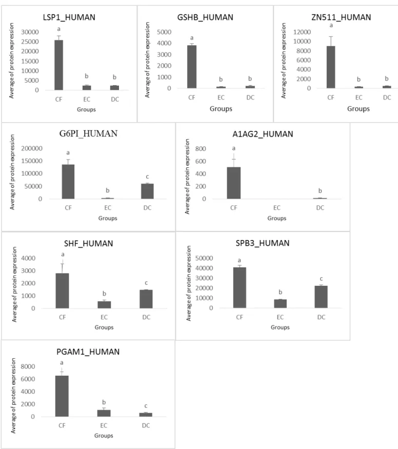
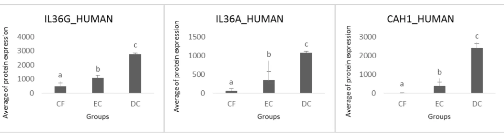
For the 1D-MSE analysis, collectively, 301 quantified proteins (0% FDR) were identified in the saliva samples and 122 proteins were statistically significant different among the three groups of children. The majority of the proteins (84) were identified in all three groups indicating a high overlap in saliva proteins. One protein was expressed only in CF and another in EC, while 11 were expressed only in CF and EC, 15 in CF and DC, 10 in EC and DC. Figure 1 shows a Venn diagram with the number of proteins from each group and their overlaps among the three groups of patients. Of the 122 proteins mentioned above, 54 proteins were at least 5 times more expressed in the comparison between the groups and were considered as candidates for biomarkers.From these 54, 18 proteins with potential mechanism of action related to dental caries were selected (Table 1).
Within this group of 18 proteins, eight were detected with greater expression in caries-free patients (Figure 2). The Lymphocyte-specific protein 1 (LSP1), Glutathione synthetase (GSHB) and Zinc finger protein 511 (ZN511) were more expressed in CF, with no statistical differences between groups EC and DC. Alpha-1-acid glycoprotein 2 (A1AG2), Serpin B3 (SPB3), Glucose-6-phosphate isomerase (G6PI) and SH2 domain-containing adapter protein F (SHF) were more expressed in the CF, but again had their increased levels in DC, and one, the Phosphoglyceric acid mutase (PGAM1), had their lowest expression levels when the caries severity was increasing.
Peroxiredoxin-4 (PRDX4) and Myeloblastin (PRTN3) were more expressed in CF and DC with no significant statistical variation (Figure 3). NK cell receptor 2B4 (CD244) has much greater expression in EC patientes than in children with no caries and with dentine caries, but the amount of such molecule in these tow groups were also significantly distinct (Figure 4). Alpha-amylase 2B (AMY2B) and Gamma-enolase (ENOG) were expressed in CF and EC, but were expressed much more in DC (Figure 5).
Figure 1. Venn diagram of salivary proteins identified in each group and across groups. GROUP I
GROUP II
GROUP III 1
1
15 10
11
84
E2AK4 SH3L2
A2GL, ACTG, AKA10, AMY1, CAPG, GDIR2, GOLM1, ISKS,
PZP, TIMP1, CAP1
1433Z, AL3A1, BPIB2, FYN, GRP78, GSHB, HOP, LSP1, LV301, PDIA1, PITX1, TRI77, ZN511, CIAO1, DOPD, EST2, PGAM1, ACTBM, G6PI, K0125, RS12, SHF, SPB3, FIBA, HPTR, ZNHI2, CD244, RIR2, HAUS4, HV304, AHSA1, CS071, FOLR1, HUWE1, IFN16, K6PL, NEUT, POTEI, AAGAB, AMY2B, CALU, ENOG, SNX12, CAH1, IL36A, IL36G, 1433G, ECM1, I36RA, NKX28, PLST, S10A7, NAA38, ACTN4, ALDOC, B2MG, CO3,
FBX50, FXYD8, HV207, HV320, IF2B, IGHG1, KV101, LEG3, MMP9, NUCB2, PAL4A, PERM, PITH1,PRDX4, PRRP, PRTN3, S10AB, SAP, STPG1,
TCO1, TFF3, TNNC2, ZN727, AIMP1, KLH13, KLHL8, SAA1 ACTS, AK1BA, HV319,
LV102, MAAT1, PLXA4, SH3L1, A1AG2, ALBU, ASPG, CAMP, FKB1A, HBB, IRF7, PRAC1
CAN2, HV303, MBD3, CATL1, DHB2, FXL20, IGHG2, KV205,
Figure 3. Expression of proteins in saliva from caries-free children (CF), children with enamel caries (EC) and dentine caries (DC). Different letters represent significant statistical variations (p < 0.05). Protein names refer to those described in Table 1.
Figure 5. Expression of proteins in saliva from caries-free children (CF), children with enamel caries (EC) and dentine caries (DC). Different letters represent significant statistical variations (p < 0.05). Protein names refer to those described in Table 1.
Figure 7. Expression of proteins in saliva from caries-free children (CF), children with enamel caries (EC) and dentine caries (DC). Different letters represent significant statistical variations (p < 0.05). Protein names refer to those described in Table 1.
Table 1. Proteins with potential mechanism of action related to dental caries
Name Abbreviation
Lymphocyte-specific protein 1 LSP1_HUMAN
Glutathione synthetase GSHB_HUMAN
Zinc finger protein 511 ZN511_HUMAN
Glucose-6-phosphate isomerase G6PI_HUMAN
Alpha-1-acid glycoprotein 2 A1AG2_HUMAN
SH2 domain-containing adapter protein F SHF_HUMAN
Serpin B3 SPB3_HUMAN
Phosphoglyceric acid mutase PGAM1_HUMAN
Peroxiredoxin-4 PRDX4_HUMAN
Myeloblastin PRTN3_HUMAN
NK cell receptor 2B4 CD244_HUMAN
Alpha-amylase 2B AMY2B_HUMAN
Gamma-enolase ENOG_HUMAN
Serum amyloid A-1 protein SAA1_HUMAN
Aminoacyl tRNA synthase complex-interacting multifunctional protein 1
AIMP1_HUMAN
Interleukin-36G IL36G_HUMAN
Interleukin-36A IL36A_HUMAN
DISCUSSION
Recent developments of mass spectrometry methodologies associated with tools of bioinformatics have opened new avenues for the characterization of protein profiles of biological samples. Using such technologies, we have conducted the first global analysis of the saliva proteome in children with early childhood caries in different stages of severity. Some attempts to conduct such analysis have already been made before [20,21] but with limited success, probably because of the use of less sensitive techniques and the lack of patients with caries at an early stage (enamel caries - ICDAS 1, 2 and 3). In the present study, thus, we have successfully employed a proteome approach to identify specific salivary biomarkers of specific types of caries in six-year old children.
The identification of saliva proteins occurred by using a very sensitive technique and preliminary standardization of sampling that was conducted in an environment inside a refrigerated and protected with organic seal bags room with specific swabs, tubes free of contamination that could interfere with the analysis, centrifugation of samples and immediate cooling to prevent degradation of proteins by temperature. Depletion of albumin and immunoglobulin G was important to avoid masking of low molecular weight proteins and enable their identification. [19]
The proteins Eukaryotic translation initiation factor (E2AK4) and SH3 domain-binding glutamic acid-rich-like protein 2 (SH3L2) were expressed exclusively in groups CF and EC, respectively (Figure 1). Although there is no report in the literature that links these proteins and caries, a deeper investigation of them can clarify their functioning as a biomarker for resistance of caries (E2AK4) and enamel caries (SH3L2).
be a protective factor for Lactobacillus spp. in saliva and thus promote dental caries. Thus, with regard to dental caries,our results suggest a protective effect for these proteins.
Of the eight proteins, four (A1AG2, SPB3, G6PI and SHF) were more expressed in the CF, but again had their increased levels in DC, and one, the PGAM1, had their expression lowest levels when the caries severity was increasing (Figure 2B and 2C). The A1AG2 appears to function in modulating the activity of the immune system during the acute-phase reaction and SPB3 acts as a protease inhibitor which also modulate the host immune response. G6PI is an enzyme involved in activation mechanisms of the host primary immune response while SHF operates in the secondary immune response. This suggests that in healthy individuals this group of proteins is well expressed by not being requested and has diminished with the onset of an infectious process (enamel caries), increasing their levels with the increase of injury (dentine caries), but without expression levels recover to levels of healthy subjects.The PGAM1 stimulates immunoglobulin production that may have an important role in innate host defense.[25] This protein appears to be related to protection of healthy subjects once its expression decreases with increasing severity of the disease up to be less expressed in children with dentine caries.
PRDX4 and PRTN3 were more expressed in CF and DC with no significant statistical variation (Figure 3). The PRXD antioxidant family comprises six isoforms that have recently been shown to have important functions in cellular antioxidant defence[26] and PRTN3 is serine protease that degrades elastin, fibronectin, laminin, vitronectin, and collagen types I, III, and IV (in vitro).[27] The most expressed of PRTN3 in DC can be explained by the dentinal degradation caused by caries Which performs its role in the lysis of collagen. However, its greatest expression in CF, as well as the relationship of PRDX4 with this disease still need to be better investigated and appear to be an unexpected result.
For the group that had enamel caries (EC), CD244 was more expressed (Figure 4).The CD244 is a transmembrane protein belonging to the Ig superfamily, which can also be expressed by CD8+ T cells. This protein may be related to the infectious process started with their increased expression early enamel lesions. For the their behavior on other groups can not be a role defined for it.