RESEARCH ARTICLE
Identification of CTLA2A, DEFB29,
WFDC15B, SERPINA1F and MUP19 as Novel
Tissue-Specific Secretory Factors in Mouse
Jibin Zhang1, Jinsoo Ahn1,2, Yeunsu Suh1, Seongsoo Hwang3, Michael E. Davis1, Kichoon Lee1*
1Department of Animal Sciences, The Ohio State University, Columbus, Ohio, United States of America,
2The Ohio State University Interdisciplinary Ph.D. Program in Nutrition, The Ohio State University, Columbus, Ohio, United States of America,3Animal Biotechnology Division, National Institute of Animal Science, RDA, Gyeonggi, Republic of Korea
Abstract
Secretory factors in animals play an important role in communication between different cells, tissues and organs. Especially, the secretory factors with specific expression in one tissue may reflect important functions and unique status of that tissue in an organism. In this study, we identified potential tissue-specific secretory factors in the fat, muscle, heart, lung, kidney and liver in the mouse by analyzing microarray data from NCBI’s Gene Expression Omnibus (GEO) public repository and searching and predicting their subcellular location in GeneCards and WoLF PSORT, and then confirmed tissue-specific expression of the genes using semi-quantitative PCR reactions. With this approach, we confirmed 11 lung, 7 liver, 2 heart, 1 heart and muscle, 7 kidney and 2 adipose and liver-specific secretory factors. Among these genes, 1 lung-specific gene - CTLA2A (cytotoxic T lymphocyte-associated protein 2 alpha), 3 kidney-specific genes - SERPINA1F (serpin peptidase inhibitor, Clade A, member 1F), WFDC15B (WAP four-disulfide core domain 15B) and DEFB29 (defensin beta 29) and 1 liver-specific gene - MUP19 (major urinary protein 19) have not been reported as secretory factors. These genes were tagged with hemagglutinin at the 3’end and then tran-siently transfected to HEK293 cells. Through protein detection in cell lysate and media using Western blotting, we verified secretion of the 5 genes and predicted the potential pathways in which they may participate in the specific tissue through data analysis of GEO profiles. In addition, alternative splicing was detected in transcripts of CTLA2A and SERPI-NA1F and the corresponding proteins were found not to be secreted in cell culture media. Identification of novel secretory factors through the current study provides a new platform to explore novel secretory factors and a general direction for further study of these genes in the future.
OPEN ACCESS
Citation:Zhang J, Ahn J, Suh Y, Hwang S, Davis ME, Lee K (2015) Identification of CTLA2A, DEFB29, WFDC15B, SERPINA1F and MUP19 as Novel Tissue-Specific Secretory Factors in Mouse. PLoS ONE 10(5): e0124962. doi:10.1371/journal. pone.0124962
Academic Editor:Jonathan Peterson, East Tennessee State University, UNITED STATES
Received:January 17, 2015
Accepted:March 19, 2015
Published:May 6, 2015
Copyright:© 2015 Zhang et al. This is an open access article distributed under the terms of the
Creative Commons Attribution License, which permits unrestricted use, distribution, and reproduction in any medium, provided the original author and source are credited.
Data Availability Statement:All relevant data are within the paper and its Supporting Information files.
Introduction
The secretory factors are a large group of proteins synthesized by ribosomes bound to rough endoplasmic reticulum (ER). During the process of protein synthesis, the proteins are directed from the cytosolic face of the ER membrane to the ER lumen by the ER signal sequences at the N-terminus of the proteins [1]. The proteins in the ER lumen are subsequently packaged into transport vesicles that fuse with the cis-Golgi vesicles. Cis-Golgi vesicles then move toward the plasma membrane and change to trans-Golgi cisternae [2]. Some secretory factors such as hor-mones are stored in secretory vesicles and are only released upon triggers of hormonal or neu-ral signals. Other secretory factors such as those found in the extracellular matrix are
continuously secreted and exist in all cell types [3].
Due to the ubiquitous property of secretory factors, they play an important role in various cells, organs and systems in animals. As increasing numbers of secretory factors are explored, functions of one cell type and tissue and connections between different cell types and tissues become further understood and complex networks in living organisms become extensively re-vealed. For example, fat tissue was once regarded only as a lipid reservoir for excess energy. However, since the discovery of leptin by Zhang et al. [4], perceptions about fat tissue have gradually changed. Leptin is a protein secreted by mature adipocytes, and can regulate food in-take and body fat mass by binding to its receptor in the hypothalamus. Since the identification of leptin, more than 100 secretory factors have been identified that are produced and released by adipose tissue [5]. Among these factors, some adipose-specific secretory proteins such as adiponectin, resistin and visfatin are involved in the immune system [6], while some proteins such as angiotensinogen [7] and plasminogen activator inhibitor type I [8] are related to vascu-lar function. Therefore, adipose tissue is now thought of, not only as a lipid-storing organ, but also as an endocrine organ that maintains intensive cross talk with other organs.
Since tissue-specific expression of one novel secretory factor usually indicates novel function of that tissue, it is necessary to explore novel tissue-specific secretory factors. Tissue specific ex-pression of various genes can be detected by microarrays with measurements of transcript abundance in various tissues. There are thousands of microarray data records from various studies with open access in the Gene Expression Omnibus (GEO) database on the NCBI web-site, providing a useful tool to explore tissue-specific genes and predict their functions [9]. In our previous study we successfully selected some novel tissue-specific genes in the human and mouse and predicted their potential function by taking advantage of this database [10]. The subcellular location of various proteins can also be found in some databases such as GeneCards [11] and UniProt [12] or predicted by WoLF PSORT [13]. These powerful bioinformatic tools greatly facilitate the selection of novel secretory factors with tissue specific expression.
The objective of this study is to identify and evaluate novel tissue-specific secretory genes in the fat, muscle, heart, lung, liver and kidney in the mouse by performing microarray data anal-ysis, literature search, protein location information search and prediction, semi-quantitative PCR analysis and Western blotting after transfection of expression vectors in cell culture. With the bioinformatics approach developed by Song et al. [10], we detected novel tissue-specific genes in the 6 tissues and filtered potential secretory genes by searching and predicting protein location. After confirmation of tissue-specific expression through semi-quantitative PCR anal-ysis, 30 genes were identified in 6 tissues in adult mice. Among them, 11 genes are lung-specif-ic; 7 genes are liver-speciflung-specif-ic; 2 genes are heart-speciflung-specif-ic; 1 gene is muscle and heart-speciflung-specif-ic; 7 genes are kidney-specific and 2 genes are adipose and liver-specific. After the literature study, 5 novel tissue-specific secretory genes were discovered: CTLA2A (cytotoxic T lymphocyte-asso-ciated protein 2 alpha) in the lung, SERPINA1F (serpin peptidase inhibitor, Clade A, member 1F), WFDC15B (WAP four-disulfide core domain 15B), DEFB29 (defensin beta 29) in the
Grants Program no. 2014-090 and 2013-044 at The Ohio State University: JA and KL,http://oardc.osu. edu/seeds/t08_pageview/Home.htm. The funders had no role in study design, data collection and analysis, decision to publish, or preparation of the manuscript.
kidney, and MUP19 (major urinary protein 19) in the liver tissue. The secretory properties of these proteins were detected by Western blot analysis using cell lysate and medium after tran-sient transfection of expression vectors to HEK293 cells. The biological activities in which these genes may participate in the specific tissues were predicted according to GEO profiles under different physiological and pathological conditions. This study provides a novel strategy combining bioinformatics tools and molecular technologies for exploration of novel tissue-spe-cific secretory genes.
Materials and Methods
Experimental Animals
Mice raised in a mouse housing facility at The Ohio State University with free access to feed were euthanized by CO2inhalation and subsequent cervical dislocation at 3 months of age.
Then white adipose tissue, muscle, heart, lung, liver and kidney were harvested (n = 3). All pro-cedures were approved by the Institutional Animal Care and Use Committee (IACUC) of The Ohio State University. After tissue collection, all of the tissues were snap-frozen immediately in liquid nitrogen and stored at -80°C for total RNA isolation.
Data Mining and Literature Search
In order to find the secretory factors that are highly expressed in the adipose tissue, muscle, heart, lung, liver and kidney in the adult mouse, the microarray expression records in GDS3142, which is one Gene Expression Omnibus DataSet (GDS) available on the NCBI web site, were analyzed following the methods proposed by Song et al. [10]. In brief, expression rec-ords for each gene in the 6 different tissues were derived and ratios were calculated between ex-pression in each tissue and average exex-pression in the other 5 tissues; so there was one list of ratios for each tissue. After ranking the genes in descending order according to each list of ra-tios, the top 200–300 genes were selected for each tissue. For genes with several spots on the microarray, only one record was kept. Then, each of these genes was checked in Genecards (http://www.genecards.org/) and UniProt (http://www.uniprot.org/) and predicted by WoLF PSORT (http://www.genscript.com/psort/wolf_psort.html) to see whether the corresponding protein was secreted or not. Finally, only the secretory genes were kept and a literature search was conducted in Pubmed (http://www.ncbi.nlm.nih.gov/pubmed) and Google Scholar (http:// scholar.google.com/) to check whether there are reports concerning high expression of each gene and secretion of the protein in certain tissues. After the 5 novel secretory factors were se-lected, their functions in specific tissues were further predicted based on analysis of various GEO profiles. Function of CTLA2A was predicted based on GDS3950, GDS4582, GDS4914 and GDS2709; function of MUP19 was predicted based on GDS1261, GDS1053, GDS279, GDS1517 and GDS1374; function of SERPINA1F was predicted based on GDS2031, GDS4316 and GDS3675; function of WFDC15B was predicted based on GDS2030, GDS4316, GDS3675, GDS2817, GDS1583 and GDS4449; and function of DEFB29 was predicted based on
GDS2031, GDS4316, GDS1583, GDS4839 and GDS3612.
cDNA Synthesis and Semi-quantitative PCR Analysis
For the genes whose tissue-specific expressions are not reported in existing publications, we conducted a semi-quantitative PCR to detect their expression in different tissues. Total RNA was isolated from the adipose tissue, muscle, heart, lung, liver and kidney of the adult mice using Trizol reagent (Invitrogen, Carlsbad, CA, USA) and then reverse-transcribed to cDNA using moloney murine leukemia virus (M-MLV) reverse transcriptase (Invitrogen) and oligo
dT. The conditions for reverse transcription were 65°C for 5 min, 37°C for 50 min and 70°C for 15 min. After the RNA was reverse-transcribed to cDNA, PCR was conducted with 1μL of cDNA, 0.2μL of 10mM deoxynucleotide triphosphate mix (dNTP), 1μL of 10×ThermopoIII (Mg-free) reaction buffer, 0.2μL of 100 mM MgSO4, 0.2 nM of each of the forward and reverse
primers, 0.05μL of Taq DNA polymerase (New England BioLabs, Ipswich, MA, USA) and nu-clease-free water up to 10μL. The cycling parameters for PCR were 95°C for 1 min, followed by 32 to 40 cycles of 94°C for 30 s, 58°C for 30 s and 72°C for 40 s with a final elongation for 10 min. To ensure that equal amounts of cDNA were added to the PCR reaction for different tissues, expression of the cyclophilin (CYC) gene was used as a control and cDNA was diluted until the PCR products of CYC showed similar brightness for different tissues in agarose gel electrophoresis. After the equalization of cDNA, PCR was conducted for the other genes. The PCR primers for all of the genes are listed inS1 Table.
Vector Construction and Cell Transfection
The subcellular locations of 5 of the selected genes were not found in the literature. To verify secretion of their encoding proteins from cells, we constructed plasmids containing target genes to transfect cells to detect the location of expressed protein. First, coding sequences of these genes were amplified with primers shown inS2 Table. For CTLA2A, 2 forward primers were used to amplify 2 transcripts with alternative start codons. Hemagglutinin (HA) tag se-quence, which is underlined in the table, was linked to the 5’end of all of the reverse primers to add the HA-tag to the C terminal of the target proteins. After gel extraction of the PCR prod-uct, the amplified sequences were ligated to the pCR2.1 vector (Invitrogen) using T4 DNA li-gase (New England BioLabs). Then, the plasmid with an insertion of each PCR product was transformed toE.colicells, which were then spread over plates containing 5-bromo-4-chloro-3-indolyl-beta-D-galacto-pyranoside (X-gal). Because the lacZ gene in the plasmid is disrupted by inserted sequences, the colonies were white, whereas, the plasmids with no insertion had in-tact lacZ genes expressingβ-galactosidase, and its reaction with X-gal stained the colony in a blue color. After selection of positive colonies and incubation in Luria-Bertani (LB) broth, plas-mids were extracted using a miniprep Kit (QIAGEN, Valencia, CA, USA) and the direction of inserted genes was verified through restriction enzyme digestion and PCR with M13 primers and the primers of inserted genes. The plasmids with correct orientation of insertion were then digested with HindIII and XhoI (New England BioLabs) and ligated to a pcDNA 3.1 vector, which was also digested with the same enzymes. The constructed vectors were then trans-formed to E. coli and insertion was verified by digestion with HindIII and XhoI and subsequent electrophoresis in 0.6% agarose gels. After confirmation of the correct ligation, the constructed vectors were extracted using a plasmid midi kit (QIAGEN).
Transient Transfection of Plasmids in HEK 293 Cells
Western Blot Analysis
After collecting the cell medium, proteins expressed in HEK293 cells were extracted using ice-cold 1× lysis buffer (125 mM Tris-HCl pH 6.8, 0.5% SDS). Proteins in the medium were precip-itated using Tricholoroacetic acid (TCA). In brief, the medium was spun down at 1,200 × g to pellet the cell debris and the supernatant was centrifuged at 16,000 × g after adding TCA (25% v/v). The protein pellet was washed 2 times with ice-cold acetone, and then resolved in 2X loading buffer. Equal amounts of proteins were loaded and separated by 10–15% SDS-PAGE and wet-transferred to polyvinylidene difluoride (PVDF) membranes (Bio-Rad, Hercules, CA). After blocking for 30 min in Tris-buffered saline with Tween-20 (TBST) containing 4% nonfat dry milk, the membranes were incubated overnight at 4°C with mouse anti-HA-tag (1:3000; Cell Signaling, Danvers, MA) antibody as a primary antibody. Horseradish peroxidase-linked anti-mouse IgG (1:5000; Cell Signaling) was used as a secondary antibody for an 1-h incuba-tion at room temperature. After detecting signals with ECL plus reagents (GE Healthcare Bio-sciences, Pittsburgh, PA), proteins were visualized by exposure of the membranes to X-ray films (GE Healthcare Biosciences).
Statistical Analysis
A mixed model (MIXED) procedure was performed for comparison among more than 2 groups using SAS software (version 9.3, SAS Institute Inc., USA). The DIFF command was used to detect significant differences between pairs of least squares means. Student’s t-test was performed in JMP 10 to compare gene expression between 2 groups. Differences with p-values lower than 0.05 were treated as significant. Statistical values are presented as least squares means with standard errors of the least squares means (SEM).
Results
Discovery of Tissue-Specific Secretory Genes
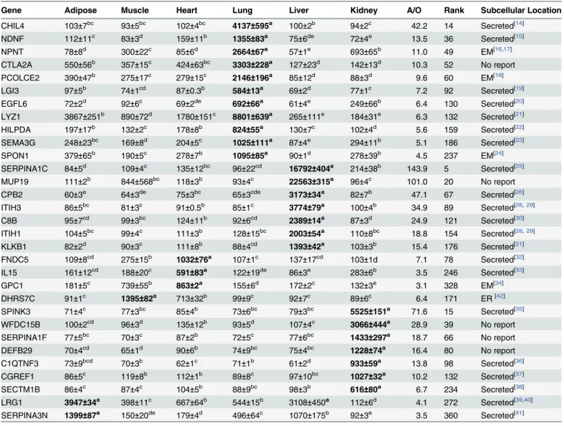
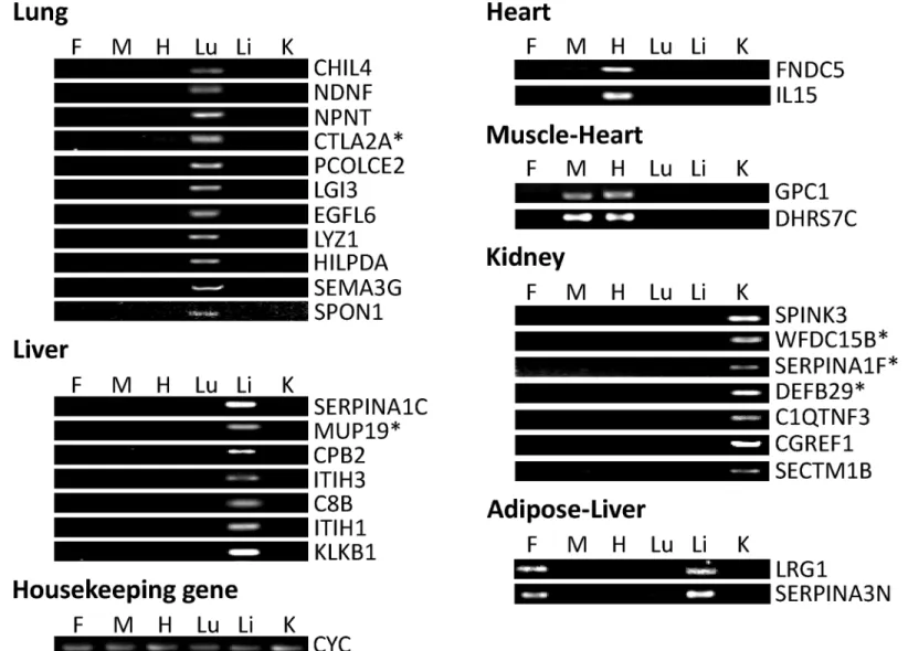
Six Excel spreadsheet files were generated with the top 200–300 genes in terms of highest ex-pression in the fat, muscle, heart, lung, liver and kidney in the adult mice based on GDS3142. After conducting searches of the Genecards and Uniprot websites, protein subcellular localiza-tion prediclocaliza-tion in WoLF PSORT and literature searches in Pubmed and Google Scholar, non-secretory genes and known non-secretory genes with reported tissue-specific expression consistent with our findings in GDS3142 were deleted and 31 genes were kept with their average expres-sion values in 6 different tissues from GDS3142 listed inTable 1. Among these genes, 11 are more highly expressed in the lung, 7 genes are more highly expressed in the liver; 2 are more highly expressed in the heart, and one—(GPC1) shows nearly equal expression in heart and muscle; one gene (DHRS7C) shows the highest expression in the muscle without much differ-ence in expression in the heart; 7 genes are expressed more highly in the kidney; 2 genes show the highest expression in adipose with little difference in expression in the liver. The expression patterns of these genes were confirmed by semi-quantitative PCR and gel electrophoresis. As shown inFig 1, both adipose-specific genes showed similar expression in adipose tissue and liver; DHRS7C and GPC1 showed similar expression in muscle and heart; whereas, all other genes only showed detectable expression in one specific tissue.
As shown inTable 1, 25 of the selected genes have been reported to produce extracellular proteins. In the lung-specific genes, CHIL4 encodes Chitinase-like protein 4, which is also called secreted Ym2 protein. It has been identified in respiratory secretions and reported to be increasingly expressed in the allergic lung [14]. Neuron-Derived Neurotrophic Factor (NDNF) was reported to be a secreted glycosylated protein by Kuang et al. [15], but its secretion was
reported in the brain and neuron cell culture rather than in the lung. NPNT encodes nephro-nectin, which is an extracellular matrix protein and has been reported to be secreted from the embryonic kidney [16] and the MC3T3-E1 cell line [17]. PCOLCE2 encodes procollagen C-en-dopeptidase enhancer 2, a collagen-binding protein that was reported to be secreted in trans-fected human 293 EBNA-1 cells [18]. Leucine-rich glioma inactivated 3 (LGI3) is a secreted leucine-rich repeat protein. It was reported to be highly expressed in the brain and, thus, has been extensively studied in the nervous system [19], but there is no report in lung. EGFL6 en-codes an epidermal growth factor–like protein, which has been reported to be secreted in an os-teoblastic-like cell culture [20]. LYZ1 encodes lysozyme, which has been reported to be
primarily secreted by serous cells of submucosal glands in the lung [21]. Hypoxia Inducible Lipid Droplet-Associated (HILPDA) was reported to be secreted in COS7 cell culture, but its Table 1. Gene expression values of selected genes.
Gene Adipose Muscle Heart Lung Liver Kidney A/O Rank Subcellular Location
CHIL4 103±7bc 93±5bc 102±4bc 4137±595a 100±2b
94±2c 42.2 14 Secreted[14] NDNF 112±11c 83±3d 159±11b 1355
±83a 75±6de 72±4e 13.5 36 Secreted[15]
NPNT 78±8d 300±22c 85±6d 2664
±67a 57±1e 693±65b 11.0 49 EM[16,17]
CTLA2A 550±56b 357±15c 424±63bc 3303
±228a 127±23d 142±13d 10.3 52 No report
PCOLCE2 390±47b 275±17c 279±15c 2146±196a 85±12d
88±3d 9.6 60 EM[18]
LGI3 97±5b 74±1cd 87±0.3b 584
±13a 69±2d 77±1c 7.2 92 Secreted[19]
EGFL6 72±2d 92±6c 69±2de 692
±66a 61±4e 249±66b 6.4 130 Secreted[20]
LYZ1 3867±251b 890±72d 1780±151c 8801
±639a 265±111e 184±31e 6.3 132 Secreted[21]
HILPDA 197±17b 132±2c 178±8b 824±55a 130±7c
102±4d 5.6 159 Secreted[22] SEMA3G 248±23bc 169±8d 204±5c 1025
±111a 87±4e 294±11b 5.1 186 Secreted[23]
SPON1 379±65b 190±5c 278±7b 1095
±85a 90±1d 278±39b 4.5 237 EM[24]
SERPINA1C 84±5d 109±4c 135±12bc 96±22cd 16792
±404a 214±38b 143.9 5 Secreted[25]
MUP19 111±2b 844±568bc 118±3b 93±4c 22563±315a 96±4c
101.0 20 No report CPB2 60±3e 64±3de 75±3bc 65±3cde 3173
±34a 82±7b 47.1 67 Secreted[26]
ITIH3 86±5bc 81±3c 91±0.5b 85±1c 3774
±79a 100±4b 34.9 89 Secreted[28,29]
C8B 95±7cd 99±3bc 124±11b 92±6cd 2389
±14a 87±3d 24.9 121 Secreted[30]
ITIH1 104±5bc 99±4c 111±3b 128±15bc 2003±54a 110±8bc
18.8 154 Secreted[28,29] KLKB1 82±2d 90±3c 111±8b 88±4cd 1393
±42a 103±3b 15.4 176 Secreted[31]
FNDC5 109±8cd 275±15b 1032
±76a 107±1c 137±17cd 103±1d 7.1 78 Secreted[32]
IL15 161±12cd 188±20c 591
±83a 122±19de 86±3e 283±6b 3.5 246 Secreted[33]
GPC1 181±5c 739±55b 863±2a 155±6d
172±2c 132±3e 3.1 328 EM[34]
DHRS7C 91±1c 1395
±82a 713±32b 99±9c 92±7c 89±6c 6.4 171 ER[42]
SPINK3 71±4c 77±3bc 85±4b 73±6bc 79±3bc 5525
±151a 71.6 15 Secreted[35]
WFDC15B 100±2cd 96±3d 135±12b 93±5d 107±4c 3066
±444a 28.9 39 No report
SERPINA1F 77±5bc 70±3c 87±2b 72±5c 77±6bc 1433±297a 18.7 66 No report
DEFB29 70±4cd 65±1d 90±6b 74±9bc 75±4bc 1228
±74a 16.4 80 No report C1QTNF3 73±9bcd 70±3b 62±1c 71±1b 61±2d 933
±59a 13.8 98 Secreted[36]
CGREF1 86±5c 119±8b 112±1b 89±8c 97±10bc 1027
±32a 10.2 132 Secreted[37]
SECTM1B 86±4c 87±4c 104±5b 88±9bc 98±3b 616±80a 6.7 234 Secreted[38]
LRG1 3947±34a 398±11c 667±64b 544±15b 3108±450a 112±6d 4.1 272 Secreted[39,40]
SERPINA3N 1399±87a 150±20de 179±4d 496±64c 1070±175b 92±3e 3.5 360 Secreted[41]
a-eDifferent superscripts within a row indicate signi
ficant difference (P<0.05).
The highest expression value for each gene is highlighted in bold font. A/O: ratio between average expression of certain gene in tissue with specific expression of that gene and expression in other tissues. EM: Extracellular matrix; ER: Endoplasmic reticulum.
expression was only reported in fetal kidney and renal cell carcinoma [22]. SEMA3G encodes semaphorin 3G-a member of murine class 3 semaphorin. It is expressed in the lung and kidney and secreted in HEK293-T cells [23]. SPON1 encodes F-spondin, which is an extracellular ma-trix protein secreted in the floor plate in the embryonic neural tube [24]. Its secretion and func-tion have been extensively studied in the nervous system. However, there are few reports about this gene in the lung.
Among the liver-specific genes, SERPINA1C encodes one isoform of murine a-1-antitryp-sin, which is produced in the liver, secreted in serum and circulated to the lung [25]. Secretion of SERPINA1C has also been reported by Paterson and Moore [26]. CPB2 encodes plasma Carboxypeptidase B2, which is secreted from the liver [27]. Both ITIH1 and ITIH3 encode one heavy chain of inter-alpha-trypsin inhibitor (ITI), which is one plasma protease inhibitor, and is synthesized and secreted in a hepatoma HepG2 cell culture [28] and COS7 cell culture [29]. C8B encodes theβsubunit of complement component C8, which is a component of the Fig 1. Expression of adult mouse gene transcripts detected by semi-quantitative PCR.Expressions of selected genes in fat (F), muscle (M), heart (H), lung (Lu), liver (Li) and kidney (K) were detected by PCR reaction and 1% agarose gel electrophoresis. Housekeeping gene—cyclophilin (CYC)—was used as a loading control. Genes without reported subcellular locations are marked with asterisks.
doi:10.1371/journal.pone.0124962.g001
membrane attack complex (MAC) and has been reported to be secreted by hepatocytes into serum. The a, b, and g subunits normally associate with each other before secretion [30]. KLKB1 encodes plasma kallikrein, which is synthesized in the liver [31].
Among heart-specific genes, FNDC5 encodes fibronectin type III domain-containing protein 5, which is a membrane protein proteolytically cleaved and secreted as a hormone peptide— Iri-sin. Irisin is mainly secreted by cardiac and skeletal muscle, with the former producing more than the latter [32]. IL15 encodes interleukin 15, whose production has been detected in various tissues. This gene produces 2 different isoforms through alternative splicing. The 21-aa signal isoform mRNA is expressed in the heart, thymus, appendix and testis, translated efficiently but not secreted. The 48-aa signal isoform mRNA is expressed in skeletal muscle, placenta, heart, lung, liver, thymus and kidney, translated less efficiently and secreted at a low level [33]. GPC1 encodes glypican-1, which is a cell membrane heparan sulfate proteoglycan. It is attached to the cell membrane by a glycosylphosphatidylinositol (GPI) linkage. The GPI anchor can be cleaved and thus lead to secretion of glypican-1 or shedding into the extracellular matrix [34].
Among the kidney-specific genes, SPINK3 encodes pancreatic secretory trypsin inhibitor in the mouse. It is produced in acinar cells in the pancreas and secreted with digestive enzymes into secretory granules [35]. However, its secretion in the kidney has not been reported. C1QTNF3 encodes C1q/TNF-related Protein-3, which is an adipokine, and, thus, has been studied extensively in adipose tissue and cell lines [36]. However, it is rarely reported in the kidney. CGREF1, which is also known as CGR11, encodes hydrophobestin. This protein has been reported to be secreted in MtT/S cell culture [37]. SECTM1B encodes secreted and trans-membrane protein 1. This protein shows a perinuclear Golgi-like pattern; its N-terminal do-main was reported to be secreted in peripheral blood leukocytes and breast cancer cell lines [38]. However, there are no reports about this gene in the kidney.
As shown by semi-quantitative PCR, both LRG1 and SERPINA3N are highly expressed in both liver and adipose tissue (Fig 1). However, they are only reported to be secreted in liver. Leucine-Rich Alpha-2-Glycoprotein (LRG1) is a plasma glycoprotein primarily produced in the liver [39]. It is also synthesized and secreted by ovarian cancer cells [40]. SERPINA3N is one isoform of murineα-1 antichymotrypsin (ACT), which has also been reported to be highly expressed in the liver. ACT is primarily synthesized and secreted by hepatocytes and released in plasma. In addition, SERPINA3N is reported to be secreted in Sertoli cell cultures [41].
Among the remaining 6 genes, DHRS7C is located in the sarcoplasmic reticulum of the heart and skeletal muscle [42], although it is predicted to be secreted by WoLF PSORT. The other 5 genes—CTLA2A in the lung, MUP19 in the liver and WFDC15B, SERPINA1F and DEFB29 in the kidney have not been reported to be secreted in currently available publications.
Identification of Alternative Splicing of CTLA2A and SERPINA1F
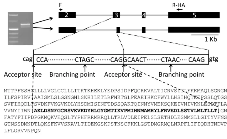
which contains an alternative acceptor site, resulting in deletion of 165 nt in mRNA and 55 amino acids in protein.
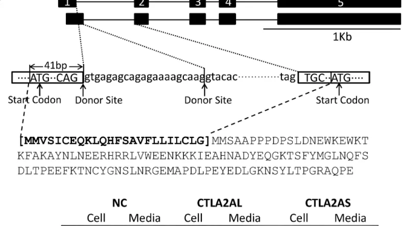
The alternative splicing in both genes fits the classical splicing model outlined by Keller and Noon [43] and Padgett et al. [44]. In the classical model, the consensus sequence at the donor site should usually be AG at the 3’end of an exon followed by GT at the beginning of the adja-cent intron. This feature is located not only at the 5’end of intron 1, but also 21 bp from the 5’
end of intron 1 of CTLA2A, making alternative splicing feasible. On the other hand, the con-sensus sequence at the acceptor site is usually AG at the 3’end of an intron followed by G at the beginning of adjacent exon. The sequence CTA/GAC/T at the branching point should be 20–50 base pairs from the 3’splice site. These features are located not only in the 3’end but also in the middle area of exon 3 in Serpina1f, which facilitates the alternative splicing. Fig 2. Different isoforms of CTLA2A and their expression in HEK293 cell culture. A,alternative splicing of CTLA2A that leads to change in protein sequence.B,cell lysates and conditioned medium from HEK293 cells transiently transfected with HA-tagged murine expression vector of long isoform (CTLA2AL) and short isoform (CTLA2AS) and empty pcDNA3.1 vector as negative control (NC) were detected using Western blotting.
doi:10.1371/journal.pone.0124962.g002
Detection of Novel Secretory Genes in HEK293 Cell Culture
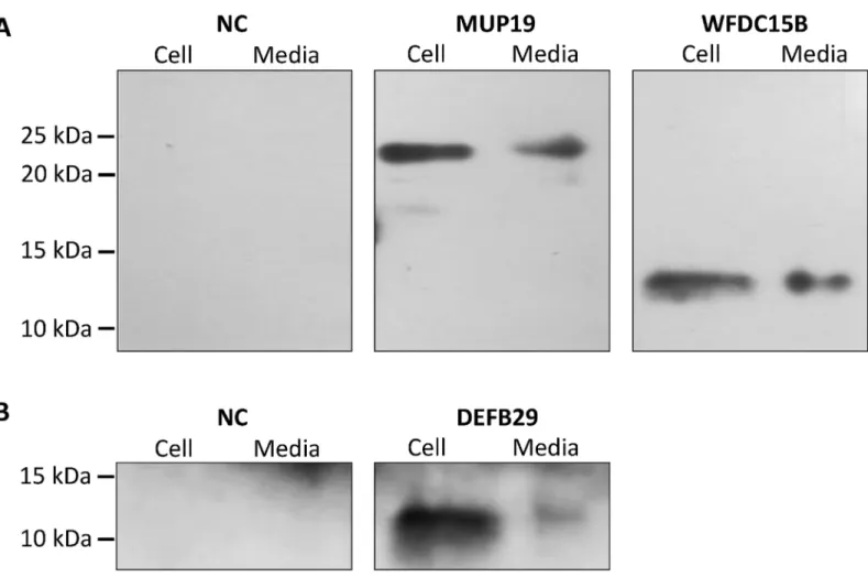
Western blotting demonstrates that CTLA2A with full-length amino acids is secreted as it was detected in both cell lysate and conditioned media. However, the short isoform with deletion of N-terminal amino acids is not secreted as predicted by WoLF PSORT (Fig 2B), because it lacks signal peptide. For SERPINA1F, the long isoform is detected in both cell lysate and conditioned media, indicating that it is secreted outside the cells. However, the short isoform, which lacks 55 amino acids in the middle region of the protein, is not secreted, because it was only detected in cell lysate (Fig 3B). WFDC15B, DEFB29 and MUP19, which do not have alternative splicing, are also detected in both cell media and lysate (Fig 4), indicating their secretion out of cells. Fig 3. Different isoforms of SERPINA1F and their expression in HEK293 cell culture. A,alternative splicing of SERPINA1F that leads to double bands in gel electrophoresis of PCR product and change in protein sequence.B,cell lysates and conditioned medium from HEK293 cells transiently transfected with HA-tagged murine expression vector of long isoform (SERPINAL) and short isoform (SERPINAS) and empty pcDNA3.1 vector as negative control (NC) were detected using Western blotting.
Finally, for the cells transfected with empty vectors, no protein was detected. This validates the successful insertion of target genes into the vector and expression of target genes in HEK293 cells.
GEO Data Analysis of Novel Secretory Genes
For the 5 novel secretory genes, there are few reports about their temporal and spatial expres-sion, as well as function, in the tissues where they are highly expressed. In order to understand potential functions of these genes, gene expressions were compared between different age groups, different tissue sections and different treatment groups or genetic lines by analyzing GEO profiles.
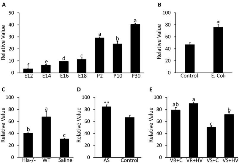
Expression of CTLA2A in the lung seems to be affected by bacterial infection and other in-juries. As can be seen fromFig 5A, GDS3950 shows that CTLA2A expression exhibits an in-creasing trend during development of the mouse lung except for a slight decrease on postnatal d 10. In particular, there is a dramatic increase in expression after the mouse is born
(P<0.0001). GDS4583 shows that expression of CTLA2A in the lungs of C57BL/6 mice
in-creased 1.6 times (P<0.05) after intratracheal instillation ofEscherichia coli(E.coli) (Fig 5B).
In another case, GDS4582 suggests that CTLA2A expression is promoted more than 2 times Fig 4. Detection of protein expression of MUP19, WFDC15B and DEFB29 in HEK293 cell culture. A,Cell lysates and conditioned medium from HEK293 cells transiently transfected with HA-tagged murine expression vector of MUP19 and WFDC15B and empty pcDNA3.1 vector as negative control (NC) were detected using Western blotting.B,Cell lysates and conditioned medium from HEK293 cells transiently transfected with HA-tagged murine expression vector of DEFB29 and empty pcDNA3.1 vector as negative control (NC) were detected using Western blotting.
doi:10.1371/journal.pone.0124962.g004
(P<0.05) 24 h after infection by wildtypeStaphylococcus aureus (S.aureus). On the other hand,
deletion of Alpha-hemolysin (Hla)—an essential lethal factor secreted byS.aureus-reduced expression of CTLA2A to a medium level (Fig 5C). In GDS4914, expression of CTLA2A in the lungs of C57BL/6 mice increased 1.3 times (P<0.01) after exposure to 50 ppm of arsenate for
90 d compared to the control group (Fig 5D). A similar change occurred in GDS2709 as well (Fig 5E). In a rat strain resistant to ventilator-associated lung injury (VALI), expression of CTLA2A is significantly higher (P<0.05) than that in a strain sensitive to VALI under the
same treatment. In the VALI sensitive strains, high tidal volume ventilation induced expression of CTLA2A 1.4 times (P<0.001) compared to the control group.
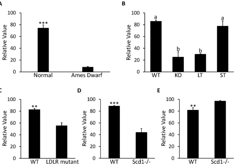
The correlation between MUP19 and the GH/IGF-1 signaling pathway is demonstrated by GDS1261 and GDS1053. According to GDS1261, expression of MUP19 was reduced more than 90% in Ames dwarf mice with mutant pituitary specific transcription factor 1 (PIT-1)—
one key regulator in the GH/IGF-1 signaling pathway (Fig 6A). In GDS1053, knockout of the Fig 5. Expression profile in lung for CTLA2A in microarray DataSets obtained from NCBI website. A,Expression in mice on embryonic day 12 (E12), 14 (E14), 16 (E16) and 18 (E18) and postnatal d 2 (P2), 10 (P10) and 30 (P30) in GDS3950 (n = 2 per time point).B,Expression in mice infected byE.coliand control group in GDS4583 (n = 3 per group).C,Expression in mice after 24-h infection byS.aureusdeficient in alpha-hemolysin (Hla-/-) and wild type S. aureus (WT) with saline treated mice as control group (n = 3 per group).D,Expression in mice after 90-d exposure to 50 ppm arsenate (AS) with untreated mice as control in GDS4914 (n = 5 per group).E,Expression in resistant rat strain (VR) to ventilator-associated lung injury (VALI) and sensitive rat strain (VS) to VALI under treatment of high tidal volume ventilation (HV) with untreated mice as control (C) group in GDS2709 (n = 3 per group). Each bar represents mean±SEM. Statistical difference is indicated by different letters (P<0.05),*(P<0.05) and**(P<0.01) above the bars.
growth hormone receptor (GHR) led to a 60% decrease in MUP19 expression (P<0.001) in the
liver of 42-d old mice. In addition, when the GHR was truncated at residue 596, there was not a significant decrease in MUP19; when GHR was truncated at residue 391, the decrease in MUP19 became 65% (P<0.0001) compared to the wild type mice (Fig 6B). Expression of
MUP19 is also affected by deficiency of low density lipoprotein (LDL) receptors. In GDS279, expression of MUP19 in the liver was repressed by 33% in C57BL/6 mice lacking LDL receptors compared to the normal mice after high fat diet treatment for 12 wk (Fig 6C). The correlation between MUP19 and Stearoyl-CoA desaturase 1 (SCD1) seems to be affected by food intake. In GDS1517, expression of MUP19 decreased 50% in livers of mice with SCD1 deficiency com-pared to wild type mice under treatment of a low fat, high carbohydrate diet (P<0.001,Fig 6D). However, in GDS1374, where the SCD1-/- mice were fed a chow diet, the expression of MUP19 was increased 1.2 times compared to the wild type mice (P<0.01,Fig 6E).
Fig 6. Expression profile in liver for MUP19 in microarray DataSets obtained from NCBI website. A,Expression in Ames Dwarf mice (n = 8) and normal mice (n = 7) with free access to standard food in GDS1261.B,Expression in 42-d old mice with growth hormone receptor knocked out (KO), truncated at amino acid residue 569 (ST) and 391 (LT) with wild type (WT) mice as control group in GDS1053 (n = 3 per group).C,Expression in C57BL/6 mouse with null mutant low-density lipoprotein receptor (LDLR) under the treatment of high fat diet for 12 wk along with WT mice as control group in GDS279 (n = 3 per group).D,Expression in mice with stearoyl-CoA desaturase 1 deficient mutants (Scd1-/-) under treatment of low-fat, high-carbohydrate diet along with WT mice as control group in GDS1517 (n = 5 per group).E,Expression in 6-wk Scd1-/- mice fed with chow diet along with WT mice as control group in GDS1374 (n = 5 per group). Each bar represents mean±SEM. Statistical difference is indicated by different letters (P<0.05),**(P<0.01) and***(P<0.001) above the bars.
doi:10.1371/journal.pone.0124962.g006
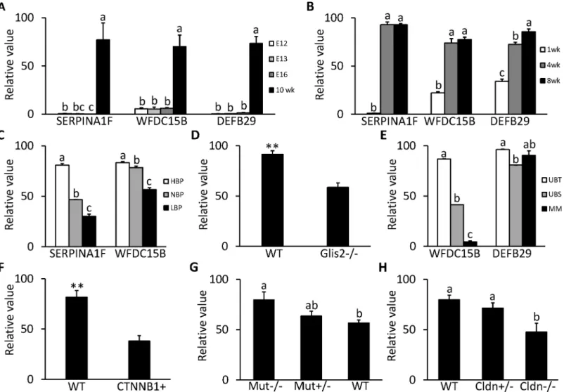
SERPINA1F, WFDC15B and DEFB29 are all highly expressed in the kidney. From GDS2031/2030 (Fig 7A) and GDS4316 (Fig 7B), it can be seen that the expressions of all 3 genes were at a very low level during the embryonic and neonatal stages and only increased after the mouse was born and stayed at a high level or kept increasing after postnatal 4 wk, indi-cating that these 3 genes are important for normal function of the developed kidney.
SERPINA1F encodes one isoform of serine peptidase inhibitor, clade A, member 1 (SER-PINA1). In GDS3675, it seems that expression of SERPINA1F is positively related to blood pressure in the kidney (Fig 7C). Expression in the hypertension mouse strain is about 2 times that of normotension mice (P<0.0001), while the expression in the hypotension mouse strain
dropped 30% compared to that of normotension mice (P<0.0001).
As was the case for SERPINA1F, expression of WFDC15B in the kidney is also positively correlated with blood pressure as shown in GDS3675 (Fig 7C). Expression in the hypertension mouse strain is about 1.1 times that of normotension mice (P<0.05), while expression in the
hypotension mouse strain also dropped 30% compared to normotension mice (P<0.0001). In
GDS2817, expression of WFDC15B is downregulated by 36% (P<0.01) in the kidney of mice
with severe kidney atrophy and fibrosis caused by loss of the GLIS2 gene compared with nor-mal mice (Fig 7D). In GDS1583, expression of WFDC15B at the ureteric bud tip is 2 times and 10 times higher respectively than in the ureteric bud stalk and the metanephric mesenchyme in the embryonic kidney (Fig 7E). In GDS4449, in kidneys of 12 d mouse embryos with defective ureteric branching morphogenesis and nephrogenesis due to overexpression ofβ-catenin, WFDC15b was downregulated by 54% (P<0.01) compared to the wild type group (Fig 7F).
As was the case for WFDC15B, DEFB29 shows higher expression in the ureteric bud tip than in the ureteric bud stalk (P<0.0001) in the mouse embryonic kidney in GDS1583, but its
expression in the metanephric mesenchyme is not significantly lower than in the ureteric bud tip (Fig 7E). In GDS4839, expression of DEFB29 is prompted by mutation of methylmalonyl-CoA mutase (MUT). In the homozygous mutant mice, expression of DEFB29 increased 1.4 times compared to that in wild type mice (P<0.05). In the heterozygotes, expression of
DEFB29 also showed an increasing tendency (Fig 7G). In GDS3612, another interesting pattern occurs in expression of DEFB29, as it seems positively correlated with expression of functional Claudin 16 (CLDN16). In mice with the homozygous mutant CLDN16, the expression of DEFB29 is 40% less than in wild type mice (P<0.05); in the heterozygous mice, expression of
DEFB29 also shows a decreasing trend (Fig 7H).
Discussion
In this study, we successfully applied the powerful approach established by Song et al. [10] to select novel tissue-specific genes in the mouse. Among the 31 tissue-specific genes selected by microarray data analysis and semi-quantitative PCR analysis, there are 3 main categories: i) genes that were reported by previous publications to be secretory genes in the tissues in which we detected high expression; ii) genes that were reported by previous publications to be secre-tory genes, but secretion had not been reported in the tissues in which we detected high expres-sion; and iii) genes that were not previously reported to be secretory genes.
Lung-Specific Genes
SEMA3G is a member of Class 3 semaphorins, which are chemorepellants for growing neurons and axons [47]. However, there is also accumulating evidence for involvement of some sema-phorins in the immune system [48]. Therefore, SEMA3G may also be involved in both the ner-vous and immune systems in the lung.
NDNF (neuron-derived neurotrophic factor), NPNT (nephronectin), PCOLCE2 (procolla-gen C-endopeptidase enhancer 2), LGI3 (leucine-rich glioma inactivated 3), EGFL6 (epidermal growth factor-like protein 6), HILPDA (hypoxia inducible lipid droplet-associated protein), and SPON1 (F-spondin) belong to category ii. They are reported to be secreted in some cell cultures or tissues, but not to be secreted or specific to the tissues in which we detected high ex-pression. NDNF was recently reported to be a regulator that promotes endothelial cell function Fig 7. Expression profile in kidney for kidney specific genes in microarray DataSets obtained from NCBI website. A, Expression of SERPINA1F, WFDC15B and DEFB29 in mice on embryonic d 12 (E12), 13 (E13) and 16 (E16) and postnatal 10 wk in GDS2030/2031 (n = 2 per time point).B, Expression of SERPINA1F, WFDC15B and DEFB29 in postnatal C57BL/6 mice at 1, 4 and 8 wk of age in GDS4316 (n = 5 per time point).C, Expression of SERPINA1F and WFDC15B in hypertensive (HBP, n = 5), normotensive (NBP, n = 5) and hypotensive (LBP, n = 4) mice strain in GDS3675.D, Expression of WFDC15B in mice with null mutant GLIS family zinc finger 2 (GLIS2-/-) and wild type (WT) mice in GDS2817 (n = 3 per group).E, Expression of WFDC15B and DEFB29 in ureteric bud tips (UBT) and stalks (UBS) and metanephric mesenchyme (MM) from embryonic d 12 mice in GDS1583 (n = 2 per section).F, Expression of WFDC15B in mice with b-catenin overexpression (CTNNB1+) and WT mice on embryonic d 12 in GDS4449 (n = 3 per group).G, Expression of DEFB29 in homozygous mice for mutant methylmalonyl-CoA mutase (Mut-/-), heterozygous mice (Mut+/-) and WT mice in GDS4839 (n = 4 per group).H, Expression of DEFB29 in WT mice (n = 4), heterozygous mice (Cldn+/-, n = 3) and homozygous mice for mutant claudin 16 (Cldn-/-, n = 4) in GDS 3612. Each bar represents mean±SEM. Statistical difference is indicated by different letters (P<0.05) and**(P<0.01) above the bars.
doi:10.1371/journal.pone.0124962.g007
and its secretion is reported to be stimulated by hypoxia in the vascular system [49]. So, it may play a similar role in the lung. NPNT is a ligand of integrin subunitα8β1, which is an impor-tant regulator that promotes branching morphogenesis in the lung [50] and kidney [51]. The interaction between NPNT and integrinα8 has proved to be important for kidney develop-ment [51]. Therefore, NPNT may play an important role in lung morphologenesis. PCOLCE2 was reported to be highly expressed in the adult human heart rather than in the lung. It is an enhancer for sufficient procollagen processing that is required for myocardial collagen deposi-tion in the adult human heart [52]. Thus, it may also play the same role in the extracellular ma-trix of lung. LGI3 is a member of the secreted leucine-rich repeat LGI family. This protein family was reported to be crucial for development and function of the nervous system [53]. LGI3 has been proven to induce neurite outgrowth in the brain [19] and promote metabolic in-flammation in adipose tissue [54]. Therefore, it may also be involved in the nervous and im-mune systems in the lung. EGFL6 was reported to promote endothelial cell migration and angiogenesis [20]. Therefore, it may be important for the endothelium of vasculature, including that in pulmonary circulation. HILPDA was reported to be a target protein of hypoxia-induc-ible factor 1 (HIF-1) and upregulated by hypoxia [55], indicating that it is involved in metabo-lism regulation and oxygen delivery in the lung. SPON1 was reported to inhibit human umbilical vein endothelial cell migration and angiogenesis [24]. Therefore, SPON1 may be im-portant in the stabilization of endothelial cells in the mature lung as well.
CTLA2A (cytotoxic T lymphocyte-associated protein 2 alpha) belongs to category iii. It is mostly expressed in cytotoxic T lymphocytes, and its expression is induced upon lymphocyte activation [56]. Therefore, it is tightly correlated with immunological response. Because of the massive surface of lung epithelium and continuous exposure to external environment, the lung has many T lymphocytes to regulate host defenses against viruses, fungal pathogens and bacte-ria [57]. Therefore, high expression of CTLA2A in the lung may be essential for normal T cell response and pulmonary host defense. As indicated by the cell culture and Western blot analy-sis, alternative splicing of CTLA2A, which can barely be detected by PCR, leads to a non-secre-tory protein without signal peptide. Therefore, the secretion of this protein is predominant with only a tiny amount maintained inside the cell. In GEO data analysis, expression of CTLA2A increased dramatically after birth due to exposure to the external environment, after infection withE.coliand wild typeS.aureus, and after long-term exposure to 50 ppm of arse-nate and high tidal volume ventilation. On the other hand, its expression is also moderately ele-vated in a rat strain that is resistant to VALI and in mice infected byS.aureuslacking lethal factor. All of these data suggest that CTLA2A plays an important role in protecting the lung from harmful invasion.
Liver-Specific Genes
adaptive immune system by mediating cell lysis [61]. KLKB1 (kallikrein B) is also a plasma protein secreted by the liver. It can stimulate release of neutrophil elastase release during blood coagulation, and also participates in production of plasmin from plasminogen, the kallikrein-kinin system, the renin-angiotensin system and the alternative complement pathway [62].
MUP19 (major urinary protein 19) belongs to category iii, although according to its name, it belongs to the MUP family, which is a group of lipocalins secreted from the liver and excret-ed to urine after circulation [63]. Its secretion and function have not been reported. Therefore, we can only speculate as to its function based on GEO data analysis and the function of other members of the family. From GEO data analysis, we predict that MUP19 is positively regulated by growth hormone (GH), since mutation in the PIT-1 gene causing reduced GH production and deletion of GHR, both dramatically reduced expression of hepatic MUP19. In addition, the intensified decrease in expression of MUP19 is also concordant with the exaggerated trunca-tion of GHR [64]. In addition, decrease of MUP19 in mice with LDL receptor deficiency indi-cates expression of MUP19 is regulated by lipid homeostasis in the liver, because mice with LDL receptor deficiency cannot intake lipids to the liver from blood circulation [65]. Finally, we predict that MUP19 is a protein regulated by lipid metabolism in liver due to modulation of its expression by dietary treatment in mice lacking SCD1. Because SCD1 is a key enzyme in lipid metabolism that catalyzes the de novo synthesis of monounsaturated fatty acids (MUPA), which are components of triglycerides, its ablation impairs not only MUFA synthesis but also lipid metabolism in the liver induced by low-fat, high-carbohydrate diets [66]. Therefore, the expression of MUP19 is reduced. However, when enough dietary MUPA can be obtained from food, there is no compensatory increase in fatty acid synthesis. Instead, the activation of lipid oxidation and reduction in triglyceride synthesis produced an anti-obesity effect [67]. There-fore, the expression of MUP19 increased. The reduction of MUP19 expression by low-fat, high-carbohydrate diets and induction of its expression by a chow diet indicates that MUP19 may play a positive role in lipid metabolism promotion and may be related to lipid deposition in the liver. This role is similar to that of MUP1, which has been extensively studied. Liver-spe-cific overexpression of MUP1 significantly reduced triglyceride levels in livers, and chronic treatment with MUP1 also reduced plasma lipids in db/db mice [68].
Heart/Muscle-Specific Genes
The 2 heart-specific genes, FNDC5 (fibronectin type III domain-containing protein 5) and IL15 (interleukin 15) are in category i. The secreted protein of FNDC5—irisin is induced after physical exercise, and increases caloric expenditure by converting white adipose tissue to brown adipose tissue, and, thus, opposes the formation of atheromata [69]. IL15 is a wide-spread cytokine regulating immune response. It can stimulate the proliferation of T cells, and promote development and function of natural killer cells. However, according to the GEO data analysis and semi-quantitative PCR analysis, IL15 is expressed more in the heart than in the other 5 tissues, indicating that it may have a special function in the heart. It has been proven that elevated circulating levels of IL15 can reduce body fat significantly and inhibit obesity [70]. Therefore, high expression of IL15 may be helpful in preventing excessive lipids in the cardiovascular system. GPC1 (glypican-1) is an important protein in both skeletal and cardiac muscle, since it is specifically expressed in both the muscle and heart. As suggested by Velle-man et al. [71], GPC1 promotes myogenic satellite cell proliferation and responsiveness of fi-broblast growth factor 2 (FGF2), which is a stimulator of muscle cell proliferation and an inhibitor of differentiation.
Kidney-Specific Genes
Except for SERPINA1F, WFDC15B and DEFB29, the other 4 kidney-specific genes belong to category ii because there are no publications about their secretion in kidney. However, the high expressions we detected in mouse kidney suggest their potential functions in the kidney. SPINK3 (serine peptidase inhibitor, kazal type 1) is highly expressed in the pancreas and has mainly been studied in the pancreas and sex glands. It is known as a protease inhibitor, a growth factor for acinar cells in the pancreas, and an inhibitor of calcium uptake on the sperm head [72]. Therefore, SPINK3 may also function as a regulator of calcium transport or as a growth factor in the kidney. C1QTNF3 (c1q and tumor necrosis factor related protein 3) is spe-cifically expressed in cartilage and kidney in adult mice [73]. It is known as an adipokine op-posing leptin [36] and an enhancer for skeletal development [73]. However, its function in the kidney is still unclear. CGREF1 (cell growth regulator with EF-hand domain 1) is a protein that mediates cell adhesion [37] and is an inhibitor of cell growth [74]. SECTM1B (secreted and transmembrane 1b) is one isoform of SECTM1. It is a chemoattractant for monocytes [75] and a stimulator for T cell activation [76], and, thus, may play an important role in the kidney immune system.
SERPINA1F, WFDC15B and DEFB29 belong to category iii. SERPINA1F was reported to be predominantly expressed in the epididymis of male mice and is reduced after castration and recovered by administration of testosterone [77]. However, our study indicates that SERPI-NA1F is also highly expressed in the kidney, although there are no reports concerning the ex-pression and function of this gene in the kidney. SERPINA1F encodes one isoform of serine peptidase inhibitor clade A member 1 (SERPINA1). Expression of SERPINA1 is up-regulated by acute kidney injury and can protect injured proximal tubule cells by reducing activity of neutrophil elastase [78]. Therefore, SERPINA1F may also play a role in protection of the kid-ney. The increase of SERPINA1F expression in a hypertension mouse strain and decrease in a hypotension mouse strain indicates that it may be involved in regulation of response to reno-vascular blood pressure. The increase in SERPINA1 excretion has been reported in patients with arterial hypertension [79]. One patient with SERPINA1 deficiency also developed uncon-trolled hypertension [80]. Therefore, SERPINA1F may play a critical anti-hypertension role in mice as does AAT in humans. This may also be attributed to its role in kidney protection since renal hypertension usually causes vascular injury and inflammatory response in the kidney. As shown in cell culture and Western blotting, the peptide translated by alternative splicing lack-ing 55 residues seems to be retained inside the HEK293 cells rather than secreted. This situa-tion is similar to that of soluble secreted endopeptidase (SEP) and cholesteryl ester transfer protein (CETP). In SEP, deletion of 23 residues following the transmembrane domain at the beginning of the luminal domain resulted in retention of the protein in ER [81]. In CETP, dele-tion of 60 amino acids in the central region also resulted in its retendele-tion within ER [82]. Ac-cording to prediction of the transmembrane protein with the TMHMM server (http://www. cbs.dtu.dk/services/TMHMM/), the deleted region in SERPINA1F also closely flanks one transmembrane domain. Therefore, the short peptide translated by alternative splicing may also stay in the ER and the retention may be also mediated by the luminal domain as SEP. In addition, inhibition of secretion of full-length protein by the short protein for CETP through complex association also provides a clue for future study of the association between the 2 iso-forms of SERPINA1F.
important for both the health of the mature kidney and development of the embryonic kidney, because its expression was reduced significantly after the occurrence of severe kidney atrophy and defective ureteric branching morphogenesis. In addition, high expression of WFDC15B in ureteric bud tip cells may indicate its contribution to development of nephrons as a secretory factor from the ureteric bud tip, because the ureteric bud tip cells play an important role in in-ducing conversion of metanephric mesenchyme into nephrons by secreted factors [84]. In ad-dition, WFDC15B may be also associated with blood pressure as it shows the same expression pattern as SERPINA1F in hypertension and hypotension mouse strains compared with the normotension strain.
DEFB29 (defensin, beta 29) is one member of theβ-defensin family, which is a group of small antibiotic proteins involved in host defense by disrupting the cytoplasmic membrane of microorganisms [85]. Therefore, DEFB29 may contribute to the immune response of infections and inflammatory disease. GEO data analysis indicates that expression of DEFB29 is increased concordantly with the loss of methylmalonyl-CoA mutase (MUT). Because MUT is an enzyme that mediates metabolism of carbon skeletons through the Krebs cycle, deficiency of MUT usu-ally causes increased oxidative stress in the renal tubule, which leads to chronic tubulointersti-tial nephritis [86]. Therefore, the increase in DEFB29 may reflect the immune response caused by renal tubular dysfunction. In addition, DEFB29 may also be related to renal Ca2+and Mg2+ homeostasis, because its expression decreases concordantly with the loss of Claudin 16 (CLDN16). As reported by Will et al. [87], CLDN16 is a critical regulator of Ca2+and Mg2+ transport in the thick ascending loop of Henle. Finally, DEFB29 may play an important role in development of the ureteric bud and metanephric mesenchyme since it is highly expressed in both structures in embryonic kidneys.
Adipose and Liver-Specific Genes
The 2 adipose and liver-specific genes, LRG1 (leucine-rich alpha-2-glycoprotein) and SERPI-NA3N (serpin peptidase inhibitor, clade A, member 3N) belong to category i, but they are only reported to be secreted from liver to plasma. LRG1 in plasma can sequester cytochrome C (Cyt C) released by apoptotic cells and protect lymphocytes from the toxic effects of Cyt C [88]. In addition, LRG1 can promote angiogenesis by regulating transforming growth factorβ(TGF-β) signaling [89]. Adipose stromal cells have been reported to alleviate tissue damage and their se-creted media has also been proven to protect neuronal cells from apoptosis [90]. In addition, preadipocytes also become relatively resistant to apoptosis during differentiation [91]. There-fore, LRG1 may be secreted in adipose tissue as a survival factor. In addition, angiogenesis is very important during adipogenesis, providing new vasculature to supply nutrients to and re-move metabolic waste from growing and proliferating adipocytes [92]. Therefore, this evidence provides support for prospective studies on the role of LRG1 in adipose tissue.
SERPINA3N is one isoform of Serpin Peptidase Inhibitor, Clade A, member 3 (SERPINA3). This protein is a typical acute phase protein that is upregulated dramatically in response to in-flammation. As an important inhibitor of leukocyte cathepsin G, SERPINA3 can prevent exces-sive or prolonged activity of cathepsin G that may lead to tissue damage at an inflammation site [93]. Therefore, secretion of SERPINA3 may also reflect function of adipose tissue related to regulation of metabolic inflammation and immune response.
From the above review of the selected genes, it can be noticed that functions of most genes in category ii are rarely studied in the lung and kidney; so, we can only speculate as to their pos-sible functions in these tissues based on studies of their functions in other tissues. However, the possibility of special functions cannot be ruled out for these genes in the lung and kidney. Therefore, further research in these tissues is important for a thorough understanding of the
functions of these genes. In general, our study developed a powerful approach to identify novel tissue-specific secretory genes by GEO data analysis, and to confirm it at the mRNA and pro-tein levels by showing predominant expression of genes in specific tissues and secretion of these proteins. With the same strategy, more novel genes sharing common cellular locations, common physiological functions, or common pathological mechanisms can be explored in other animals, tissues and protein groups. In addition, the prediction of gene function by inte-grating valuable microarray data and the literature is also an efficient way to identify novel se-cretory genes related to different systems such as immune response, developmental regulation and homeostasis regulation.
Supporting Information
S1 Table. Primer sequences for semi-quantitative PCR.
(DOCX)
S2 Table. Primer sequences for amplification of insertions into expression vectors.
(DOCX)
Acknowledgments
We appreciate the contribution of Michelle E. Milligan in proofreading and modifying the manuscript.
Author Contributions
Conceived and designed the experiments: JZ KL. Performed the experiments: JZ JA YS KL. An-alyzed the data: JZ JA KL. Contributed reagents/materials/analysis tools: SH. Wrote the paper: JZ JA MD KL.
References
1. Corsi AK, Schekman R. Mechanism of polypeptide translocation into the endoplasmic reticulum. J Biol Chem. 1996; 271: 30299–30302. PMID:8939984
2. Glick B, Malhotra V. The curious status of the Golgi apparatus. Cell. 1988; 95:883–889.
3. Lodish H, Berk A, Zipursky SL, Matsudaira P, Baltimore D, Darnell J. Section 17.3, Overview of the Se-cretory Pathway. In: Molecular Cell Biology. 4th ed. New York: W. H. Freeman; 2000. Available:http:// www.ncbi.nlm.nih.gov/books/NBK21471/.
4. Zhang Y, Proenca R, Maffei M, Barone M, Leopold L, Friedman JM. Positional cloning of the mouse obese gene and its human homologue. Nature. 1994; 372: 425–432. PMID:7984236
5. Hauner H. Secretory factors from human adipose tissue and their functional role. Proc Nutr Soc. 2005; 64: 163–169. PMID:15960861
6. Lago F, Dieguez C, Gomez-Reino J, Gualillo O. Adipokines as emerging mediators of immune re-sponse and inflammation. Nat Clin Pract Rheumatol. 2007; 3: 716–724. PMID:18037931
7. Massiéra F, Bloch-Faure M, Ceiler D, Murakami K, Fukamizu A, Gasc JM, et al. (2001) Adipose angio-tensinogen is involved in adipose tissue growth and blood pressure regulation. FASEB J 15: 2727–
2729. PMID:11606482
8. Alessi MC, Peiretti F, Morange P, Henry M, Nalbone G, Juhan-Vague I. Production of plasminogen acti-vator inhibitor 1 by human adipose tissue: possible link between visceral fat accumulation and vascular disease. Diabetes. 1997; 46: 860–867. PMID:9133556
9. Barrett T, Suzek TO, Troup DB, Wilhite SE, Ngau WC, Ledoux P, et al. NCBI GEO: mining millions of expression profiles—database and tools. Nucleic Acids Res. 2005; 33: D562–566. PMID:15608262
11. Rebhan M, Chalifa-Caspi V, Prilusky J, Lancet D. GeneCards: a novel functional genomics compendi-um with automated data mining and query reformulation support. Bioinformatics. 1998; 14: 656–664. PMID:9789091
12. Wu CH, Apweiler R, Bairoch A, Natale DA, Barker WC, Boeckmann B, et al. The Universal Protein Re-source (UniProt): an expanding universe of protein information. Nucleic Acids Res. 2006; 34: D187–
191. PMID:16381842
13. Horton P, Park KJ, Obayashi T, Fujita N, Harada H, Adams-Collier CJ, et al. WoLF PSORT: protein lo-calization predictor. Nucleic Acids Res. 2007; 35: W585–587. PMID:17517783
14. Webb DC, McKenzie ANJ, Foster PS. Expression of the Ym2 lectbinding protein is dependent on in-terleukin (IL)-4 and IL-13 signal transduction. J Biol Chem. 2001; 276: 41969–41976. PMID:11553626
15. Kuang X, Zhao X, Xu H, Shi Y, Deng J, Sun G. Spatio-temporal expression of a novel neuron-derived neurotrophic factor (NDNF) in mouse brains during development. BMC Neurosci. 2010; 11:137. doi: 10.1186/1471-2202-11-137PMID:20969804
16. Brandenberger R, Schmidt A, Linton J, Wang D, Backus C, Denda S, et al. Identification and characteri-zation of a novel extracellular matrix protein nephronectin that is associated with integrinα8β1 in the embryonic kidney. J Cell Biol. 2001; 154: 447–458. PMID:11470831
17. Kahai S, Lee S, Lee DY, Yang J, Li M, Wang CH, et al. MicroRNA miR-378 regulates nephronectin ex-pression modulating osteoblast differentiation by targeting GalNT-7. PLoS One. 2009; 4: e7535. doi: 10.1371/journal.pone.0007535PMID:19844573
18. Steiglitz BM, Keene DR, Greenspan DS. PCOLCE2 encodes a functional procollagen C-proteinase en-hancer (PCPE2) that is a collagen-binding protein differing in distribution of expression and post-trans-lational modification from the previously described PCPE1. J Biol Chem. 2002; 277: 49820–49830. PMID:12393877
19. Park WJ, Lim YY, Kwon NS, Beak KJ, Kim DS, Yun HY. Leucine-rich glioma inactivated 3 induces neur-ite outgrowth through akt and focal adhesion kinase. Neurochem Res. 2010; 35: 789–796. doi:10. 1007/s11064-010-0136-0PMID:20162351
20. Chim SM, Qin A, Tickner J, Pavlos N, Davey T, Wang H, et al. EGFL6 promotes endothelial cell migra-tion and angiogenesis through the activamigra-tion of extracellular signal-regulated kinase. J Biol Chem. 2011; 286: 22035–22046. doi:10.1074/jbc.M110.187633PMID:21531721
21. Dajani R, Zhang Y, Taft PJ, Travis SM, Starner TD, Olsen A, et al. Lysozyme secretion by submucosal glands protects the airway from bacterial infection. Am J Respir Cell Mol Biol. 2005; 32: 548–552. PMID:15746432
22. Togashi A, Katagirl T, Ashida S, Fujioka T, Maruyama O, Wakumoto Y, et al. Hypoxia-inducible protein 2 (HIG2), a novel diagnostic marker for renal cell carcinoma and potential target for molecular therapy. Cancer Res. 2005; 65: 4817–4826. PMID:15930302
23. Taniguchi M, Masuda T, Fukaya M, Kataoka H, Mishina M, Yaginuma H, et al. Identification and charac-terization of a novel member of murine semaphorin family. Genes Cells. 2005; 10: 785–792. PMID: 16098142
24. Klar A, Baldassare M, Jessell TM. F-spondin: a gene expressed at high levels in the floor plate encodes a secreted protein that promotes neural cell adhesion and neurite extension. Cell. 1992; 69: 95–110. PMID:1555244
25. Song S, Morgan M, Ellis T, Poirier A, Chesnut K, Wang J, et al. Sustained secretion of human alpha-1-antitrypsin from murine muscle transduced with adeno-associated virus vectors. Proc Natl Acad Sci USA. 1998; 95: 14384–14388. PMID:9826709
26. Paterson T, Moore S. The expression and characterization of five recombinant murine a1-protease in-hibitor proteins. Biochem Biophys Res Commun. 1996; 219: 64–69. PMID:8619829
27. Eaton DL, Malloy BE, Tsai SP, Henzel W, Drayna D. Isolation, molecular cloning, and partial characteri-zation of a novel carboxypeptidase B from human plasma. J Biol Chem. 1991; 266: 21833–21838. PMID:1939207
28. Bourguignon J, Sesboüé R, Diarra-Mehrpour M, Daveau M, Martin JP. Human inter-alpha-trypsin inhib-itor. Synthesis and maturation in hepatoma HepG2 cells. Biochem J. 1989; 261: 305–308. PMID: 2476114
29. Martin-Vandelet N, Paris S, Bourguignon J, Sesboue R, Martin JP, Diarra-Mehrpour M. Assembly and secretion of recombinant chains of human inter-a-trypsin inhibitor in COS-7 cells. Eur J Biochem. 1999; 259: 476–484. PMID:9914530
30. Ng SC, Sodetz JM. Biosynthesis of C8 by hepatocytes. Differential expression and intracellular associ-ation of the alpha- gamma- and beta-subunits. J Immunol. 1987; 139: 3021–3027. PMID:3312411
31. Borges DR, Webster ME, Guimaraes JA, Prado JL. Synthesis of prekallikrein and metabolism of plas-ma kallikrein by perfused rat liver. Biochem Pharplas-macol. 1981; 30: 1065–1069. PMID:6455122
32. Kuloglu T, Aydin S, Eren MN, Yilmaz M, Sahin I, Kalayci M, et al. Irisin: A potentially candidate marker for myocardial infarction. Peptides. 2014; 55: 85–91. doi:10.1016/j.peptides.2014.02.008PMID: 24576483
33. Fehniger TA, Caligiuri MA. Interleukin 15: biology and relevance to human disease. Blood. 2001; 97: 14–32. PMID:11133738
34. Velleman SG, Song Y, Shin J, McFarland DC. Modulation of turkey myogenic satellite cell differentia-tion through the shedding of glypican-1. Comp Biochem Physiol A Mol Integr Physiol. 2013; 164: 36–
43. doi:10.1016/j.cbpa.2012.10.007PMID:23069913
35. Wang J, Ohmuraya M, Hirota M, Baba H, Zhao G, Takeya M, et al. Expression pattern of serine prote-ase inhibitor kazal type 3 (Spink3) during mouse embryonic development. Histochem Cell Biol. 2008; 130: 387–397. doi:10.1007/s00418-008-0425-8PMID:18386042
36. Peterson JM, Wei Z, Wong GW. C1q/TNF-related Protein-3 (CTRP3), a novel adipokine that regulates hepatic glucose output. J Biol Chem. 2010; 285: 39691–39701. doi:10.1074/jbc.M110.180695PMID: 20952387
37. Devnath S, Kataoka T, Miura K, Kusuda M, Kitamura K, Kumada Y, et al. Cgr11 encodes a secretory protein involved in cell adhesion. Eur J Cell Biol. 2009; 88: 521–529. doi:10.1016/j.ejcb.2009.04.003 PMID:19473726
38. Slentz-Kesler KA, Hale LP, Kaufman RE. Identification and characterization of K12 (SECTM1), a novel human gene that encodes a golgi-associated protein with transmembrane and secreted isoforms. Ge-nomics. 1998; 47: 327–340. PMID:9480746
39. Shirai R, Hirano F, Ohkura N, Ikeda K, Inoue S. Up-regulation of the expression of leucine-richα 2-gly-coprotein in hepatocytes by the mediators of acute-phase response. Biochem Biophys Res Commun. 2009; 382: 776–779. doi:10.1016/j.bbrc.2009.03.104PMID:19324010
40. Anderson JD, Boylan KL, Jemmerson R, Geller MA, Misemer B, Harrington KM, et al. Leucine-rich alpha-2-glycoprotein-1 is upregulated in sera and tumors of ovarian cancer patients. J Ovarian Res. 2010; 3:21. doi:10.1186/1757-2215-3-21PMID:20831812
41. Sipione S, Simmen KC, Lord SJ, Motyka B, Ewen C, Shostak I, et al. Identification of a novel human granzyme B inhibitor secreted by cultured sertoli cells. J Immunol. 2006; 177: 5051–5058. PMID: 17015688
42. Lu B, Tigchelaar W, Ruifrok WP, van Gilst WH, de Boer RA, Sillje HH. DHRS7c, a novel cardiomyo-cyte-expressed gene that is down-regulated by adrenergic stimulation and in heart failure. Eur J Heart Fail. 2012; 14: 5–13. doi:10.1093/eurjhf/hfr152PMID:22143674
43. Keller EB, Noon WA. Intron splicing: a conserved internal signal in introns of animal pre-mRNAs. Proc Natl Acad Sci USA. 1984; 81: 7417–7420. PMID:6209716
44. Padgett RA, Grabowski PJ, Konarska MM, Seiler S, Sharp PA. Splicing of messenger RNA precursors. Annu Rev Biochem. 1986; 55: 1119–1150. PMID:2943217
45. Kzhyshkowska J, Gratchev A, Goerdt S. Human chitinases and chitinase-like proteins as indicators for inflammation and cancer. Biomark Insights. 2007; 2: 128–146. PMID:19662198
46. Song HM, Jang AS, Ahn MH, Takizawa H, Lee SH, Kwon JH, et al. Ym1 and Ym2 expression in a mouse model exposed to diesel exhaust particles. Environ Toxicol. 2008; 23: 110–116. doi:10.1002/ tox.20319PMID:18214922
47. Tran TS, Kolodkin AL, Bharadwaj R. Semaphorin regulation of cellular morphology. Annu Rev Cell Dev Biol. 2007; 23: 263–292. PMID:17539753
48. Kikutani H, Kumanogoh A. Semaphorins in interactions between T cells and antigen-presenting cells. Nat Rev Immunol. 2003; 3: 159–167. PMID:12563299
49. Ohashi K, Enomoto T, Joki Y, Shibata R, Ogura Y, Kataoka Y, et al. Neuron-derived neurotrophic factor functions as a novel modulator that enhances endothelial cell function and revascularization processes. J Biol Chem. 2014; 289: 14132–14144. doi:10.1074/jbc.M114.555789PMID:24706764
50. Benjamin JT, Gaston DC, Halloran BA, Schnapp LM, Zent R, Prince LS. The role of integrinα8β1 in fetal lung morphogenesis and injury. Dev Biol. 2009; 335: 407–417. doi:10.1016/j.ydbio.2009.09.021 PMID:19769957
51. Kim HY, Nelson CM. Extracellular matrix and cytoskeletal dynamics during branching morphogenesis. Organogenesis. 2012; 8: 56–64. doi:10.4161/org.19813PMID:22609561
52. Baicu CF, Zhang Y, Van Laer AO, Renaud L, Zile MR, Bradshaw AD. Effects of the absence of procolla-gen C-endopeptidase enhancer-2 on myocardial collaprocolla-gen accumulation in chronic pressure overload. Am J Physiol Heart Circ Physiol. 2012; 303: H234–240. doi:10.1152/ajpheart.00227.2012PMID: 22610170