RESEARCH ARTICLE
Analyses of the Pathways Involved in
Early-and Late-Phase Induction of IFN-Beta during
C
.
muridarum
Infection of Oviduct Epithelial
Cells
Sishun Hu1,2, Kristen L. Hosey1, Wilbert A. Derbigny1*
1Department of Microbiology and Immunology, Indiana University School of Medicine, Indianapolis, Indiana, United States of America,2College of Veterinary Medicine, Huazhong Agricultural University, Wuhan, People’s Republic of China
Abstract
We previously reported that the IFN-βsecreted byChlamydia muridarum-infected murine oviduct epithelial cells (OE cells) was mostly dependent on the TLR3 signaling pathway. To further characterize the mechanisms of IFN-βsynthesis duringChlamydiainfection of OE cellsin vitro, we utilized specific inhibitory drugs to clarify the roles of IRF3 and NF-κB on both early- and late-phaseC.muridaruminfections. Our results showed that the pathways involved in the early-phase of IFN-βproduction were distinct from that in the late-phase of IFN-βproduction. Disruption of IRF3 activation using an inhibitor of TBK-1 at early-phase
Chlamydiainfection had a significant impact on the overall synthesis of IFN-β; however, dis-ruption of IRF3 activation at late times during infection had no effect. Interestingly, inhibition of NF-κB early duringChlamydiainfection also had a negative effect on IFN-βproduction; however, its impact was not significant. Our data show that the transcription factor IRF7 was induced late duringChlamydiainfection, which is indicative of a positive feedback mecha-nism of IFN-βsynthesis late during infection. In contrast, IRF7 appears to play little or no role in the early synthesis of IFN-βduringChlamydiainfection. Finally, we demonstrate that antibiotics that target chlamydial DNA replication are much more effective at reducing IFN-β
synthesis during infection versus antibiotics that target chlamydial transcription. These re-sults provide evidence that early- and late-phase IFN-βproduction have distinct signaling pathways inChlamydia-infected OE cells, and suggest thatChlamydiaDNA replication might provide a link to the currently unknown chlamydial PAMP for TLR3.
Background
Epithelial cells lining the genital tract are the major cell type productively infected with
Chlamydiaduring genital tract infections. The acute host response toChlamydiais primarily initiated and sustained by these infected epithelial cells, resulting in an array of innate-immune OPEN ACCESS
Citation:Hu S, Hosey KL, Derbigny WA (2015) Analyses of the Pathways Involved in Early- and Late-Phase Induction of IFN-Beta duringC. muridarumInfection of Oviduct Epithelial Cells. PLoS
ONE 10(3): e0119235. doi:10.1371/journal. pone.0119235
Academic Editor:David M. Ojcius, University of California Merced, UNITED STATES
Received:May 9, 2014
Accepted:January 26, 2015
Published:March 23, 2015
Copyright:© 2015 Hu et al. This is an open access article distributed under the terms of theCreative Commons Attribution License, which permits unrestricted use, distribution, and reproduction in any medium, provided the original author and source are credited.
Data Availability Statement:All relevant data are within the paper and its Supporting Information files.
Funding:This work was supported by the National Institutes of Health (http://nih.gov/) grants 1K22AI072072-01A1 (WAD and KLH) and R25GM079657-04 (KLH). The funders had no role in study design, data collection and analysis, decision to publish, or preparation of the manuscript.
cytokines and chemokines with chemo-attractant and pro-inflammatory functions being se-creted in the genital tract [1,2]. Consistent with that paradigm, we previously reported that cloned murine oviduct epithelial (OE) cell lines responded toC. muridaruminfection by se-creting a plethora of inflammatory cytokines and chemokines into the supernatants, and that the acute inflammatory cytokines such as IL-6 and GM-CSF were triggered in a TLR2-depen-dent manner [3,4]. We subsequently showed that theC. muridarum-infected OE cells secreted substantial amounts of interferon beta (IFN-β); however, its synthesis was largely MyD88-in-dependent, requiring TRIF, IRF3, and TLR3 [5,6].
IFN-βis an immunomodulatory type-I interferon that plays an important role in the switch from innate to adaptive immunity [7].Chlamydiainduces IFN-βexpression in a variety of cell types including macrophages, fibroblast, endothelial, and epithelial cells [8–13]. Our previous investigations into the specific role of IFN-βinduced duringC.muridaruminfection of OE cells revealed that IFN-βmodulates the transcription of several other cytokines and chemo-kines induced duringChlamydiainfection, and that IFN-βcan restrictC.muridarum replica-tion in TLR3-deficient OE cells [14]. Our findings in OE cells corroborate the investigations of others that demonstrate an important role for epithelial cells in theChlamydia-induced synthe-ses of innate-immune mediators into the genital tracts during infection, and that the Chlamyd-ia-induced IFN-βis an important modulator of the immune response.
In the present study, we further examined theChlamydia-induced synthesis of IFN-βin OE cells in an attempt to better understand the mechanism(s) by which IFN-βis synthesized in these cells during the course of infection. Our goal was to further clarify the roles of the impor-tant signaling mediates IRF3, IRF7, and NF-κB inChlamydia-induced IFN-βsynthesis at vari-ous times post-infection, in an effort to ascertain the relative contributions of their respective signaling pathways to the overall IFN-βresponse in OE cells. Finally, we examined the roles of bacterial DNA replication and bacterial gene transcription in theChlamydia-induced IFN-β
synthesis, as we seek to identify the currently unknown chlamydial pathogen-associated molec-ular pattern (PAMP) that binds to and stimulates TLR3.
Materials and Methods
Cell culture and
C.
muridarum
infection
Derivation of the Bm1.11 cloned oviduct epithelial cell line has been described previously [4]. The cloned OE cell lines are grown at 37°C in a 5% CO2humidified incubator and maintained
in epithelial cell media: 1:1 DMEM:F12K (Sigma-Aldrich, St. Louis, MO), supplemented with 10% HyClone fetal bovine serum (Thermo Scientific, Rockford, IL), 2mM l-alanyl-l-glutamine (GlutaMAX I; Life Technologies/Invitrogen, Carlsbad, CA), 5μg/ml of bovine insulin, and 12.5 ng/ml recombinant human fibroblast growth factor-7 (keratinocyte growth factor; Sigma-Aldrich, St. Louis, MO) as previously described [4,6]. The cells were seeded in 24-well tissue culture plates and used when they reached 70–90% confluence.
min as previously described [16]. To ensure that treated EBs were completely inactivated, via-bility was tested on McCoy cells as described above. No recoverable IFU was found after incu-bation of inactivated EBs on McCoy cells for a period of 30 h (data not shown).
Antimicrobial susceptibility testing
Antimicrobial susceptibility testing ofC.muridarumin Bm1.11 OE cells to the antibiotics ofloxacin and rifampicin was carried out using similar methodology for the minimum inhibito-ry concentration (MIC) testing as previously described [17]. Briefly, Bm1.11 cells were grown to confluence in 48-well plates before being infected with 10 IFU/ cell ofC.muridarumand centrifuged at 1,200 rpm (200 x g) as described above. At 2 h of post-infection, culture wells were supplemented with DMSO-dissolved antibiotic in various concentrations. The cells were allowed to incubate in the presence of the antibiotic at 37°C in a CO2incubator until 18h PI.
After 18 h incubation, the medium containing antibiotic was replaced with fresh antibiotic-free media and allowed to incubate for an additional 12 h in a 37°C CO2incubator. At 30 h
post-in-fection, cells were harvested and the cell pellets were stored at−80°C till use.C.muridarum ti-ters were determined on fresh McCoy cell monolayers as previously described [18].
Inhibitor treatments of
C.
muridarum-infected OE cells
OE cells were infected withC.muridarumand at various times post-infection, the media was either replaced and/or supplemented with either 0.01μg/ml of rifampicin (MP Biomedicals, Santa Ana, CA), 0.1μg/ml ofloxacin (Sigma-Aldrich, St. Louis, MO), 10 x IC50 (110 nM) of the TBK-1 inhibitor BX-795 (Sigma-Aldrich, St. Louis, MO), or 75μM of the NF-κB inhibitor JSH-23 (Sigma-Aldrich, St. Louis, MO). All reagents were dissolved in DMSO. The cells were allowed to incubate in the presence of these inhibitors for various time intervals. Supernatants were subjected to analyses of theChlamydia-induced IFN-βsynthesis by IFN-βspecific ELISA. The concentrations used for the inhibitors were determined empirically as the minimum amount that can be used to significantly inhibit the respective pathways without significantly affecting overall chlamydial replication.
Preparation of
Chlamydia-infection conditioned medium
Bm1.11 OE cells were grown to confluence in 48-well plates before being infected with 10 IFU/ cell ofC.muridarumas described above. For 4–8h time-point: either sterile PBS or cyclohexi-mide dissolved in PBS was added to the cell supernatants to achieve a 1μM cycloheximide con-centration immediately after centrifugation (0h post-infection), and cells were incubated at 37°C in 5% CO2as described above. The supernatants containing cycloheximide were removed
at 4h post-infection, and replaced with fresh epithelial-cell medium.Chlamydia-infected OE cells were returned to the 37°C CO2incubator until 8h post-infection when the conditioned
su-pernatants were collected and filtered through 22μM filter (Nalgene; Rochester NY) before being added to fresh Bm1.11 monolayers. For 18–22h time-point: either sterile PBS or cyclo-heximide dissolved in PBS was added to theChlamydia-infected OE cell supernatants achieve a 1μM concentration at 14h post-infection, the OE cells were returned to the 37°C tissue culture incubator, and the supernatants containing cycloheximide were removed at 18h post-infection and replaced with fresh epithelial cell media. Cells were returned to the 37°C tissue culture in-cubator until 22h post-infection, and conditioned supernatants were collected and filtered through 22uM filter before being added to fresh Bm1.11 monolayers. Conditioned superna-tants were incubated with the uninfected Bm1.11 monolayers for 4 hours before cells were har-vested for RT-qPCR analyses.
IFN-
β
and IFNAR1 neutralization
For all experiments, OE cells were either mock-treated or infected with 10 IFU/ cellC. muri-darumfor at least 1 h before the culture medium was supplemented with either antibody. The neutralizing antibodies and isotype controls were diluted and stored according to manufactur-ers’instructions. To neutralize IFN-βactivity, cell supernatants were supplemented with either 0.1 or 1μg/ml of rabbit anti-IFN-βpolyclonal antibody (Thermo Scientific, Rockford, IL), or the corresponding IgG control antibody (Thermo Scientific, Rockford, IL). The manufacturer’s recommended range for neutralization is listed as 0.2μg/ml-1.0μg/ml for this product. In the experiments to ascertain the impact of neutralizing IFN-βfunction at specific time-points on the total amounts ofChlamydia-induced IFN-βat 24 hours post-infection, either anti-IFN-βor IgG control antibody was added to the supernatants of infected cells at the specified time-point, followed by gentle swirling of the 24-well plate before returning to the 37°C tissue-cul-ture incubator for the remainder of the 24h infection. Identical control wells were included for each time-point that were also infected withChlamydia; however, the supernatants were in-stead collected at the specified time, and the amount of IFN-βat that time-point was measured by ELISA (see below). The amount of IFN-βsynthesized pre- and post-addition of antibody was calculated by subtracting the IFN-βsecreted in the control experiments, from the total amounts secreted at 24h in the experiments when antibody was added. To examine the impact of neutralizing IFN-βfunction on the transcription rates of gene products of signaling compo-nents of the type-1 IFN signaling pathway, we added 1ug/ml of either anti-IFN-βor IgG con-trol antibody, and the antibody was allowed to remain in the cultures until the specified time-point in which the cellular RNA was harvested and analyzed by RT-qPCR.
To block the interferon-alpha receptor 1 (IFNAR1) mediated autocrine-paracrine signaling of type-1 IFN, we used a neutralizing monoclonal antibody against mouse IFNAR1 and the corresponding mouse IgG1 isotype control that were purchased from Biolegend (San Diego, CA). For neutralization of IFNAR1, we used either 1μg/ml of IFNAR1 neutralizing antibody or 1μg/ml of the isotype control antibody. The antibody was added to the cell supernatants at either 1 hour post-infection, or 1 hour after being mock-infected with epithelial cell medium lacking viableChlamydia(recombinant IFN-βexperiments) as described previously in [19]. Recombinant murine IFN-βwas purchased from R&D Systems (Minneapolis, MN). Recombi-nant murine IFN-βwas reconstituted, stored, and used at 50 U/ml in epithelial medium as de-scribed [14]. The recombinant IFN-βwas allowed to remain in the cultures until the cells were analyzed for transcription of either IFNα-2 or IFN-βby RT-qPCR. Untreated cells and su-crose-phosphate-glutamate (SPG) buffer-only treatment were used as uninfected and mock-in-fected controls, respectively.
Gene expression analysis by real-time quantitative PCR
CA) for targeting specific genes are listed inTable 1. Each gene’s mRNA quantification was normalized to the amount of theβ-actinhousekeeping gene. Relative expression and fold change was calculated using 2−ΔΔCTmethod as previously described [21].
Transfection of poly-IC into Bm1.11 cells
Transfections of poly-IC (Sigma-Aldrich; St. Louis, MO) were done by complexing either 10, 25, or 50μg/ml poly-IC to Lipofectamine 2000 reagent (Invitrogen; Grand Island, NY) in serum-free media as previously described [5]. After 30 min incubation at room temperature, the poly-IC complexes were added to Bm1.11 cells for 1 h. After 1 h, the transfection medium was removed and replaced with fresh epithelial cell media, or fresh epithelial media supple-mented with either BX-795 or JSH-23. IFN-βsecreted into the medium after 24 h post-trans-fection was measured by ELISA.
RNA interference
Mouse Silencer small interfering RNA (siRNA) (catalog no. AM16708) and the negative con-trol siRNA (catalog no. AM4611) were obtained from Thermo Scientific. Bm1.11 OE cells and RAW264.7 macrophages were transfected with siRNA using Lipofectamine 2000 according to the manufacturer’s instructions. At 24 h post-transfection, the cells were used for further exper-iments and the level of IRF7 gene expression was determined by RT-qPCR.
Table 1. Primers for RT-qPCR.
Sense Primer Antisense Primer
IFN-β 5ʹ˗AAGAGTTACACTGCCTTTGCCATC˗3ʹ 5ʹ˗CACTGTCTGCTGGTGGAGTTCATC˗3ʹ
IFNα-2 5ʹ˗AGCAGATCCAGAAGGCTCAA˗3ʹ 5ʹ˗CATTCCAAGCAGCAGATGAA˗3ʹ
IFNα-4 5ʹ˗TTCTGCAATGACCTCCATCA˗3ʹ 5ʹ˗TATGTCCTCACAGCCAGCAG˗3ʹ
STAT1 5ʹ˗CGGAGTCGGAGGCCCTAAT˗3ʹ 5ʹ˗ACAGCAGGTGCTTCTTAATGAG˗3ʹ
STAT2 5ʹ˗TTTGGCTACCTGGATTGAAGAC˗3ʹ 5ʹ˗GGCTGAATTTTCGCAAGTTATGC˗3ʹ
IRF3 5’-TGGACGAGAGCCGAACGAGGTT-3’ 5’-CGAACTCCCATTGTTCCTCAGC-3’ IRF7 5ʹ˗CAATTCAGGGGATCCAGTTG˗3ʹ 5ʹ˗AGCATTGCTGAGGCTCACTT˗3ʹ
β-actin 5ʹ˗GGCTGTATTCCCCTCCATCG˗3ʹ 5ʹ˗CCAGTTGGTAACAATGCCATGT˗3ʹ
IFNAR1 5’-ACACAGTTTCGTGTCAGAGCA-3’ 5’-AACGGATCAACCTCATTCCAC-3’
TLR1 5’-GTGAATGCAGTTGGTGAAGAAC-3’ 5’-GCTCATTGTGGGACAAATCCAA-3’ TLR2 5’-CTTGTTTCTGAGTGTAGGGGCT-3’ 5’-CGAACCAGGAGGAAGATAAACT-3’ TLR3 50-CCCGTTCCCAACTTTGTAGATG-30 50-TGCCAAATACTCCCTTTGTTGAA-30
TLR5 5’-CAGTATCAGCTGATGAGACATGAG-3’ 5’-GACAGTACCGCAATAGGGATGG-3’ TLR6 5’-TACGGAGCCTTGATTTCCATGT-3’ 5’-TGGACCTCTGGTGAGTTCTGAT-3’ IL-6 5’-CTATACCACTTCACAAGTCGGAGG-3’ 5’-TGCACAACTCTTTTCTCATTTCC3–3’
GM-CSF 5’-GCCATCAAAGAAGCCCTGAA-3’ 5’-GCGGGTCTGCACACATGTTA-3’
CCL4 5’-CCCACTTCCTGCTGTTTCTC-3’ 5’-GAGGAGGCCTCTCCTGAAGT-3’ CCL5 5’-ACTCCCTGCTGCTTTGCCTAC-3’ 5’-GAGGTTCCTTCGAGTGACA-3’
CXCL9 5’-GAGTTCGAGGAACCCTAGTGATAAGGAA-3’ 5’-AGGTTTGATCTCCGTTCTTCAGTGTAGC-3’
CXCL10 5’-CGATGACGGGCCAGTGAGAATG-3’ 5’-TCAACACGTGGGCAGGATAGGCT-3’
IL-1α 5’-AGGAGAGCCGGGTGACAGTA-3’ 5’-AACTCAGCCGTCTCTTCTTCAGA-3’ TNFα 5’-TGTGGCTTCGACCTCTACCTC-3’ 5’-GCCGAGAAAGGCTGCTTG-3’ Omp1 5’-GCCGCTTTGAGTTCTGCTTCCTC-3’ 5’-CCAAGTGGTGCAAGGATCGCA-3’
16S rRNA 5’-GCAAGTCGAACGGAGCAATT-3’ 5’-ACCCTTCCGCCACTAAACA-3’
doi:10.1371/journal.pone.0119235.t001
Western blotting
Bm1.11 cells were plated in 24-well tissue culture plates and grown to confluency. Subsequent-ly, these cells were either si-RNA treated, mock-infected, orC. muridarum-infected for 24 h. After removal of the cell supernatants, the cells were gently washed with PBS. Soluble proteins were then extracted with a lysis buffer containing 50 mM Tris-HCl (pH 7.4), 150 mM NaCl, 1% Triton X-100, 0.1% SDS purchased from Imgenex (San Diego, CA), and a mixture of prote-ase inhibitor cocktail at 1μl/ml (Sigma); while incubated on ice for 30 minutes. Cell lysates were clarified by centrifugation at 14,000 rpm (2400 × g) and proteins were quantified and sep-arated by SDS-PAGE as previously described [5]. After proteins were transferred to nitrocellu-lose transfer membranes (Bio-Rad), the transfer membranes were blocked according to manufacturer’s protocol with a protein-free Tris-buffered saline (pH 7.4) containing 0.05% Tween-20 blocking buffer purchased from Thermo-scientific, Pierce (Rockland, IL). The pro-teins were stained by immunoblotting with either a 1:1000 dilution of a murine antibody against theC. muridarummajor outer membrane protein (MOMP) [22], a 1:1000 dilution of anti-IRF7 antibody [EPR4718] (Abcam; Cambridge, MA), or 1:5000 dilution of anti-β-actin monoclonal antibody (clone AC-15; Sigma-Aldrich). Protein-antibody complexes were de-tected by secondary blotting with a 1:10,000 dilution of horseradish peroxidase-conjugated goat anti-mouse polyclonal antibody (Thermo scientific, Pierce). Proteins were visualized using the ECL western blotting substrate (Thermo scientific, Pierce) as described in the manu-facturer’s protocol.
ELISA measurement of IFN-
β
and IL-6
Peptidoglycan (PGN-EC) from Escherichia coli serotype O111:B4 (125 endotoxin units (EU)/ mg), ODN1826, and ODN1826 (control) were purchased from InVivogen, (San Diego, CA) Bm1.1 OE cells and RAW 264.7 macrophages were either mock-treated, treated with peptido-glycan (PGN), transfected with poly-IC, or infected with 10 IFU/ cellC.muridarum. Cells were incubated in the presence or absence of specific chemical inhibitors to measure the impact of the inhibitors/ infection on cytokine synthesis starting at 1 or 2h post-treatment/ post-infec-tion. Supernatants were harvested for measuring IFN-βand IL-6 using an in-house ELISA as previously described [3,5]. The lower range of the assay sensitivity was 10 pg/ml for the IFN-β
ELISA, and 50 pg/ml for the IL-6 ELISA. All experiments were repeated at least three times, and significance was determined using the statistical analyses described below.
Statistical Analyses
Statistical analysis was carried out using Student'st-test. Thepvalues<0.05 were considered
statistically significant (p<0.05;p<0.01). For RT-qPCR data, relative expression and fold
change was calculated using 2−ΔΔCTmethod as previously described [21].
Results
A factor secreted early into the supernatants during infection is important
for optimal IFN-
β
synthesis in
C.
muridarum-infected OE cells
IFN-βsignaling pathways (Figs.1and2). In order to first identify the time-point during the course of infection that was most critical for the optimal synthesis of IFN-β, Bm1.1 OE cells were infected withC.muridarumat 10 IFU /ml, the supernatants were harvested from the cells at various time points post-infection, and ELISA was used for measuring the amount of IFN-β
synthesized up till that time (0-Xh). Fresh medium was added to the cells, and residual amount of IFN-βsecreted into the supernatants up until 24 h PI was also measured by ELISA (Xh-24h).
As shown inFig. 1A, IFN-βwas detected in the supernatants as early as 4 h post-infection (4 h), and steadily increased in concentration up until 24 h PI, which corroborates the results of our earlier studies [5]. However, replacing the medium at either 6 h or 8 h post-infection ap-peared to have the greatest impact on the residual amount of IFN-βsecreted into the superna-tants of these cells (X-24h).Fig. 1Bshow results of a similar experiment examining the IFN-β
synthesized within the first 12-hours of infection, whereby the media was replaced with fresh media at 4, 6, 8, or 10 h post-infection, and only the residual IFN-βsynthesis (up until 12 h) was measured. As indicated, replacing the media at either 4 h or 6 h post-infection appeared to have the greatest impact on the residual amount of IFN-βsecreted into the supernatants within
Fig 1. Kinetics of IFN-βproduction inC.muridarum-infected Bm1.1 OE cells. (A)Bm1.11 cells were infected with 10 IFU/ cellC.muridarumto measure the amount of IFN-βsecreted into the supernatants for 24 hours. The supernatants were collected from the infected cells at each time point listed (denoted as 0-Xh), and replaced with fresh medium for the remainder of the 24 hour infection (denoted as X-24h). Total IFN-β
secreted in the supernatants of the two phases was measured by ELISA.(B)Bm1.11 cells were infected with 10 IFU/cellC.muridarumto measure only the residual amount of IFN-βsecreted into the supernatants after the media was replaced. Each time interval shows residual amounts of IFN-βsynthesis up until 12hr post-infection. Horizontal bars represent mean±SD of cytokine levels from experiments performed in triplicate.
Results are representative data from one of three independent experiments.**=p<0.01 compared to 24h C.muridarum infection control.
doi:10.1371/journal.pone.0119235.g001
the first 12 h-infection. Interestingly, replacing the media at 10 h post-infection had virtually no impact on the overall amount of residual IFN-βsecreted into the supernatants within the first 12 h-infection withChlamydia, suggesting that the cells are properly‘conditioned’for op-timalChlamydia-induced IFN-βwithin the first 10 h of infection. Our data imply that there is a secreted factor within the first 4–8 h post-infection that is important for the proper‘ condi-tioning’of the OE cells for optimalChlamydia-induced IFN-βsynthesis.
To test our hypothesis thatChlamydiainfection induces the synthesis of secreted factors that can acclimatize cells for the optimal expression of innate-immune factors such as IFN-β, we next examined the effect thatChlamydia-infection conditioned media has on the transcrip-tion rates of several gene products found in signaling pathways of innate-immune mediators in uninfected OE cells [3]. Supernatants were removed fromC.muridaruminfected OE cells for 4 hour intervals representing either early (4–8h PI) or late (18–22h PI) infection. The condi-tioned supernatants were filtered through 22μm filters and allowed incubate with uninfected Bm1.11 cells for 4hrs before monolayers were harvested for RNA isolation (SeeMaterials and Methods).S1 Fig.shows RT-qPCR results of several genes found in the type-1 IFN signaling pathway (S1A Fig.), of various inflammatory mediators known to be induced during Chlamyd-iainfection (S1B Fig.), and of the various TLRs known to be expressed and functional in the Bm1.11 OE cells [3] (S1C Fig.). As shown, most of the candidate genes were induced by the conditioned media removed fromChlamydia-infected OE cells representing both early- and late-times post-infection. Our data show that the majority of these genes were induced to higher levels when incubated with conditioned supernatants from late infection, with TLR3 and IFNα-2 being notable exceptions. OE cells that were infected withC.muridarumin the presence of the eukaryotic transcription inhibitor cycloheximide resulted in conditioned media that were able to induce expression of the candidate genes, but at substantially reduced levels. Collectively, these data support our hypothesis of a factor secreted into the supernatants of
Chlamydiainfected cells that can enhance the synthesis of innate immune modulators by OE cellsin vitro. Results from OE cells infected in media containing cycloheximide suggest that these secreted factors are likely of cellular origin.
IFN-
β
induced early during
Chlamydia
infection requires TLR3 signaling,
but does not involve IFNAR
Our data suggest that there is a factor secreted early duringChlamydiainfection that is impor-tant for optimal levels of IFN-βsynthesis throughout the remaining course of infection. One possible explanation is that IFN-βitself is the secreted factor that induces its own expression in an autocrine-paracrine manner early duringChlamydiainfection. We examined the kinetics of IFN-βgene induction to ascertain if there was a disproportionally higher induction of the
IFN-βgene early duringChlamydiainfection to support this theory.Fig. 2Ashows the RT-qPCR re-sults of the IFN-βgene throughout the 24 h course ofChlamydiainfection of the Bm1.11 OE cells. As shown, the IFN-βgene expression was induced at 4 h post-infection, and the expres-sion steadily increased until reaching its highest levels at 16 h. These results echo the findings of the IFN-βprotein data (Fig. 1) that show that the IFN-βprotein is secreted at the lowest lev-els early during infection, but that it was secreted at steadily increasing levlev-els as the
infection progresses.
role for TLR3 in the synthesis of IFN-βearly duringChlamydiainfection of OE cells as was previously reported [6,14]. TLR3 transcription appears to decrease later on during the infec-tion, which is indicative that TLR3 likely plays a lesser role in the pathogenesis ofChlamydia
infected OE cells at late times during the infection. These data corroborate our previous find-ings that suggest TLR3 plays a role in the early synthesis of IFN-βduringChlamydiainfection of OE cells [14,19].
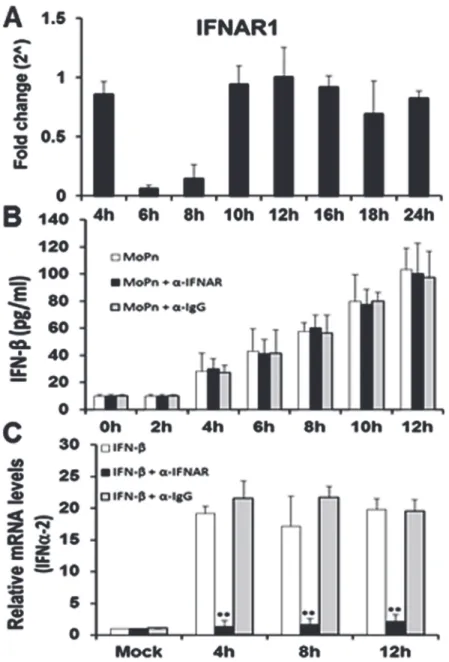
We next examined the function and transcription profile of the interferon-alpha/beta recep-tor-1 (IFNAR1) in the Bm1.11 OE cells throughout the course ofChlamydiainfection. As shown inFig. 3A, IFNAR1 transcription remains consistent throughout the course of Chla-mydiainfection; however, there was a significant down-regulation in the transcription of IFNAR1 between 6 h and 8 h post-infection. The down-regulation of IFNAR1 transcription ap-pears to require viableChlamydiareplication, since we were unable to noticeably impact IFNAR1 gene expression using heat-killedC.muridarum(S2 Fig.). The data reveal that the IFNAR1 receptor is at its lowest expression level during the 6–8 h time frame that we hypothe-size a factor is secreted into the supernatants to promote the optimal synthesis of IFN-βduring
Chlamydiainfection.Fig. 3Bshows that blocking IFNAR1 signaling with IFNAR1-specific neutralizing antibody had no impact on the overall amounts of IFN-βsecreted into superna-tants of Bm1.11 OE cells during the first 12 h ofC.muridaruminfection. These data corrobo-rate our transcription data (Fig. 3A), which suggest that IFNAR1 is likely not involved in the
Chlamydia-induced synthesis of IFN-βbetween 6 h and 8 h post infection. Control
Fig 2. Gene expression levels of IFN-βand TLR3 during the course ofC.muridaruminfection.Bm1.11 cells were infected with 10 IFU/ cellC.muridarum, and the gene expression levels of:(A)IFN-βand(B)TLR3 were measured by RT-qPCR after total cell mRNA was harvested at each time-point indicated.The results shown are representative of three independent experiments; Fold change is compared to Mock-infected controls.
doi:10.1371/journal.pone.0119235.g002
experiments using uninfected Bm1.11 OE cells demonstrate that the IFNAR1-specific neutral-izing antibody was effective in blocking the induction of the IFNα-2 gene transcription when uninfected OE cells were incubated in media supplemented with exogenous, recombinant mu-rine IFN-β(Fig. 3C). As shown, the recombinant murine IFN-βsubstantially induced IFNα-2
Fig 3. IFNAR1 early gene expression and function during the course ofC.muridaruminfection. (A)
Bm1.11 cells were infected with 10 IFU/ cellC.muridarumand the transcription levels of IFNAR1 was measured by RT-qPCR after total cell mRNA was harvested at each time-point indicated.(B)Bm1.11 cells were infected with 10 IFU/cellC.muridarumin the presence or absence of either the IFNAR1-specific antibody (denoted asα-IFNAR) or the isotype control (denoted asα-IgG) at 1h post-infection, and the amount of IFN-βsecreted into the supernatants during the first 12 h of infection was measured by ELISA.(C)Bm1.11 cells were incubated for 1h in media alone or in media containing either the IFNAR1-specific antibody or the isotype control, before adding 50U/ml recombinant murine IFN-β. Total cell mRNA was harvested after cells were exposed to recombinant IFN-βfor an additional 12h, and IFNα-2 transcription was measured by RT-qPCR at the time-points listed.The results shown are representative of three independent experiments; Fold change and relative mRNA levels are compared to Mock-infected controls;**= p<0.01 when compared to cells treated with recombinant IFN-βalone.
transcription in these cells throughout the 12 h incubation period; however, the IFNα-2 gene induction was significantly reduced in the presence of the IFNAR1 neutralizing antibody. In contrast, IFNα-2 transcription was not affected by the presence of the IgG control antibody. Collectively, our data suggest that IFN-βis likely not the secreted factor between 6–8 h that is required for optimalC.muridarum-induced IFN-βsynthesis in OE cells; however, the cyclo-heximide results ofS1 Fig.suggest that it is likely a cell-derived factor. Additionally, we show that IFNAR1 is indeed functional in its role in the autocrine-paracrine induction of type-1 IFNs in the OE cells. However, our data suggest an unlikelihood that the unidentified factor se-creted into the supernatants between 6–8 h post-infection will require the IFNAR1 receptor.
Chlamydia-induced IFN-
β
is essential for optimal IFN-
β
synthesis late
during infection of OE cells
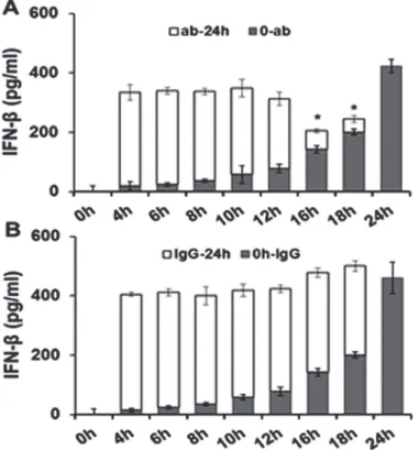
To ascertain which phase ofC.muridaruminfection in OE cells is the most likely time in which IFN-βacts in an autocrine-paracrine manner to induce optimal levels of IFN-β, we ex-amined the levels of IFN-βsynthesis when neutralizing antibody to IFN-βwas added at differ-ent intervals (Fig. 4). OE cells were infected with 10 IFU/cellC.muridarumand the medium was supplemented with either 0.1μg/ml IFN-βneutralizing antibody (Fig. 4A), or 0.1μg/ml IgG control antibody (Fig. 4B) at various times post-infection. The antibody amount used was
Fig 4. Disruption of IFN-βautocrine-paracrine pathways with neutralizing antibody.Bm1.11 cells were infected with 10 IFU/ cellC.muridarumto measure the amount of IFN-βsecreted into the supernatants for 24 hours by ELISA. The supernatants were supplemented at each time-point listed with either 0.1μg/ml of:(A)
IFN-βneutralizing antibody, or(B)isotype control antibody. The amount of IFN-βsecreted into the
supernatants after the addition of antibody (ab/IgG-24h) was estimated by subtracting out the amount of
IFN-βsecreted in control experiments in which supernatants were instead collected and assayed for IFN-β
synthesis at the same time-point (seeMaterials and Methods).Results are representative data from one of three independent experiments.*=p<0.05compared to 24h C.muridarum infection control;0-ab/IgG denotes“0hr PI until time antibody added”; ab/IgG-24h denotes“time antibody added until 24h PI”.
doi:10.1371/journal.pone.0119235.g004
titrated empirically using a customized version of the IFN-βneutralization assay [23], to a con-centration that would neutralize only a portion of the total amount of IFN-βsynthesized within 24 hours inC.muridaruminfected OE cells. Results of the RT-qPCR based IFN-β neutraliza-tion assay revealed that 0.1μg/ml concentration of the IFN-βneutralizing antibody was suffi-cient to block 20–35% of IFN-βfunction (data not shown). Our goal was to measure the effectiveness of IFN-βneutralizing antibody in blocking the autocrine-paracrine induction of IFN-βduringChlamydiainfection, and to ascertain which time-point during infection was the most important autocrine-paracrine induction. In this regard, we measured the total amount of IFN-βsecreted into supernatants ofC.muridarum-infected OE cells at 24h post-infection after the media was supplemented at various time-points with 0.1μg/ml concentration of either the IFN-βneutralizing antibody, or the corresponding IgG control.
As shown inFig. 4A, the total amounts of IFN-βremain consistent until 16 h post-infection when IFN-βneutralizing antibody was added and the total amount of IFN-βsecreted into the supernatants began to significantly decrease. Closer examination of the early-infection data in-dicated that the total amount of IFN-βsecreted is not significantly changed by the addition of the neutralization antibody up through the 12 h post-infection. These data corroborate our previous hypothesis that the IFN-βsynthesized early during infection plays little or no role augmenting the synthesis of itself, and that the mechanism for the early synthesis of IFN-β dur-ingChlamydiainfection likely does not involve autocrine-paracrine pathways. In contrast, neutralizing antibody added late duringC.muridaruminfection appeared to have a more pro-found impact on the synthesis of IFN-βlate during infection. Because we were able to block IFN-β’s ability to induce its own synthesis at late times and not at early times, our data suggest a heavier reliance on the autocrine-paracrine mechanisms late. These findings support our hy-pothesis that these autocrine-paracrine pathways are likely most active after the 12 h
time point.
Late-stage IFN-
β
is synthesized via pathways that involve IRF7 in
Chlamydia-infected OE cells
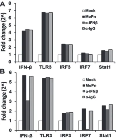
To further delineate the pathways involved in early vs late stage IFN-βsynthesis, we examined the transcription levels of IFN-β, TLR3, IRF3, IRF7 and STAT1; which are proteins known to be required in theChlamydia-induced synthesis of IFN-β[5,12,24–28]. As shown inFig. 5A, OE cells were infected withC.muridarum, and the media was replaced with fresh media con-taining 2μg/ml of either IFN-βneutralizing antibody or IgG control antibody at 4 h post-infec-tion. The cells were harvested at 12 h and assessed for the transcription levels of the various genes involved in theChlamydia-induced IFN-βsynthesis pathways. As indicated,C. muri-daruminfection substantially induced the early transcription levels of IFN-βand TLR3; while Stat1 and IRF7 were not induced in any appreciable manner. Interestingly, we saw only a mod-erate induction of the IRF3 gene early during infection and the levels remained consistent throughout infection; however, there were no changes in the transcription of any of these genes after the addition of either IFN-βneutralizing antibody or IgG control.
down-regulation of IRF7 gene transcription, contrasting the early infection IRF7 results. We also saw diminished levels of IFN-βtranscription when OE cells were incubated in the medium contain-ing IFN-βneutralizing antibody when compared toC.muridarum-only reactions, suggesting a positive feedback loop for IFN-βby inducing IRF7 in OE cells. We saw no appreciable changes in the transcription rates of any of these genes early or late when media was replaced with fresh media containing the IgG control antibody.
We further examined the role of IRF7 in the IFN-βresponse to ascertain whether IRF7 pro-tein had any direct role inChlamydia-induced synthesis of IFN-βin OE cells (S3 Fig.). As shown inS3A Fig., we again showed thatC.muridaruminfection was able to induce transcrip-tion of IRF7 more substantially at late times during infectranscrip-tion. As indicated, we were able to sig-nificantly reduce IRF7 mRNA using IRF7-specific si-RNA, which lead to diminished levels of total IRF7 protein and a significant decrease in IFN-βsynthesis levels at late times during Chla-mydiainfection (S3B Fig.andS3C Fig.) Collectively, these data implicate a role for IRF7 in the late infectionChlamydia-induced synthesis of IFN-βin OE cells, and corroborates our previous finding that IFN-βplays a role in inducing itself late duringChlamydiainfection.
Fig 5.C.muridarum-induced IFN-βaffects the gene expression levels of components found in the type-1 IFN signaling pathway.Bm1.1 cells were infected with 10 IFU/ cellC.muridarum, and the gene expression levels of IFN-β, TLR3, IRF3, IRF7, and Stat1 were measured by RT-qPCR after total cell mRNA was harvested at either early (12h) or late (24h) times post infection.(A)Transcription results of Bm1.11 OE cells harvested at 12h PI to measure the impact on transcription of candidate genes after cells were
incubated from 4-to-12h PI in culture medium containing 1μg/ml of either IFN-βneutralizing antibody (α-IFNβ) or isotype control antibody (α-IgG).(B)Transcription results of these genes after cells were incubated in culture medium containing either antibody from 12-to-24h PI.The results shown are representative of three independent experiments; Fold change is compared to Mock-infected controls.
doi:10.1371/journal.pone.0119235.g005
Inhibition of IRF3 but not NF-
κ
B significantly impacts the early synthesis
of IFN-
β
in
C.
muridarum
infected OE cells
To confirm our hypothesis that IRF3 is involved in the early synthesis of IFN-βduring Chla-mydiainfection of OE cells, we examined the impact of specific inhibitors of IRF3 and NF-κB on total IFN-βsecretion. BX-795 is a potent and relatively specific inhibitor of IκB kinaseε
(IKKε), phosphoinositide 3-dependent kinase 1 (PDK1), and TANK-binding kinase 1 (TBK1) [29,30]. BX-795 disrupts the function of TBK1 and IKKε, which then blocks the phosphoryla-tion, nuclear translocaphosphoryla-tion, and transcriptional activity of IRF3 [29]. JSH-23 is a potent inhibi-tor of NF-κB nuclear translocation, which does not affect the degradation of IκB kinases [31].
C.muridarum-infected Bm1.11 OE cells were allowed to incubate for various hours post-infec-tion before adding either BX-795 (IRF3 inhibitor) or the NF-κB inhibitor JSH-23. Supernatants were harvested from the infected OE cells at 24h PI, and subjected to an ELISA assay to mea-sure the total amounts of IFN-βsecreted.
As shown inFig. 6A, the addition of BX-795 resulted in significant decreases in the levels of IFN-βsecretion when added between 4 and 10 h post-infection, while having little effect when the compound was added to the cells mid- and late-infection. Blocking NF-κB activity with JSH-23 seemed to have a minor effect on total IFN-βsynthesis inC.muridarum-infected OE cells when added at 4 h post-infection; however, we routinely noticed only a minimal (but non-significant) decrease in IFN-βsecreted when JSH-23 was added. In contrast, disrupting NF-κB activity with JSH-23 appeared to dramatically reduce theChlamydia-induced synthesis of IL-6; however, IL-6 production was not affected by the addition of the IRF3 inhibitor BX-795 (Fig. 6B). We previously reported that IL-6 was induced via TLR2-dependent pathways inC.
muridarum-infected OE cells [3], and our findings suggest a more critical role for NF-κB in the
Chlamydia-induced synthesis of IL-6 throughout the course of infection. Positive control reac-tions (Fig. 6C) show that when added to the media at the identical inhibitory concentrations utilized in theC.muridaruminfected OE cells, both BX-795 and JSH-23 were equally able to significantly reduce the amount of IFN-βsecreted by Bm1.11 OE cells that were transfected with the TLR3 agonist poly-IC [32]. Negative control reactions using DMSO show no signifi-cant impact on the secretion of IFN-βduringChlamydiainfection of OE cells (Fig. 6A), and we observed no significant effect on overallC.muridarumreplication in the cells treated with ei-ther inhibitor or DMSO alone (data not shown).
Data from these experiments, in which we used BX-795 as a known inhibitor of PDK1, TBK1, and IKKε, showed significant reductions in early synthesis of IFN-βduringChlamydia
infection. These findings suggest a disruption in IRF3 phosphorylation since TBK1 and IKKε
are known to phosphorylate and regulate the function of IRF3 [29]. However, because IKKε
and TBK1 also have some function in the phosphorylation and activation of IRF7 in human lung epithelial cells[33,34], it is possible that the disruption inChlamydia-induced IFN-β syn-thesis is due to unwanted inhibition of IRF7 pathways by BX-795. IRF7 has been shown to be the major IRF functioning in type-1 IFN induced via TLR9 signaling [35,36]. Since Bm1.11 OE cells lack TLR9 and do not respond to the TLR9 agonist ODN 1826 [3], we used RAW 264.7 cells to specifically activate IRF7 via TLR9 signaling using ODN 1826.S4A Fig.shows thatC.
IFN-βis likely due to disruption of IRF3 function in the early stages of theChlamydia -induced IFN-βsynthesis, and that IRF7 is likely not involved in the IFN-βsynthesis early during infection.
Fig 6. Inhibition of IRF3 and NF-κB affects early synthesis of IFN-βinduced duringChlamydia
infection. (A)Bm1.1 cells were infected with 10 IFU/ cellC.muridarumup until the indicated time-points when the media was supplemented with either the IRF3 inhibitor BX-795, the NF-κB inhibitor JSH-23, or the solubilizing agent DMSO as a negative control. Cells were allowed to incubate in the presence of each inhibitor from the time indicated until cell supernatants were harvested at 24h PI, and IFN-βsecreted was measured by ELISA.(B)C.muridarum-infected Bm1.11 cells were allowed to incubate in the presence of each inhibitor from the time indicated until cell supernatants were harvested at 24h PI, and IL-6 secreted was measured by ELISA.(C)Uninfected Bm1.11 cells were either DMSO-treated, transfected with 10, 25, or 50μg/ml poly-IC, or transfected with poly-IC prior adding the inhibitors BX-795 and JSH-23 to the cells 1h post-transfection. IFN-βsecreted into the supernatants 24 h post transfection was measured by ELISA.The results shown are representative of three independent experiments.*=p<0.05;**= p<0.01; NS = not statistically significant compared to poly-IC alone (C),or 24h C.muridarum infection control without inhibitor (A and B; denoted as MoPn).
doi:10.1371/journal.pone.0119235.g006
Transient inhibition of bacterial DNA replication decreases IFN-
β
secretion in
C.
muridarum-infected OE cells
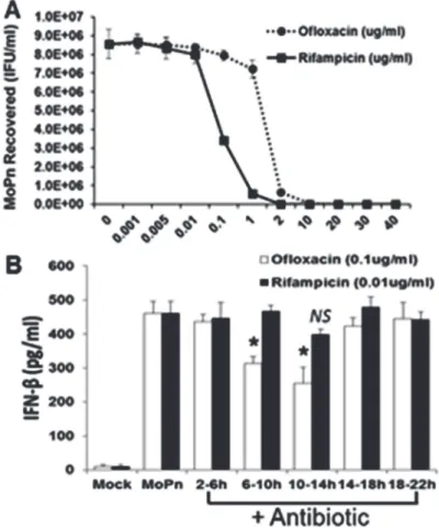
We reported a major role for TLR3 in theC.muridarum-induced synthesis of IFN-β[6,14]. However, the exact chlamydial PAMP that binds to and stimulates TLR3 when the OE cells are infected withC.muridarumhas not been identified. We hypothesized that either bacterial DNA or RNA were the likely PAMP(s) that binds to TLR3 duringChlamydiainfection of OE cells. We utilized the bacterial DNA replication inhibitor ofloxacin [37], and the bacterial tran-scription inhibitor rifampicin [38] to ascertain which of these cell-cycle events had the greatest impact onChlamydia-induced IFN-βsynthesis in OE cells. Both ofloxacin and rifampicin are quite effective in the inhibition of overall chlamydial replication [39]. However, we sought to ascertain the optimal dose for these experiments whereby there was only minimal inhibition of the overall replication ofC.muridarumin OE cells, even if there was a considerable decrease in either chlamydia DNA or RNA. Our goal was to ensure that decreases observed in IFN-β syn-thesis was due more to decrease in the respective nucleic acids, and less due to decreases in progeny totals. We infected Bm1.11 OE cells with 10 IFU/cellC.muridarumand, at 2 h post-infection, we replaced the medium with fresh medium supplemented with increasing concen-trations of each antibiotic. The cells were allowed to incubate until 18 h post-infection before the supernatants containing antibiotic were removed and replaced with antibiotic-free fresh media. The cells were then harvested at 30 h post-infection and theChlamydiatiters were as-sessed using McCoy cell monolayers (SeeMaterials and Methods).
As shown inFig. 7A, rifampicin was a much more potent inhibitor ofChlamydiareplication whereas we began to see a measurable decrease inChlamydiareplication starting at the 0.01μg/ml concentration. We saw almost complete suppression ofC.muridarumreplication at the 2μg/ml concentration or higher. In contrast, we did not see any significant reduction in C.muridarumreplication until we reached the 1μg/ml ofloxacin concentration. We did not see complete inhibition ofC.muridarumreplication until the 10μg/ml concentration of oflox-acin.S5 Fig.demonstrates the corresponding reductions in chlamydial DNA and RNA when OE cells were infected in the presence of increasing doses of the respective antibiotic. We chose 0.01μg/ml and 0.1μg/ml concentrations of rifampicin and ofloxacin, respectively, as a subopti-mal inhibitory dose for these experiments, whereby we begin to see only minor but equal re-ductions in chlamydial replication in these cells.
We infected Bm1.11 OE cells with 10 IFU/cellC.muridarumbefore removing (and saving) the supernatants, and replacing it with fresh media containing either 0.01μg/ml rifampicin or 0.1μg/ml ofloxacin at various time points of post-infection. The cells were incubated in the presence of antibiotic for 4 h intervals before the supernatants containing the antibiotic was re-moved, saved, and replaced with the original media that was removed prior to the addition of antibiotic. The cells were then allowed to incubate until 24 h post-infection, and the amount of IFN-βsecreted into the combined supernatants (w and w/o antibiotic) was determined by ELISA. As shown inFig. 7B, both antibiotics had varying impact on overall amounts of Chla-mydia-induced IFN-βsynthesis when the antibiotics were added between 2–18 h post-infec-tion. However, we saw substantial and significant reductions in overall IFN-βlevels when ofloxacin was added at the 6–10 h and the 10–14 h time points. We did not see a significant re-duction inChlamydia-induced IFN-βsynthesis when rifampicin was added to the cells, though we did see a slight but non-significant decrease at the 10–14 h time point. There was no impact in the DMSO control reactions onChlamydia-induced IFN-βsynthesis (not shown).
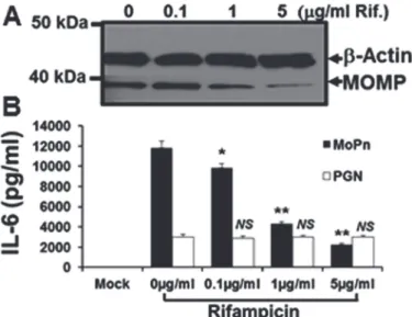
expressed decreasing amounts of the chlamydial major outer membrane protein (MOMP) at 24 h post-infection, when incubated in medium supplemented with increasing amounts of ri-fampicin. However, despite utilizing rifampicin concentrations that correspond to 500X the concentration used in theChlamydia-induced IFN-βexperiments (seeFig. 7B); there was no equivalent reduction in the transcription of the host-cell structural proteinβ-actin.Fig. 8B shows that rifampicin had no immunomodulatory effect on the host-cell synthesis of IL-6 when uninfected Bm1.11 OE cells were induced for 24 h in medium supplemented with 10μg/ ml of the purified TLR2 agonistE.colipeptidoglycan (PGN) [41]. In contrast, there was a dose-dependent reduction in the amount of TLR2-dependent IL-6 [3] detected in the superna-tants at 24 h in theC.muridarum-infected Bm1.11 OE cells. The reduction in the synthesis of IL-6 seen in theChlamydia-infected OE cells (but not observed in the PGN treated uninfected OE cells), suggest that the diminished synthesis of IL-6 at high-dose concentrations of rifampi-cin are most likely due to substantially reduced transcription of the chlamydial TLR2 PAMPs during infection. Taken together, our data show that inhibition of chlamydial DNA replication
Fig 7. Role of bacterial DNA replication and bacterial transcription inChlamydiainduced IFN-β synthesis. (A)Bm1.11 cells were infected with 10 IFU/ cellC.muridarumand cells were incubated in the presence of increasing concentrations of either rifampicin or ofloxacin starting at 2h PI. The medium was replaced with antibiotic-free medium at 18h PI, cells were harvested at 30h PI, andC.muridarumtiters (IFU/ ml) were measured on McCoy cell monolayers as described in Materials and Methods.(B)Bm1.11 cells were infected with 10 IFU/ cellC.muridarumand cells were incubated in the presence of either 0.01μg/ml rifampicin or 0.1μg/ml ofloxacin for each 4h interval indicated, before cell supernatants were harvested at 24h PI and IFN-βsecreted was measured by ELISA.The results shown are representative of three independent experiments.*=p<0.05; NS = not statistically significant compared to 24h C.muridarum infection control without antibiotic (denoted as MoPn).
doi:10.1371/journal.pone.0119235.g007
has a much more dramatic impact on theChlamydia-induced synthesis of IFN-βin OE cells than does the bacterial transcription. In effort to identify a putative chlamydial PAMP that triggers the TLR3-dependent IFN-βsynthesis, our results are highly suggestive that the TLR3 PAMP inC.muridarum-infected OE cells is derived from products ofChlamydiaDNA replication.
Discussion
Our research focuses on the immunopathogenesis ofChlamydiainfection and the overall con-tribution to the innate-immune response of epithelial cells lining the lumen of oviduct tissue. We have described the roles of TLR signaling in the synthesis of inflammatory cytokines, and understanding the mechanisms that lead to damage of the genital tract epithelium is para-mount to development of therapeutic measures to prevent the sequelae of chronic genital tract
Chlamydiainfections. Using the OE cells as a model to studyin vitro Chlamydiainfections, we showed thatC.muridaruminduces IFN-βin a mostly TLR3-dependent manner, and we were the first to demonstrate TLR3-specific immune response toChlamydiainfectionin vitro[6]. Our subsequent investigation into TLR3-dependent responses toChlamydiainfection in OE cells revealed that TLR3 stimulation resulted in the synthesis of a plethora of other inflammato-ry mediators, and that the TLR3-dependent IFN-βupregulated the gene expression of several of these innate-immune factors [14].
The mechanism by whichChlamydiainduces IFN-βsynthesis is an area of intense investi-gation. It has been shown thatChlamydiacan induce IFN-βexpression in a variety of cell types including macrophages, fibroblast, endothelial, and epithelial cells [8–13]. TheChlamydia -in-duced IFN-βsynthesis in peritoneal macrophages was shown to be MYD88-dependent and likely signals through TLRs 7, 8, or 9 [11]. More recent results demonstrate thatChlamydia -in-duced IFN-βsynthesis is independent of TLR signaling in multiple cell types, and strongly
Fig 8. Rifampicin does not affect host-cell mechanisms.Bm1.11 cells were either mock-treated, treated with 10μg/ml of the purifiedE.colipeptidoglycan (PGN), or infected with 10 IFU/ cellC.muridarum, and cells were incubated in the presence of increasing concentrations of rifampicin starting at 2h post-treatment/ infection. After 24 h post-treatment/ infection, cell lysates and supernatants were harvested for:(A) western-blot analyses for theChlamydia-specific major outer membrane protein (MOMP) and cellularβ-actin.(B)
ELISA analyses of PGN-induced versusChlamydia-infection specific cytokine expression.The results shown are representative of three independent experiments.*=p<0.05;**= p<0.01; NS = not statistically significant compared to control cells without rifampicin (0μg/ml); MoPn denotes Chlamydia infection.
associated with the newly described stimulator of IFN gene (STING) protein [28,42]. Though the relationship between TLR3 signaling and STING has not been investigated in OE cells, it is a strong possibility that these pathways intersect duringChlamydiainfection in these cells. The apparent differences observed in the pathways to IFN-βsynthesis highlight the possibility that cell-type specific mechanisms of IFN-βsynthesis are stimulated duringChlamydiainfection. To this regard, we sought to ascertain the contribution of other signaling pathways contribut-ing to the synthesis of IFN-βduringC.muridaruminfection of OE cells. In this report, we demonstrate thatChlamydia-induced IFN-βproduction in OE cells follows a biphasic pattern of expression, which involves the coordination of distinct signaling pathways for early and late infection synthesis of IFN-βthat is similar to mechanisms described by others [28,43–47]. Our data proposes that the initial wave of IFN-βsynthesized early duringChlamydiainfection is TLR3-dependent and occurs through pathways involving IRF3; while late stage IFN-β synthe-sis is triggered by the type-1 IFN secreted into the supernatants, and signals through pathways that require IRF7.
We first showed that there was a factor secreted into the supernatants early duringC. muri-daruminfection of OE cells infection that is critical for‘conditioning’the cells for the optimal induction of IFN-β. In order to identify this putative factor, we first examined whether IFN-β
itself was this secreted factor since it is known that IFN-βcan induce itself in an autocrine-paracrine manner [43,48]. However, our data indicate that IFN-βwas not likely the secreted factor early during infection, though we have not ruled out the possibility of it having a role in inducing itself late during infection. Data from experiments in which OE cells were infected in the presence of cycloheximide show that inhibition of eukaryotic transcription has a negative impact on the synthesis of this putative secreted factor, and thus suggest this factor is derived from the OE cell machinery.
We showed that IFNAR1 was at its lowest expression levels between 6–8 h post-infection, and that inhibition of IFNAR1 mediated signaling with IFNAR1-specific neutralizing antibody had no effect onChlamydia-induced synthesis of IFN-βduring the first twelve hours post in-fection. In contrast, the IFNAR1-neutralizing antibody had a profound effect in disrupting the gene transcription of IFNα-2 when uninfected OE cells were induced with purified recombi-nant murine IFN-β. Our findings indicate that the IFNAR1 mediated pathway is indeed func-tional in OE cells, and that type-1 IFN can be induced through this pathway; however, data from these and our other ongoing investigation [19], show that IFNAR1 mediated signaling differentially induces the various type-1 IFNs throughout the course ofChlamydiainfection in OE cells. The exact mechanism for the differential induction of type-1 IFNs through IFNAR1 signaling in OE cells is not known, though we hypothesize that it likely depends on the cyto-kine composition of the supernatants at that particular time during infection [49]. We surmise there must be some host cell orChlamydia-induced component early duringChlamydia infec-tion that either causes the preferential inducinfec-tion of IFNαthrough IFNAR1, or shuts off the ability of this pathway to induce IFN-β.
One possible candidate for the secreted factor that is critical for the optimal synthesis of IFN-βduring Chlamydia infection of OE cells is cyclic-di-AMP (c-di-AMP). Cyclic-di-AMP was shown to be secreted by the intracellular bacteriaListeria monocytogenesand is a major contributor in inducing IFN-β[50]. In that study, it was shown that cyclic-di-AMP coordinates bacterial growth, cell wall stability, and the stress response; while also playing a crucial role in the establishment of bacterial infection. The specific cell-sensor for this cyclic dinucleotide in the IFN-βresponse has not yet been identified, but it was demonstrated that this pathway does function through downstream adaptor molecule STING. Subsequent to that study, it was dem-onstrated thatChlamydiasynthesizes cyclic di-AMP, and that this metabolite was a prominent ligand for STING-mediated activation of type-1 IFN responses late during infection [42]. In
that study, c-di-AMP was shown to be present in the cells of primary mouse lung fibroblasts and HEK293T late duringC.trachomatisinfections. It is not known whether the c-di-AMP can be detected in the culture medium duringChlamydiainfection as in theListeriastudies, but because of its relationship to the endoplasmic reticulum (ER) membrane protein STING, it can be postulated that this di-nucleotide can be secreted from the cell through ER networks. Further studies are needed to ascertain whether c-di-AMP is synthesized duringC.muridarum
infection of OE cells, and whether its presence in the supernatants contribute to the overall lev-els of IFN-βsynthesis duringChlamydiainfection in OE cells.
We showed in IFN-βneutralization experiments that autocrine-paracrine synthesis of
IFN-βinC.muridarum-infected cells occurs late during infection. In addition, we showed that this autocrine-paracrine induction occurs through pathways that likely signals through IRF7. Con-versely, our data shows that IRF3 likely plays a larger role early duringChlamydiainfection in OE cells. IRF3 and IRF7 are key activators of the transcriptional induction of IFNsαandβ
genes [36,47]. Both IRF3 and IRF7 play distinct and essential roles in the IFN-α/βresponse to virus infection [35,36]. In these and other studies, IRF3 was shown to be an important compo-nent of the immediate-early response to virus infection [51], while IRF7 is involved in the late induction phase of IFN expression during viral infection [45,46]. The importance of IRF3 and IRF7 in regulating the early and late phases of IFN expression during viral infection was dem-onstrated through the generation of IRF3 and IRF7 knock-out mice [47].
The requirement for activation of IRF3 and IRF7 in the IFN-βresponse to bacterial infec-tion has been demonstrated inListeria[52],C.pneumonia[53], andNeisseria meningitidis
[54]. However, the exact temporal relationship between these IRFs has not been clearly defined in these studies. The roles for IRF3 and IRF7 have been examined inC.muridaruminfection, and it was shown that there was an initial IRF3-dependent IFN-βsecretion that forms a posi-tive feedback loop by inducing IRF7; which is then required for maximal IFN-βexpression dur-ing chlamydial infection of murine peritoneal macrophages [28]. Because the investigators in those studies demonstrated such a high dependence on STING during chlamydial induction of IFN-β, it was concluded that TLR signaling, MyD88, and TRIF were all dispensable for IFN-β
upregulation during chlamydial infection of peritoneal macrophages.
Our data showed that IRF3 was involved early in theChlamydia-induced synthesis of
IFN-β, and that inhibiting IRF3 phosphorylation early with BX795 resulted in substantial reductions in overall IFN-βsynthesis. Interestingly, we did not observe any effect on overall IFN-β synthe-sis when BX-795 was added late duringChlamydiainfection, which suggest that IRF3 was not involved in the late-infection synthesis of IFN-β. BX-795 functions by inhibiting the auto-phosphorylation of IKKε, TBK1, and PDK1; and thereby preventing the nuclear translocation, phosphorylation, and transcriptional activity of IRF3 [29,30]. Although IKKεand TBK1 also have function in the phosphorylation and activation of IRF7 in human lung epithelial cells [34], there have been no reports detailing a disruption of IRF7 function in cells treated with BX-795, and we too were unable to disrupt IRF7-specific signaling pathways using this inhibi-tor in RAW264.7 cells (seeS4 Fig.). The fact that BX-795 can quite effectively disrupt the func-tion of IRF3, yet not have any impact on the funcfunc-tion of IRF7, suggest that IKKεand TBK1 regulates these transcription factors via different mechanisms [33]. Because we were not able to disrupt late-infection synthesis of IFN-βin theChlamydia-infected OE cells with BX-795, our results propose that the late-infection IFN-βsynthesis involves the contributions from IRFs other than IRF3; which supports the observations of Pratneret al, demonstrating a role for IRF7 late duringChlamydiainfection of peritoneal macrophages [28].
infection. In contrast, there were no changes in the gene expression levels of IRF7 when IFN-β
neutralizing antibody was added to the cells early during infection; which is indicative of IRF7 playing little to no role in the synthesis of IFN-βearly duringC.muridaruminfection. These results were corroborated in supplemental experiments where we showed significant reduc-tions inC.muridarum-induced IFN-βsynthesis late during infection in gene-knockdown stud-ies using IRF7-specific si-RNA. As also suggested by the IFN-βneutralization results,
disrupting IRF7 gene expression early during infection had little impact on IFN-βsynthesis, which implies that IRF7 is not involved early in this immune response. Collectively, our find-ings in OE cells corroborate the results shown in peritoneal macrophages by Pratneret al[28], as we demonstrate an early role for IRF3 and a late role for IRF7 during theChlamydia -in-duced synthesis of IFN-β. However, the early IFN-βresponse toChlamydiainfection in OE cells is largely TLR3-dependent, which differs from the findings in peritoneal macrophages and implicates divergent pathways to IFN-βsynthesis that converge and activate IRF3 in these two cell-types. The differential utilization of distinct IRF proteins duringC.muridarum infec-tion of OE cells likely lead to an ordered inducinfec-tion of IFN-βsynthesis, resulting in tight control of these cytokines through a positive feedback mechanism [55].
We investigated the role of NF-κB in theChlamydia-induced synthesis of IFN-βin OE cells, and we showed that inhibiting NF-κB activity with JSH-23 appeared to have only a minor ef-fect on total IFN-βsynthesized, but only when used early during infection. We routinely saw measurable decreases in overall IFN-βsynthesis when JSH-23 was added at the 4hr PI time-point; however, the amount of decrease in the overall IFN-βsecreted never quite reached statis-tical significance in our analyses. Interestingly, JSH-23 was much more effective in disrupting IFN-βsynthesis in Bm1.11 cells that were transfected with poly-IC (seeFig. 6C). We previously reported that Bm1.11 cells did not synthesize IFN-βwhen poly-IC was added directly to the media, despite the robust expression of TLR3 in these cells [5]. The reason for the lack of re-sponse to extracellular poly-IC is poorly understood; however, we postulated that it may have been due to poly-IC’s inability to effectively enter into the BM1.11 cell endosome, due to poorly functioning micropinocytic pathways in the OE cells when compared to other cell types [56]. We found that transfecting the cells with poly-IC induced a very strong IFN-βresponse; how-ever, the response was largely TRIF-independent [5]. The TRIF-independent induction of IFN-βby transfected poly-IC implicates other cytoplasmic receptors in OE cells that trigger type-1 IFN synthesis in response to double-stranded RNA (ds-RNA) such as RIG-I and MDA5 pathways [57–60]. Because both NF-κB and IRF3 play prominent roles in the synthesis of type-1 IFN in both the RIG-I and the MDA5 signaling pathway, it is conceivable that inhibitors of NF-κB and IRF3 would also disrupt signaling through these pathways. The relative contri-butions for both NF-κB and IRF3 might differ in RIG-I and MDA5 pathways when compared to TLR3 signaling pathways, which could possibly explain why inhibiting the function of
NF-κB in these pathways has a much more dramatic effect on IFN-βsynthesis in the poly-IC trans-fected OE cells.
In contrast to what we observed regarding theChlamydia-induced IFN-βresponse, control reactions measuring IL-6 production in OE cells revealed that there were significant reductions inChlamydia-induced synthesis of IL-6 throughout the course of infection in the JSH-23 treat-ed cells. Collectively, our data implicate a much more impactful role for NF-κB in the synthesis of IL-6 duringChlamydiainfection of OE cells, and that NF-κB pathways play a lessor role in the early infection induction of IFN-β. The significance of the relatively minor role for NF-κB in the TLR3-dependent synthesis of IFN-βis unclear; though, it is possible that it reflects the temporal relationship of when NF-κB and the IRF3 heterodimer are assembled into the IFN-β
enhanceosome complex [61]. Investigations into assembly of the individual proteins into the human IFN-βenhanceosome complex suggest that NF-κB is assembled into the complex by
binding to the positive regulatory domain II (PRDII) binding site of the IFN-βenhancer ele-ment, prior to the incorporation of the IRF3 heterodimer to the PRDI and III binding sites [62]. We have not examined the roles of IRF3 and NF-κB prior to the 4hr time point to ascer-tain whether NF-κB plays a more impactful role very early duringChlamydiainfection of the OE cells. Future studies are necessary to address the possibility that NF-κB plays a more signifi-cant role in optimal IFN-βsynthesis duringChlamydiainfection at time points earlier than the 4hr PI, and to address the hypothesis that NF-κB is more heavily recruited to the enhanceo-some complex prior to the 4hr time point.
It is well known that TLR3 recognizes ds-RNA during viral infection to induce type-I inter-feron synthesis in a variety of different cell types [32,57]. Others have identified an inosine-containing single-stranded RNA PAMP that induces TLR3-dependent responses to respiratory syncytial virus (RSV) infections [63]. Although we have demonstrated a TLR3-dependent
IFN-βresponse toC.muridarumin the OE cells [6,14], the actual chlamydial PAMP that stimulates the TLR3-dependent responses is currently unknown. Because there is no known ds-RNA moi-ety associated with the chlamydial structure, we hypothesized that it was either an unconven-tional TLR3 PAMP presented duringC.muridaruminfection (such as bacterial DNA or RNA), or thatC.muridaruminduced a cellular ds-RNA during infection that served as a TLR3 PAMP. Our data demonstrate that inhibition of bacterial DNA replication at early-to-mid in-fection had the most dramatic impact on overallChlamydia-induced IFN-βsynthesis in OE cells, while inhibition of bacterial transcription had virtually no impact on cellular IFN-β syn-thesis. The significance of this finding is unclear; however, it would support the hypothesis that
C.muridarumDNA replication produces a PAMP that augments TLR3-dependent IFN-β syn-thesis in these cells. Our data underscores the possibility of chlamydial DNA being that uncon-ventional TLR3 PAMP that is presented during the course ofC.muridaruminfection in the OE cells, or that the chlamydial DNA enhances overall IFN-βsynthesis through other path-ways the involve cellular DNA sensors that can signal type-1 IFN responses via STING as an al-ternative mechanism [42,64]. Studies are underway that will further address the putative role ofChlamydiaDNA as a possible TLR3 PAMP in OE cells.
Supporting Information
S1 Fig.ChlamydiaConditioned media induces transcription in uninfected OE cells. Super-natants were removed fromC.muridaruminfected Bm1.11 OE cells that were incubated in presence or absence of cycloheximide for the 4 hour intervals indicated. The conditioned su-pernatants were added to uninfected Bm1.11 cells for 4 h before monolayers were harvested for RNA isolation to examine effect on transcription rates of:(A)components of type-1 IFN sig-naling pathways,(B)various inflammatory mediators, and(C)TLRs known to be expressed and functional in Bm1.11 OE cells.The results shown are representative of three independent ex-periments.Cyclo = 1μM cycloheximide.
(TIF)
S2 Fig. Effect of non-viableC.muridarumon IFNAR1 gene transcription.Bm1.11 cells were either mock-infected or infected with 10 IFU/ cellC.muridarumthat has been heat-inac-tivated at 56°C for 30 minutes (seeMaterials and Methods). Gene expression levels of IFNAR1 was measured by RT-qPCR after total cell mRNA was harvested at 24 h post-infection.The re-sults shown are representative of three independent experiments; MoPn (HK) = heat killed C.
S3 Fig. IRF7 has a role in theChlamydia-induced synthesis of IFN-βat late times during in-fection.Bm1.1 cells were infected with 10 IFU/ cellC.muridarum24 h after transfection with either si-RNA specific for IRF7(si-IRF7), scrambled control si-RNA(si-SCR), or lipofecta-mine-only control(MoPn).(A)RT-qPCR results showing the gene expression levels of IRF7 after total cell mRNA was harvested at either early (12h) or late (24h) times post infection.(B) Western blot analysis showing IRF7 protein expression at 12 and 24 h post-infection.(C) Chla-mydia-induced IFN-βlevels in the supernatants of each group were determined by ELISA at 12 and 24 h post-infection.The results shown are representative of three independent experiments.
Statistical significance was determined by comparing the treatment conditions of si-IRF7–
trans-fected Bm1.11 cells with Bm1.11 cells transfected with lipofectamine-only.=p<0.01.
(TIF)
S4 Fig. Role of IRF7 in TLR9-dependent IFN-βsynthesis. (A)ELISA showing IFN-βlevels detected in supernatants from RAW 264.7 cells infected with 10 IFU/ cellC.muridarum,or
stimulated with either the TLR9 agonist ODN1826 or ODN1826 control.(B)ELISA showing IFN-βsynthesis in RAW 264.7 cells that were stimulated with ODN1826, or stimulated with ODN1826 24 h after transfection with either si-RNA specific for IRF7(si-IRF7)or scrambled control si-RNA(si-SCR).(C)IFN-βwas measured in RAW 264.7 cells that were stimulated with ODN1826 in the absence or presence of increasing concentrations of the IRF3 inhibitor BX-795.The results shown are representative of three independent experiments.Statistical signif-icance was determined by comparing the specified condition versus RAW264.7 cells treated with ODN1826;= p<0.05; NS = not statistically significant.
(TIF)
S5 Fig. Inhibition of bacterial DNA replication and bacterial transcription inChlamydia -infected OE cells.Bm1.1 cells were infected with 10 IFU/ cellC.muridarumand cells were in-cubated in the presence of increasing concentrations of either rifampicin or ofloxacin starting at 2h PI. The medium was replaced with antibiotic-free medium at 18h PI, cells were harvested at 30h PI for analysis of chlamydial gene transcription and DNA replication.(A)Quantitative PCR using primers specific foromp1to measure chlamydial DNA replication at the 30 h time-point.(B)RT-qPCR showing transcription of theChlamydia-specific 16S rRNA versus the cel-lularβ-Actin gene.The results shown are representative of three independent experiments. (TIF)
Acknowledgments
We thank Dr. Byron Batteiger and Mr. James Williams for graciously providing theC. muri-darumspecific anti-MOMP antibody. We gratefully acknowledge the thoughtful critiques of Dr. Qigui (Andy) Yu that were instrumental to the evolution of this manuscript. We also thank Awahngie Akwo for his technical assistance.
Author Contributions
Conceived and designed the experiments: SH KLH WAD. Performed the experiments: SH KLH WAD. Analyzed the data: SH WAD. Contributed reagents/materials/analysis tools: WAD. Wrote the paper: SH WAD.
References
1. Kagnoff MF, Eckmann L (1997) Epithelial cells as sensors for microbial infection. J Clin Invest 100: 6– 10. PMID:9202050