A New Genus of Aplodontid Rodent (Mammalia,
Rodentia) from the Late Oligocene of Northern Junggar
Basin, China
Shundong Bi1,2*, Jin Meng3, Sarah McLean2, Wenyu Wu1, Xijun Ni1, Jie Ye1
1Key Laboratory of Evolutionary Systematics of Vertebrates, Institute of Vertebrate Paleontology and Paleoanthropology, Chinese Academy of Sciences, Beijing, China, 2Department of Biology, Indiana University of Pennsylvania, Indiana, Pennsylvania, United States of America,3Division of Paleontology, American Museum of Natural History, New York, United States of America
Abstract
A new genus and species of aplodontid rodent,Proansomys dureensis, from the late Oligocene of the northern Junggar Basin of China is described. The new genus is referred to as Ansomyinae because the ectoloph on the upper cheek teeth, although not fully crested, has attained the same characteristic bucket-handle-shaped configuration as other members of the subfamily. It represents the earliest record of the subfamily yet discovered in Asia and is more plesiomorphic than species of the genusAnsomysin having a partly crested ectoloph, a lower degree of lophodonty, and less complex tooth basins (lacking accessory lophules).Proansomyshas transitional features betweenProsciurusandAnsomys, suggesting that the Ansomyinae derived from a group of aplodontids related toProsciurus, as did other advanced aplodontid rodents. This provides new light on the paleobiogeography of the Ansomyinae.
Citation:Bi S, Meng J, McLean S, Wu W, Ni X, et al. (2013) A New Genus of Aplodontid Rodent (Mammalia, Rodentia) from the Late Oligocene of Northern Junggar Basin, China. PLoS ONE 8(1): e52625. doi:10.1371/journal.pone.0052625
Editor:Alistair Robert Evans, Monash University, Australia
ReceivedMay 25, 2012;AcceptedNovember 19, 2012;PublishedJanuary 24, 2013
Copyright:ß2013 Bi et al. This is an open-access article distributed under the terms of the Creative Commons Attribution License, which permits unrestricted use, distribution, and reproduction in any medium, provided the original author and source are credited.
Funding:This research has been funded by the Chinese Academy of Sciences (CAS) Key Project (XDB03020500), the National Nature Science Foundation of China (KA212201), the CAS 100-Talent Project, and the CAS Fossil Excavation and Preparation Fund. Meng is also supported by funds from the American Museum of Natural History. The funders had no role in study design, data collection and analysis, decision to publish, or preparation of the manuscript.
Competing Interests:The authors have declared that no competing interests exist.
* E-mail: sbi@iup.edu
Introduction
Mountain beavers of the subfamily Ansomyinae are small-sized aplodontid rodents, characterized by a bucket-handle shaped ectoloph on their upper cheek teeth (in occlusal view the buccal margin of the ectoloph undulates as does the grip on the handle of a bucket). Until recently the subfamily comprised only the type genusAnsomys and was found primarily from the late Oligocene and the middle Miocene of Eurasia [1,2,3,4]. In the last few years, however, several new taxa have been discovered from the Oligocene and Miocene of North America [5,6,7]. Though found across the Holarctic region, the evolutionary origins and biogeography of the group are still poorly known because of relatively few records and poor representation of some taxa [5]. In this study, we describe a new genus and species of the basal subfamily Ansomyinae from the late Oligocene Tieersihabahe and Saerduoyila localities in northern Junggar basin of China. The specimens represent the earliest record of the subfamily in Asia and provide new information on the early history of the Ansomyinae.
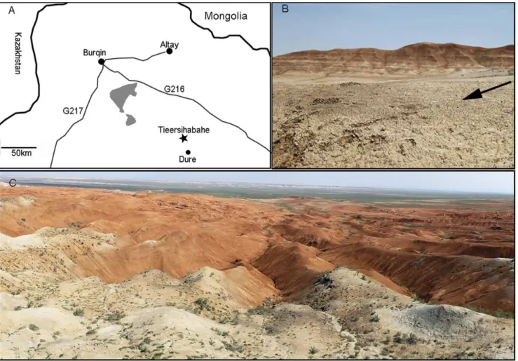
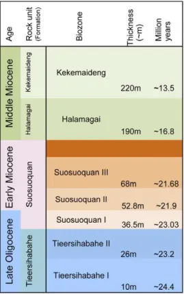
Tieersihabahe section in northern Junggar basin of China is renowned for its continuous sequences from the late Oligocene to middle Miocene that contains the Oligocene/Miocene boundary (Figure 1A–C). The fauna and stratigraphy have been widely studied by a number of researchers [8,9,10,11,12], and Meng et al. [13] presented a comprehensive biostratigraphy and magnetos-tratigraphy of the section and recognized five mammal assemblage zones for the lower Tieersihabahe and Suosuoquan formations.
The biozones are, in ascending order from the older to younger, Tieersihabahe faunal assemblage Zones I and II, Suosuoquan faunal assemblage Zones I, II, and III (Figure 2). The majority of specimens described here, collected during the 1998, 1999, 2000, 2002, and 2004 expeditions, were from the Tieersihabahe faunal assemblage Zone I (T-I) and were identified as Ansomyinae gen.et sp. nov in faunal lists by Ye et al. [10]. The age of the fauna is biostratigraphically and paleomagnetically correlative to the late Oligocene of 24.4,24.15 Ma [13]. The fossils were also found in 2000 from Saerduoyila, approximately 50 km northwest of Tieersihabahe section in the Halamagai area. The constitution of Saerduoyila fauna suggests that it was equivalent to the Tieersihabahe faunal assemblage Zone I.
Materials and Methods
Dental terminology used follows that of [14]. Teeth were measured using a Nikon SMZ 8 microscope set at 206 magnifi-cation; measurements were recorded to the nearest 0.01 mm. The SEM photographs of teeth were taken from uncoated specimens using a Hitachi SEM at the American Museum of Natural History.
Nomenclatural acts
The electronic edition of this article conforms to the require-ments of the amended International Code of Zoological Nomen-clature, and hence the new names contained herein are available under that Code from the electronic edition of this article. This published work and the nomenclatural acts it contains have been registered in ZooBank, the online registration system for the ICZN. The ZooBank LSIDs (Life Science Identifiers) can be resolved and the associated information viewed through any standard web browser by appending the LSID to the prefix ‘‘http://zoobank.org/’’. The LSID for this publication is: urn: lsid:zoobank.org:pub:7060CE3B-7EFB-4987-A8EE-50691F5D1F38. The electronic edition of this work was published in a journal with an ISSN, and has been archived and is available from the following digital repositories: PubMed Central, LOCKSS.
Results
Systematic paleontology
Order RODENTIA Bowdich, 1821
Family APLODONTIDAE Trouessart, 1897 Subfamily ANSOMYINAE Qiu, 1987
PROANSOMYS, gen. nov.
urn:lsid:zoobank.org:act:3CE4A9A6-380D-4E1E-A135-04A4BB3FF6C7
Type Species. Proansomys dureensis, sp. nov. (Figures 3–4) Diagnosis. Cheek teeth brachydont without accessory lo-phules; ectoloph on P4-M1/2 with a bucket-handled shape; mesostyle on P4-M1/2 single, not fully crested to close the central valley; metaconid anteroposteriorly compressed but with distinct cusp on lower molars; hypolophid attached to the hypoconulid rather than the ectolophid on p4, but complete and connected to the ectolophid on lower molars.
Differs from Ansomys in having a single mesostyle, a partly crested ectoloph, less developed parastyle on P4, a straight protoloph on most upper molars, weakly developed mesostylid on p4 and m1, an incomplete hypolophid on p4, a lower degree of lophodonty, and less complex tooth basins (lacking accessory lophules). Differs fromProsciurusin development of bucket-handle shaped ectoloph on P4 and M1/2, absence of the hypocone on upper cheek teeth, and presence of a complete hypolophid on lower molars. The cheek teeth of Proansomys are also generally more lophate than those ofProsciurus, but less lophate than those of
Ansomys.
Etymology. Pro-, Latin ‘‘before’’, implying the more primitive morphology of the new taxon in comparison withAnsomys. Figure 1. Location and overview of the Tieersihabahe locality.A, Location of the Tieersihabahe and Saerduoyila localities; B, close up of Tieersihabahe Formation where the fossils were found; C, Broad expanse of Tieersihabahe Section in the northern Junggar Basin. The black arrow in B above indicates the Tieersihabahe Formation.
Proansomys dureensissp. nov.
urn:lsid:zoobank.org:act:631ADBC0-C9D5-4727-A0A1-FA73E71C1CE1
(Figures 3–4; Table 1)
Type Specimen. IVPP V18534.5, left M1 or M2 (Figure 3B). Referred Specimens. Thirty-seven specimens from site XJ98023: IVPP V18533.1-3, 3 left P4; V18533.4, a right P4; V18533.5-6, 2 left M1/2; V18533.7-11, 5 right M1/2; V18533.12-16, 5 left M3; V18533.17-18, 2 right M3; V18533.19-20, 2 left dp4; V18533.21, a right dp4; V18533.22-23, 2 left p4; V18533.24-27, 4 right p4; V18533.28-29, 2 left m1; V18533.30, a left m2; V18533.31, a right m2; V18533.32-34, 3 left m3; V18533.35-37, 3 right m3. Twenty-seven specimens from site XJ98035: V18534.1; a right dP4; V18534.2; a left P4; V18534.3, a right P4; V18534.4-6, 3 left M1/2; V18534.7-9, 3 right M1/2; V18534.10-12, 3 right M3; V18534.13, a left dp4; V18534.14-15, 2 dp4; V18534.16-19, 4 left p4; V18534.20, a right fragmentary mandible with p4-m1; V18534.21, a right p4; V18534.22-23, 2 left m1; V18534.24-25, 2 left m2; V18534.26, a left m3; V18534.27, a right m3. Two specimens from site XJ20004: V18535.1, a left m2; V18535.2, a left m3. Three specimens from site XJ200208: V18536.1, a left dP4; V18536.2, a right dP4; V18536.3, a left p4. Nine specimens from site XJ200209: V18537.1, a left P4; V18537.2, a left M1/2; V18537.3-4, 2 right M1/2; V18537.5, a left M3; V18537.6, a right m1, V18537.7-8, 2 right m2; V18537.9, a right m3. One specimen from site XJ200207: V18538.1, a left fragmentary mandible with m2–m3.
Localities and age. XJ98023, XJ98035, XJ200208, XJ200209, and XJ20004, late Oligocene Tieersihabahe
Forma-tion; XJ200207, late Oligocene, base of the Suosuoquan Formation, northern Junggar Basin, China.
Etymology. dure, name of the town near the type locality. Diagnosis. As for the genus.
Description
All specimens are isolated brachydont teeth. At present, we prefer to interpret these specimens as from a single taxon due to small sample size, while acknowledging that some variations seen in the morphology of upper molars may indicate that more than one species is possibly present. In unworn specimens, major cusps are higher and cuspate with poorly developed lophs compared to those of Ansomys. The cheek teeth have smooth enamel basins, lacking accessory crests or lophules. Two small buccal roots and one strong lingual root support the upper cheek teeth. The lower molars have three roots, one under the trigonid and two under the talonid. The p4 has two roots in four specimens, and three roots in five specimens.
The P4 is triangular in outline (Figure 3A). The anterocone is large and widely separated from the protoloph by a broad valley. The parastyle and anterostyle are both present; the parastyle is much smaller than the anterocone, with the developed buccal cingulums; the anterostyle is a minute cuspule present just posterolingually at the base of the anterocone. The protocone is very prominent, with short, steep anterior and posterior arms. The conules are large relative to the buccal cusps, the protoconule being slightly larger than the metaconule. Both the protoloph and metaloph are low and zigzag shaped; the protoloph runs from the paracone to the protoconule, then bends posteriorly to converge with the metaloph buccal to the protocone. There is no hypocone. The paracone is larger than the metacone, both having a flat labial surface. The single mesostyle is connected with the paracone by a strong loph, but does not close the central valley between the metacone and the mesostyle. The ecotolph slightly bulges buccally, forming bucket-handle shaped crest.
The M1 and M2 are morphologically indistinguishable. They are sub-rectangular, much wider than long (Table 1). The anterior cingulum is strong, forming a high anterior edge of the tooth. The crescentic protocone is large with anterior and posterior crests, the crests continue with the anterior and posterior cingulums, respectively. The paracone, metacone, and metaconule are somewhat more buccolingually compressed than the counterparts of P4. The buccal surface of the paracone and metacone are flat. The protoloph is straight, running from the paracone toward the protocone, the protoconule is barely discernible and incorporated in the protoloph in eight out of thirteen specimens (Figure 3B); it is zigzagged with distinct protoconule in five out of thirteen specimens (Figure 3D). The metaloph runs from the metacone to metaconule, then bends anteriorly to converge with the protoloph just lingual to the protoconule. In ten M1/2s, the mesostyle is anteroposteriorly elongate, but not closing the central valley, whereas in three M1/2s, there is no distinct mesostyle.
The M3 is large relative to the preceding molars (Table 1). It is circular with slightly narrower posterior end (Figure 3C). Com-pared to M1/2, the anterior cingulum is more prominent. The posterior end of the tooth is reduced, the metacone is incorporated into the metaloph, and the metaconule is shaped as an anteroposteriorly elongate crest attached to the posterior margin. The ectoloph is not developed, and there is no mesostyle.
The DP4 is similar to P4 in morphology, but is smaller, lower-crowned, with less prominent cusps (Figure 3E). Unlike in P4, the metaloph is straight, running from the metacone toward the metaconule.
Figure 2. A summary of stratigraphic positions and age estimates for major mammal assemblages of Tieersihabahe Section in the northern Junggar Basin, China.
doi:10.1371/journal.pone.0052625.g002
The p4 is sub-triangular in outline with tapered anterior end (Figure 4B). The protoconid and metaconid are set close together, with the metaconid distinctly higher than the protoconid. The metaconid is slightly anterior to the protoconid and is separated from the latter by a narrow notch. No anteroconid is present but there is a small cuspule at the anterior corner of the tooth on two p4s. The metastylid crest is short or absent; when present, it occurs as the posterolingual slope of the metaconid. The small mesostylid is much lower than the metaconid, with no buccal crest. The ectolophid is low, extending from the protoconid to the hypoconid, and has a large triangular mesoconid with a short buccal crest. The entoconid is located slightly anterior to the hypoconid, from which the hypolophid first extends labially, and then bends posteriorly to the hypoconulid rather than the ectolophid. The hypoconulid is distinct and separated from the hypoconid by a V-shaped posterolophid.
The m1 was preservedin situin V18534.20. The posterior width of m1 is much greater than the anterior width, largely because of
the posterobucally protruding hypoconid (Figure 4C). The anterior cingulum is strong, forming the anterior margin of the tooth between the protoconid and the metaconid. The metaconid, just lingual to the midline, is a distinct anteroposteriorly compressed cusp in unworn specimens, and, with wear, it is incorporated with the anterior cingulum. The metalophid crest II extends only half the distance from the protoconid to the metaconid and is absent at the metaconid side. The metastylid crest is absent. A distinct mesostylid is present posterolingual to the metaconid with a short, low crest that extends only a short distance toward the center of the tooth. The mesoconid is located at the middle of the low ectolophid and lacks a buccal mesolophid or has a very short one. The hypolophid is complete, running from the entoconid to the ectolophid posterior to the mesoconid. The hypoconulid is prominent, connected with the hypolophid by an anteriorly directed loph.
The m2 is similar to m1 but the metaconid is shifted more lingually, making it wider anteriorly than m1 (Figure 4D). The Figure 3. Upper dentition of Proansomys dureensis, sp. nov. A, IVPP V18534.3, RP4; B, V18534.5, LM1/2; C, V18533.12, LM3; D, V18537.3, RM1/2; E, V 18536.1, LDP4.
metastylid crest is more developed and joins the mesostylid along the anterior lingual border. The mesostylid is pronounced, with a longer internal crest oriented transversely that ends at about half way across the tooth. The hypoconulid is separated from the hypolophid by a narrow transversely oriented valley.
The m3 is the longest of the lower cheek teeth (Table 1). The trigonid of m3 is much like that of m2, but the posterior end is rounded and narrower (Figure 4E). The hypoconid is placed right at the posterolabial corner of the tooth, not labially protruding, unlike m1 and m2, and the hypoconulid is reduced.
The dp4 is similar to p4 in morphology, but is slender and lower-crowned, with lower cusp height (Figure 4A). The entoconid is less defined, confluent with prominent hypoconulid at the base, making the talonid basin broader.
Phylogenetic analysis
To evaluate the phylogenetic position ofProansomys dureensis, we plot it to the character matrix of Hopkins [5] with addition ofA.
cyanotephrus [6]. Plesispermophilus atavus and P. angustidens were excluded from the analysis because they represent a different clade from the Ansomyinae (see discussion below). Additionally, A. cruciferandA. shantungensiswere excluded from the analysis because they are represented by only a single tooth, respectively.Proansomys dureensiswas coded as follows: 01111 10000 00000 11001 01110 00001 0. Heuristic searches in PAUP* version 4.0 were conducted with all characters under equal weight. 1000 replicates of random taxon addition result in three most parsimonious trees (MPTs) (Figure 5). The tree length is 42 steps, with the consistency index being 0.738 and the retention index 0.667. The strict consensus of the three most parsimonious trees is also illustrated in figure 5.
Proansomys dureensis occurs as the basal taxon of the Ansomyinae clade in all three MPTs.
Comparisons with species ofAnsomys
Ansomys shantungensisis known only from one isolated m1 [3], so that its comparison with our samples is difficult.Proansomys dureensis
Figure 4. Lower dentition ofProansomys dureensis, sp. nov. A, IVPP V18533.19, Ldp4; B, V18534.16, Lp4; C, V18534.22, Lm1; D, V18535.1, Lm2; E, V18534.27, Rm3.
doi:10.1371/journal.pone.0052625.g004
most closely resembles A. shantungensisin morphology of the m1. Both taxa have an anteroposteriorly compressed metaconid, an incomplete metalophulid II, a weak mesostylid, a posterolabially extended hypoconid, and a complete hypolophid on m1. However, though A. shantungensis has fewer crenulations than other species ofAnsomys, it has already begun to develop accessory lophules in the basins of the teeth.Proansomys dureensishas a smooth enamel basin that lacks accessory lophules.
Proansomys dureensisoverlaps with the upper end of A. orientalis
size range, being slightly larger [1]. They both have lophate dentition, the anterostyle is present on P4, the mesostyle is distinct, the metalophid crest II extends lingually to the center of the teeth, the metaconid is crestlike, and the hypolophid is complete on lower molars. However, the cheek teeth ofProansomys dureensisare more cuspate and less lophate. The occlusal pattern is very simple, lacking the complexity of accessory lophules present in Ansomys. The mesostyle on P4 and M1/2 is enlarged to form a bucket-handle shaped ectoloph as inA. orientalis but not fully crested to close the central valley. The protoloph runs straight from the paracone toward the protocone, and the protoconule is indistin-guishable as part of a continuous protoloph on most upper molars. In contrast, the protoloph of upper molars is zigzag-shaped and the protoconule is distinct inA. orientalis. The hypolophid on dp4 and p4 is incomplete and bends posteriorly to the hypoconulid, but is complete and extends transversely to the ectolophid in A. orientalis.The m3 is relatively anteroposteriorly longer than that of
A. orientalis.
Ansomys shanwangensiswas described from the middle Miocene Shanwang Formation of Shandong Province, based on one compressed skeleton [2]. Ansomys shanwangensis is comparable to
Proansomys dureensisin size, but can be easily distinguished from it.
Ansomys shanwangensis is unique in having double protolophules, strong development of the ectoloph, developed mesoloph, complexity of accessory lophules, strong development of
lopho-donty, transversely elongated mesostylids on p4 and m1, and prominent entoconid on m3.
Proansomys dureensis is distinct from all other known species of
Ansomysby its significantly larger size, a partly crested ectoloph, the absence of accessory lophules, lower degree of lophodonty, and the straight protoloph with crest-like protoconule on most upper molars.
Discussion
Proansomys dureensis is assigned to Ansomyinae because the ectoloph of upper cheek teeth, although not fully crested, has attained the bucket-handle shaped configuration that is the most important diagnostic character of the subfamily as its name implies. Other synapomorphies uniting Proansomys with the Ansomyinae are P4 anterostyle doubled, labial faces of the paracone and metacone flat, lingual crest of metaconule joining protoconule, hypocone absent, m2 metaconid anteroposteriorly compressed, and basal part of hypoconid posterolabially expand-ed. Cladistic analysis positsProansomysas the most basal ansomyine (Figure 5). It is more plesiomorphic than species of Ansomys in having a partly crested ectoloph on upper cheek teeth, less complex dental pattern without the accessory lophules, a lesser degree of lophodonty, metaconid on m1 lingually prominent, main cusps of lower teeth not anteroposteriorly compressed, and an incomplete hypolophid on p4. Therefore, we consider this justifies its status as a new genus.
The prosciurines have been traditionally considered a para-phyletic stem group of aplodontids that gave rise to all later aplodontids [15]. On the one hand,Proansomysstill retains many generalized prosciurine features, such as straight protoloph with barely discernible protoconule on molars, single mesostyle, poorly developed ectoloph, the hypolophid bending posteriorly to join with posterolophid on p4, and weakly developed mesostylid on p4 and m1. On the other hand, it displays the apomorphic state of other characters (the lophodonty of the cheek teeth and development of the ectoloph on upper molars), but only to a moderate degree compared to Ansomys so that Proansomys is morphologically intermediate betweenProsciurusandAnsomys. The combination of the primitive prosciurine and derived ansomyine characters in Proansomys suggests that the Ansomyinae derived from a prosciurine species, as did other advanced aplodontids.
Plesispermophilus was considered as the possible candidate for ancestral Ansomyinae [1,5]. However, the discovery ofProansomys
suggests that the Ansomyinae do not seem to have evolved from
Plesispermophilus because this new species, an evident stem Ansomyinae, is more primitive thanPlesispermophilus in having a not fully crested ectoloph and less complex tooth basin. Additionally,Proansomyslacks the buccal extension of mesoconid and the anterior extension of the hypoconid as in the species of
Ansomys. In contrast,Plesispermophilushas a fossetid that is united with the buccal extension of mesoconid and the anterior extension of the hypoconid. This feature may also indicate that the Ansomyinae and Plesispermophilus represent distinct clades from each other.
Ansomys shantungensiswas the oldest known Asian representative of the subfamily. It was recovered from a drill core in Shandong and was placed in the middle or late Oligocene on the basis of evolutionary stage as illustrated by North American and European prosciurines [3]. As mentioned above, it is most comparable to
Proansomysin morphology of the m1, but is slightly more derived. This further indicates thatA. shantungensismay be late Oligocene or early Miocene in age. In addition, Proansomys is 3,000 km
Table 1.Measurements in millimeters of the teeth of Proansomys dureensis, sp. nov.
Tooth Mean Range
dP4 anteroposterior length 1.83 1.73–2.00
transverse width 1.92 1.76–2.11
P4 anteroposterior length 2.09 1.85–2.28
transverse width 2.19 2.17–2.62
M1/2 anteroposterior length 1.75 1.51–1.95
transverse width 2.34 2.10–2.56
M3 anteroposterior length 1.99 1.89–2.11
transverse width 2.15 1.99–2.41
dp4 anteroposterior length 1.85 1.65–2.16
transverse width 1.54 1.41–1.66
p4 anteroposterior length 2.03 1.58–2.40
transverse width 2.04 1.63–2.30
m1 anteroposterior length 1.95 1.85–2.11
transverse width 1.92 1.73–2.23
m2 anteroposterior length 2.00 1.94–2.08
transverse width 2.00 1.80–2.16
m3 anteroposterior length 2.18 1.98–2.49
transverse width 1.78 1.65–2.03
northwest of A. shantungensis, suggesting that the subfamily was already geographically widespread during late Oligocene time.
Two alternate hypotheses have been proposed for the paleobiogeography of theAnsomys.Whereas Hopkins [5] proposed
a European origin and a series of migrations between Europe, Asia, and North America, Korth [6] proposed an origin in North America and a single migration to Eurasia in the early Miocene. Now, recognition of the lineage (Prosciurus-Proansomys-Ansomys) Figure 5. Three most parsimonious trees (MPTs 1–3) and their strict consensus tree.
doi:10.1371/journal.pone.0052625.g005
provides new insights into the early history of the ansomyines. Although the earliest record of ansomyines wasA. cyanotephrusfrom the early late Oligocene of North America [6],Proansomys is the most primitive member of the subfamily and appears derived from
Prosciurus-like forms. Given thatProsciuruswas common in the early Oligocene in Mongolia [16], the ansomyines may have originated in Asia some time prior to the late Oligocene, then dispersed throughout Eurasia and North America. The Ansomyinae may have arrived in North America prior to the early late Oligocene becauseA. cyanotephruswas known from the early late Oligocene of South Dakota. Once again, more complete fossil discoveries will be necessary to make reliable inferences about the ansomyine paleobiogeographical relations.
Acknowledgments
We thank Zhuding Qiu for providing access to his collection; Jianfeng Su, Shaoyuan Wu, and Xinwei Zheng for field assistance. We thank Lawrence J. Flynn and two anonymous referees for their comments which helped improve the manuscript.
Author Contributions
Conceived and designed the experiments: SB JM. Performed the experiments: SB JM SM WW. Analyzed the data: SB SM JM WW. Contributed reagents/materials/analysis tools: SM XN JY. Wrote the paper: SB.
References
1. Qiu Z (1987) The Aragonian vertebrate fauna of Xiacaowan, Jiangsu;7, Aplodontidae (Rodentia, Mammalia). Vertebrata PalAsiatica 25: 283–296. 2. Qiu Z, Sun B (1988) New fossil micromammals from Shanwang, Shandong.
Vertebrata PalAsiatica 26: 50–58.
3. Rensberger JM, Li CK (1986) A New Prosciurine Rodent from Shantung Province, China. Journal of Paleontology 60: 763–771.
4. Lopatin AV (1997) The first find of Ansomys (Aplodontidae, Rodentia, Mammalia) in the Miocene of Kazakhstan. Palaeontological Journal 31: 667– 670.
5. Hopkins SSB (2004) Phylogeny and biogeography of the genusAnsomysQiu, 1987 (Mammalia: Rodentia: Aplodontidae) and description of a new species from the Barstovian (Mid-Miocene) of Montana. Journal of Paleontology 78: 731–740.
6. Korth WW (2007) A new species ofAnsomys(Rodentia, Aplodontidae) from the late Oligocene (latest Whitney-earliest Arikareean) of South Dakota. Journal of Vertebrate Paleontology 27: 740–743.
7. Kelly TS, Korth WW (2005) A new species ofAnsomys(Rodentia; Aplodontidae) from the late Hemingfordian (early Miocene) of northwestern Nevada. Paludicola 5: 85–91.
8. Tong Y, Qi T, Ye J, Meng J, Ya D (1990) Tertiary stratigraphy of the north of Junggar Basin, Xinjiang. Vertebrata PalAsiatica 28: 59–70.
9. Ye J, Wu W, Meng J (2001a) Tertiary stratigraphy in the Ulungur River Area of the Northern Junggar basin of Xinjiang. Journal of Stratigraphy 25: 193–200. 10. Ye J, Wu W, Meng J (2003) Oligocene/Miocene Beds and Faunas from
Tieersihabahe in the Northern Junggar Basin of Xinjiang. Bulletin of the American Meseum of Natural History 279: 568–585.
11. Ye J, Wu W, Meng J (2001b) The age of Tertiary strata and mammal faunas in Ulungur River Area of Xinjiang. Journal of Stratigraphy 25: 283–287. 12. Sun J, Ye J, Wu W, Ni X, Bi S, et al. (2010) Late Oligocene–Miocene
mid-latitude aridification and wind patterns in the Asian interior. Geology 38: 515– 518.
13. Meng J, Ye J, Wu W, Yue L, Ni X (2006) A recommended boundary stratotype section for Xiejian Stage from northern Junggur Basin: implications to related bio-chronostratigraphy and environmental changes. Vertebrata PalAsiatica 44: 205–236.
14. Hopkins SSB (2008) Phylogeny and evolutionary history of the Aplodontoidea (Mammalia: Rodentia). Zoological Journal of the Linnean Society 153: 769–838. 15. Korth WW (1989) Aplodontid rodents (Mammalia) from the Oligocene (Orellan and Whitneyan) Brule Formation, Nebraska. Journal of Vertebrate Paleontology 9: 400–414.