Density
Ibiara Correia de Lima Almeida Paz
Departamento de Produção e Exploração Animal - Faculdade de Medicina Veterinária e Zootecnia, UNESP
CP 560
18.618-000 - Botucatu, SP, Brazil. Telephone: +55 +14 3811 7189 Fax: +55 +14 3811 7185
E-mail: [email protected] [email protected] Mail Address
Keywords
bone, bone mineral density, optical radiographic densitometry, tibial dyschondroplasia Almeida Paz ICL1
Mendes AA2
Takita TS3
Vulcano LC3
Guerra PC4
Wescheler FS2
Garcia RG1
1 Graduate student in Animal Science,
Programa de Pós-Graduação em Zootenica, Faculdade de Medicina Veterinária e Zootecnia, UNESP - Botucatu, SP, Brazil.
2 Professor, Departamento de Produção e
Exploração Animal, Faculdade de Medicina Veterinária e Zootecnia, UNESP - Botucatu, SP, Brazil.
3 Professor, Departamento de Reprodução e
Radiologia Veterinária, Faculdade de Medicina Veterinária e Zootecnia, UNESP -Botucatu, SP, Brazil.
4 Graduate student in Animal Science,
Faculdade de Medicina Veterinária e Zootecnia, UNESP - Botucatu, SP, Brazil. Author(s)
Arrived: february / 2004 Approved: october / 2004
ABSTRACT
This study was carried out at Faculdade de Medicina Veterinária e Zootecnia, UNESP, Botucatu-SP, Brazil. The aim was to establish the normal values of bone mineral density (BMD) expressed in millimeters of aluminum in the tibia of broiler chickens using optical densitometry of radiographs. Four hundred Cobb male chicks were reared from 1 to 40 days of age, when 40 of them were selected and the right femur-tibia articulation was radiographed. Radiographs were taken with the X-ray equipment calibrated for 45 kvp and 3.2 mAs and a focus-to-film distance of 90 cm. An aluminum phantom ASTM-6063 consisting of 20 ladder steps with graduate density was placed parallel to the area to be radiographed and used as a densitometry reference standard. Radiograph images were analyzed using the software ATHENA SIA. The proximal growth plate of the right tibia epiphysis was used as the standard reading region. The inclination axis of the reading window was 0 and the window was 10 mm high and 40-45 mm wide, depending on the bone size. Optical densitometry values of the radiographs ranged from 1.46 to 1.77 mmAl, and the coefficient of variation was 9.93%. It was concluded that densitometry values beyond the range established in the present study might indicate the presence of bone alteration in the tibia of broilers.
INTRODUCTION
Tibial dyschondroplasia is a disease that affects birds with accelerated growth. The condition is characterized by the presence of an unmineralized cartilage mass that extends distally from the growth plate of the metaphysis at the proximal end of the tibia and, occasionally, to the distal end of the tibia, proximal end of the tarsometatarsus, and proximal ends of femur and humerus, which results in bone deformity and lameness (Mendonça Jr., 1990, Gonzales & Macari, 2000). Histologically, dyschondroplasia is identified as a mass of avascular cartilage with excess of hypertrophic chondrocytes, which results probably from the inability of maturing chondrocytes to differentiate completely (Poulos et al., 1978; Berry et al., 1996).
Bone resorption takes place in regions where osteoclasts differentiate from the monocyte-macrophage system under the influence of some agents; mobile and mononucleated pre-osteoclasts are separated from the bone resorption area by the remaining osteoclasts. Osteoblasts probably do not initiate a pathological process of bone resorption; rather, it is induced by prostaglandins and cytokines, such as the osteoclast activation factor that is released by inflammatory cells, mainly macrophages. Alterations on the bone matrix and on the bone mineral due to injury are a result from changes in the activity of osteoblasts, osteoclasts and osteocytes. Altered cell activity may result in atrophy,
Acknowledgements
cell degeneration and death, leading to hyperplasia, hypertrophy or unbalance between bone synthesis and resorption. Necrosis is associated to inflammatory processes and can result in compression caused by the inflammatory exudate. During such stage of the repairing process of necrosis, the bone may seem radiodense, probably due to deposition of new bone tissue on the necrotic end and mineralization of the dead osteocytes (Pharr & Bargai, 1997; Cruess & Dumont, 1985).
Some techniques are available for the characterization and assessment of such alterations. Evaluation of tibial dyschondroplasia in dead birds is generally based in the histopathological examination of the cartilage. In live birds, dyschondroplasia may be identified by radiography after the second week of age (Cruickshank & Sim, 1986; Lynch et al., 1992). Lesions generally recede after the fourth or fifth weeks of age. Bone mineral density (BMD) is a biophysical parameter with great experimental and clinical importance that might help to better understand and evaluate the process of bone mineral deposition (Louzada, 1994). The technique of optical densitometry using radiographs may be used for the sequential analysis of the alterations that occur in the bone tissue and is more precise than other techniques. Nevertheless, the technique is not widely accepted because standardization is difficult (Louzada, 1997). Moreover, few animal species have standards of bone mineral density already assessed and defined by optical densitometry using radiographs. Therefore, the standardization of this technique is imperative and animal science professionals would largely benefit from it.
MATERIAL AND METHODS
The field experiment was conducted at the experimental poultry houses from Faculdade de Medicina Veterinária e Zootecnia, UNESP (FMVZ-UNESP), Botucatu, Brazil. Four hundred Cobb male chicks with one day of age were reared at a density of 10 birds/m2 in an open-sided poultry house with wood
shavings as litter material. Water and food were given ad libitum throughout the experimental period, which was divided in initial phase (1 to 14 days of age) and final phase (15 to 40 days of age).
Optical Densitometry of Radiographs
The literature presents no available data of a reliable method for measuring the bone mineral density on the proximal tibia epiphysis in broiler chickens. Therefore,
a technique was standardized based on the method of optical densitometry using radiographs. The technique was assessed concerning the following parameters: the reading window used to take the X-rays, animal positioning, and shape and anatomic region to be used in the analysis of the radiography.
Forty birds were selected and taken to the Veterinary Hospital in FMVZ-UNESP-Botucatu. The proximal end of the tibia and the distal end of the femur were radiographed using a green X-ray film placed in an 18x24-cm cassette equipped with rare earth screens. All radiographs were taken using X-ray films from the same lot number. The animals were positioned in ventral decubitus and the right leg was stretched onto the cassette for an anteroposterior projection. Routine radiological procedures were used and film development and fixation were carried out in a standard automatic processing apparatus.
The most appropriate radiographic technique was achieved with the X-ray apparatus placed at a focus-to-film distance of 90 cm and calibrated for 45 kVp and 3.2 mAs. A calibration aluminum wedge was placed onto the cassette, 3.0 cm from the region that would be radiographed and parallel to it, to be used as a densitometry reference standard. The wedge was comprised of 20 squares with 15 x 15 mm and increasing thickness. The first step was 0.5 mm thick; thickness increased 0.5 mm up to the 10th step and 1.0 mm from
the 11th to the 20th step.
The readings of bone mineral density (BMD) of the tibia proximal end were made with a HP Scanjet 6C scanner (Hewlett Packard, 2001) and were expressed in mm Aluminium (mm Al). The scanner was connected to a HP Scanjet 6C overhead adaptor. Radiographic images were digitalized with the software HP Deskscan and saved in files for further analysis (Leal, 2002).
The stored images were recaptured using the software developed by ATHENA SIA and the most suitable region was selected for the study. After the readings of the study region and the phantom were performed, optical density values were expressed by the software in a grayscale from 1 to 256. Based on the means of gray level obtained for the phantom (in mm Al), it was then possible to establish the mean gray levels of the study region, i.e., the proximal end of the tibia.
Figure 1 Radiograph of the right tibia from a broiler chicken at 39 days of age.
Figure 2 Region of the tibia selected for analysis.
mm according to bone size. Besides, a perfect visualization of the fibula was considered as the horizontal standard for fine adjustments of the reading area.
The best reading window height was established in a series of tests made using three different heights: 30mm, 15mm and 10mm. Imprecise values were obtained in the larger reading windows, i.e., higher than 10 mm, and it was not possible to establish a correlation between the readings and tibial dyschondroplasia.
For each height of the reading window that was evaluated, the Spearmans rank correlation coefficient was calculated as the ratio between the readings of bone mineral density (BMD) and the histological score. The significance of the correlation coefficients was evaluated at p<0.01. The coefficient of correlation between each of the three window sizes and the bone mineral density was separated by the method of Steiger (1980), considering the absolute values of the correlation coefficients. For each histological score, a range of bone mineral density values was calculated. Figures 1, 2, 3, 4 and 5 show the procedures used for BMD readings.
BMD was evaluated in regions where the dyschondroplastic cartilage was already established and in regions where the lesion was in development. Therefore, images were selected so that the bones would show visible lesions of tibial dyschondroplasia in the radiography. Such evaluations were performed using the standards established for the reading window and for the inclination axis of the reading window (10 mm and zero, respectively). Nevertheless, the width of the reading window was changed so that assessment would be made first in the region with the already established dyschondroplastic cartilage, and then in the region where the dyschondroplastic cartilage was developing (boundaries of the lesion). The mean values obtained for each region were compared by Tukeys test at 1% of significance.
Histopathological evaluation
After the birds were slaughtered, the sampled tibias were dissected and decalcified for 16 days with 3% nitric acid and 14% formalin. Decalcifying solution was substituted at each 2 days. The material was then dehydrated in a series of alcohols with increasing concentration and placed in xylol. Fragments were embedded in paraffin and 7ìm cuts were made. The histological cuts were placed onto glass slides and stained with hematoxylin-eosin (HE).
RESULTS AND DISCUSSION
The 10mm-high reading window resulted in the best correlation between the histological lesion score and BMD values expressed in mm Al when compared to the techniques that used greater heights of the reading
Figure 3 Positioning for the readings and delimitation of the reading window.
Figure 4 - Fine adjustment of the reading window using the fibula as the horizontal standard.
Figure 5 Image generated by the software ATHENA-SIA for calculation of bone mineral density.
The present study permitted to establish confidence intervals for BMD values as a function of the histological scores of tibial dyschondroplasia in male broilers at 40 days of age. BMD values in normal bones ranged between 1.46 and 1.77 mm Al, and higher values were established for bones with lesions. Louzada (1997) have reported higher mean BMD values (1.77-1.96 mmAl) for the central region of the tibia diaphysis in broiler window. The method that used the 10mm-reading window was considered as the most indicated for the analysis of bone mineral density in millimeters of Aluminum (mm Al) in broiler chickens. On the other hand, the results of bone mineral density given by the techniques with reading windows with 15 mm and 30 mm were not consistent because there was a selection of areas that were areas of bone marrow or without lesions. The correlations between histological lesion score and BMD obtained for each reading window height are shown in Table 1.
Table 1 Spearmans rank correlation coefficients between the histological score and bone mineral density (BMD) values using three different heights of reading window.
BMD BMD BMD
Window Height 10mm 15mm 30mm
by bone sclerosis and causes an increase in the mean BMD of the evaluated bone structure (Table3). According to Pharr & Bargai (1997) and Cruess & Dumont (1985), bone regions that have lesions might seem denser in radiographs during deposition of new bone tissue, or yet, due to the mineralization of the dead osteocytes, and consequently show elevate BMD readings.
Concluding, it was possible to establish ranges of BMD values for the tibia in live broiler chickens as a function of the histological scores of tibial dyschondroplasia by the technique of optical densitometry of radiographs when a reading window with 10 mm of height was used. BMD values increased gradually with higher histological scores of the bones, as a result of the higher density values detected in the boundaries of the dyschondroplastic cartilage.
REFERENCES
Berry JL, Farquharson C, Whitehead CC, Mawer EB. Growth plate chondrocyte vitamin D receptor number and affinity are reduced in avian tibial dyschondroplastic lesions. Bone 1996; 19 (2):197-203.
Cruess RL, Dumont J. Healing of bone. In: Newton CD, Nunamaker DM, editor. Textbook of small animal orthopaedics. Philadelphia: J.B. Lippincott Company; 1985. p. 35-63.
Cruickshank J, Sim J. Morphometric and radiographic characterization of tibial bone of broiler chickens with twisted leg disorders. Avian Diseases 1986; 30:699-708.
Gonzales E, Macari M. Doenças das aves. Campinas (SP): FACTA; 2000.
Hewlett Packard. Scanjet 6C HP, com adaptador para transparência Scanjet 6C HP; 2001.
Leal ACR. Determinação dos valores normais da densidade mineral óssea (dmo) da extremidade distal do rádio em cães por meio da técnica de densitometria óptica em imagens radiográficas: correlação entre o peso, sexo e idade. [MS Thesis]. Botucatu (SP):Universidade Estadual Paulista; 2002.
Louzada MJQ. Otimização da técnica de densitometria óptica em imagens radiográficas de peças ósseas. Estudo in Vitro. [Phd chickens; in that study, however, birds were 53 days
old and a Macbeth TP528 densitometer was used to do the measurements.
Bones showing histological lesion score of 1, i.e., with minor lesions, presented BMD values between 1.80 and 2.31 mmAl. BMD values ranging from 2.34 and 2.91 mmAl were established for bones with histological score 2, whereas values ranging from 2.96 to 3.23 mmAl were associated with bones showing severe lesions (score 3). Histological scores of tibial dyschondroplasia lesions and the respective confidence intervals for tibia BMD values in broiler chickens are summarized in Table 2.
Table 2 Histological score of tibial dyschondroplasia lesions and the respective ranges of bone mineral density (BMD, mm Al).
Histological Score BMD (mmAl)
0 1.46 1.77 (± 0.03)A* 1 1.80 2.31 (± 0.04)B 2 2.34 2.91 (± 0.07)C 3 2.96 3.32 (± 0.05)D * Values followed by different letters are statistically different (p<0.05).
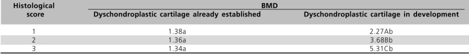
A closer analysis of the BMD readings in the regions with already established dyschondroplastic cartilage or regions in which the dyschondroplastic lesion was developing demonstrated that the increased BMD mean values in injured bones result from the higher values that are seen in the lesion regions bounded by bone sclerosis, since the established dyschondroplastic cartilage shows fairly small BMD values. It is worth noting that the technique used in the present study proposes the reading of the entire bone epiphysis region, because the localization of the lesion is not easily determined in some images and/or lesion scores. Besides, the analysis of the whole epiphysis permits to obtain BMD values that are calculated as the mean values of the regions with implanted cartilage and of the boundaries of the lesion, which is characterized
Table 3 Bone mineral density (BMD) in the proximal epiphysis of tibias with dyschondroplastic cartilage already established or in development.
Histological BMD
score Dyschondroplastic cartilage already established Dyschondroplastic cartilage in development
1 1.38a 2.27Ab
2 1.36a 3.68Bb
3 1.34a 5.31Cb
Thesis] Campinas (SP):Universidade Estadual de Campinas; 1994.
Louzada MJQ. Densidade de peças ósseas de frangos. Estudo pela densitometria óptica radiográfica. Veterinária e Zootecnia 1997; 9:95-109.
Lynch M, Thorp BH, Whitehead CC. Avian tibial dyschondroplasia as a cause of bone deformity. Avian Pathology 1992; 21:275-285.
Mendonça Jr CX. Deformidades das pernas em frangos de corte. In: Conferência Apinco de Ciência e Tecnologia Avícola; 1990; Campinas; São Paulo. Brasil. Campinas: APINCO; 1990. p.29-41.
Pharr JW, Bargai U. Radiology. In: Greenough PR, Weaver AD, editors. Lameness in cattle 3th ed. Philadelphia:W.B. Saunders Company; 1997. p. 24-40.
Poulos PW, Reiland S, Elwinger K. Skeletal lesions in the broiler, with special reference to dyschondroplasia (osteochondrosis). Acta Radiologica 1978; 358 Suppl:229-75.
Steiger JH. Tests for comparing elements of correlation matrix. Psychological Bulletin 1980; 87:245-251.
Takita TS. Efeito do genótipo, do ambiente e da interação genótipo
x ambiente na incidência de discondroplasia tibial em frangos de